Acute effects of physiotherapeutic respiratory
man-euvers in critically ill patients with craniocerebral
trauma
Manoel Luiz de Cerqueira Neto,IA´lvaro Vieira Moura,ITelma Cristina Fontes Cerqueira,II Esperidia˜o Elias Aquim,IIIA´lvaro Rea´-Neto,IVMirella Cristine Oliveira,VWalderi Monteiro da Silva Ju´nior,VIValter J. Santana-Filho,VIRosana Herminia ScolaVII
IUniversidade Federal do Parana´, Department of Internal Medicine, Curitiba/PR, Brazil.IIUniversidade Tiradentes, Departamento de Fisioterapia, Aracaju´/ SE, Brazil.IIIFaculdade Inspirar, Curitiba/PR, Brazil.IVUniversidade Federal do Parana´, Centro de Estudos e Pesquisa em Terapia Intensiva, Curitiba/PR, Brazil.VHospital do Trabalhador, Curitiba/PR, Brazil.VIUniversidade Federal do Sergipe, Departamento de Fisioterapia, Aracaju´/SE, Brazil.VIIUniversidade Federal do Parana´, Hospital de Clı´nicas, Division of Neurology/Neuromuscular Diseases, Curitiba/PR, Brazil.
OBJECTIVE:To evaluate the effects of physiotherapeutic respiratory maneuvers on cerebral and cardiovascular hemodynamics and blood gas variables.
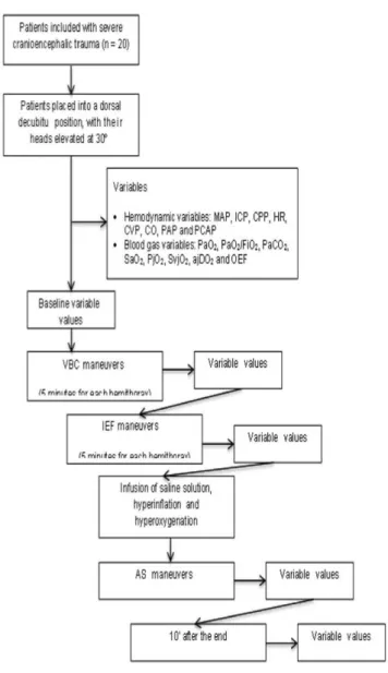
METHOD:A descriptive, longitudinal, prospective, nonrandomized clinical trial that included 20 critical patients with severe craniocerebral trauma who were receiving mechanical ventilation and who were admitted to the intensive care unit. Each patient was subjected to the physiotherapeutic maneuvers of vibrocompression and increased manual expiratory flow (5 minutes on each hemithorax), along with subsequent airway suctioning with prior instillation of saline solution, hyperinflation and hyperoxygenation. Variables related to cardiovascular and cerebral hemodynamics and blood gas variables were recorded after each vibrocompression, increased manual expiratory flow and airway suctioning maneuver and 10 minutes after the end of airway suctioning.
RESULTS:The hemodynamic and blood gas variables were maintained during vibrocompression and increased manual expiratory flow maneuvers; however, there were increases in mean arterial pressure, intracranial pressure, heart rate, pulmonary arterial pressure and pulmonary capillary pressure during airway suctioning. All of the values returned to baseline 10 minutes after the end of airway suctioning.
CONCLUSION: Respiratory physiotherapy can be safely performed on patients with severe craniocerebral trauma. Additional caution must be taken when performing airway suctioning because this technique alters cerebral and cardiovascular hemodynamics, even in sedated and paralyzed patients.
KEYWORDS: Physical Therapy Modalities; Craniocerebral Trauma; Intensive Care.
TRIAL REGISTRATION: This trial was not registered because enrollment began prior to July 1, 2005.
Neto ML, Moura AV, Cerqueira TC, Aquim EE, Rea´-Neto A´ , Oliveira MC, et al. Acute effects of physiotherapeutic respiratory maneuvers in critically ill patients with craniocerebral trauma. Clinics. 2013;68(9):1210-1214.
Received for publication onJanuary 29, 2013;First review completed onMarch 5, 2013;Accepted for publication onApril 22, 2013 E-mail: [email protected]
Tel.: 55 79 3217–5928
& INTRODUCTION
Physiotherapeutic interventions during motor and cardi-orespiratory care are considered to be important compo-nents in the management of patients admitted into intensive
care units (1–3). These interventions offer short- and medium-term benefits for the patients’ recovery (4).
Particularly in patients with severe craniocerebral trauma, the application of different physiotherapeutic interventions in the intensive care unit, whether singly or in combination, results in adaptations in various systems that can lead to clinically significant changes in respiratory function and cardiovascular and cerebral hemodynamics (3,5,6). The continuous monitoring of hemodynamic and respiratory parameters in critical patients provides additional safety while performing therapy and during patient follow-up, rendering therapy safer and more effective and thereby reducing the factors that can potentially contribute to adverse events (6).
Copyrightß2013CLINICS– This is an Open Access article distributed under the terms of the Creative Commons Attribution Non-Commercial License (http:// creativecommons.org/licenses/by-nc/3.0/) which permits unrestricted non-commercial use, distribution, and reproduction in any medium, provided the original work is properly cited.
No potential conflict of interest was reported.
Therefore, this study focused on assessing the effects of the physiotherapeutic respiratory maneuvers of vibrocompression (VBC), increased expiratory flow manual (IEFM) and airway suctioning (AS) on hemodynamic variables (mean arterial pressure [MAP], intracranial pres-sure [ICP], cerebral perfusion prespres-sure [CPP], heart rate [HR], central venous pressure [CVP], cardiac output [CO], pulmonary artery pressure [PAP] and pulmonary capillary pressure [PCAP]) and on blood gas variables (arterial oxygen tension [PaO2], the ratio of the arterial oxygen
tension to the fractional inspired oxygen [PaO2/FiO2],
arterial carbon dioxide tension [PaCO2], arterial saturation
[SaO2], jugular oxygen tension [PjO2], jugular bulb venous
oxygen saturation [SvjO2], the arterio-jugular difference in
the oxygen content [AJDO2] and cerebral extraction of
oxygen [CEO2]) in patients with severe craniocerebral
trauma admitted into the intensive care unit.
& MATERIALS AND METHODS
Design
A descriptive, longitudinal, prospective, nonrandomized clinical trial was quantitatively conducted and was approved by the research and ethics committee of the Hospital de Clı´nicas at the Federal University of Parana´ (protocol number 065EXT019/2002-11), and family mem-bers of the subjects provided informed consent.
Participants, therapists and centers
The study included consecutive male and female adult patients with severe craniocerebral trauma (Glasgow Coma Scale#8), who were selected within the first 48 hours after they were admitted into the intensive care unit. The patients included in the study presented no radiological alterations in their lungs, were sedated with propofol (Ramsay scale of 6), were paralyzed with pancuronium bromide, had their cough reflexes inhibited, were intubated and received mechanical ventilation in the assisted/controlled volume control ventilation mode (which was estimated at 8 ml/kg of body weight) with a positive end-expiratory pressure of 5 mmHg and an inspired oxygen fraction of 40%. Patients presenting any of the following criteria were excluded from the study: hemodynamic instability with a MAP of less than 60 mmHg; a CPP of less than 50 mmHg; pulmonary contusions; fractured ribs; or an undrained pneumothorax.
Intervention and outcome measurements
The patients were subjected to intra-arterial catheteriza-tion for MAP monitoring; arterial blood sample colleccatheteriza-tion for blood gas analysis (PaO2, PaO2/FiO2, PaCO2, SaO2);
intraventricular catheterization to measure the ICP and calculate the CPP; and pulmonary artery catheterization (Swan-Ganz) to monitor the CVP, CO, PAP and PCAP. All of the catheters were connected to a multi-parameter monitor, through which the HR was also monitored. A jugular-bulb catheter was also placed to collect venous blood samples for blood gas analysis, to assess PjO2 and
SvjO2and to calculate AJDO2and CEO2. Each patient was
then placed into a dorsal decubitus position, with his or her head elevated at 30
˚
, and all of the variables were assessed to establish baseline values. The physiotherapeutic maneu-vers of VBC and IEFM (5 minutes for each hemithorax) were subsequently applied for 10 minutes each. VBC was performed first to modify the physical properties of themucus, resulting in reduced viscosity due to the thixotropy provided by this maneuver. The IEFM maneuvers followed and were responsible for mobilizing, moving and eliminat-ing secretions from the periphery toward the trachea via increased expiratory flow. AS was then performed but was preceded by a 5-ml instillation of saline solution (0.9%) and three hyperinflations and hyperoxygenations using a manual resuscitator connected to an O2 source (flow of
10 l/s). A disposable, sterile probe with a valve was used for 10 to 15 seconds during this procedure, which was only performed once. To avoid cumulative effects, an interval of 5 minutes was established between physiotherapeutic maneuvers (7). Figure 1 provides design and flow of the participants through the trial.
Data analysis
The data were analyzed using statistical analysis software (Statistica). When comparing the procedures across time points, the null hypothesis of equal means at all of the time points was testedvs. the alternative hypothesis of at least
one time point with a different mean. Repeated measures
analysis of variance was used for this analysis. If the null hypothesis was rejected, the time points were subjected to pair-wise comparisons using the Newman-Keuls test. When comparing only two time points, Student’s t-test for analyzing dependent variables was used. The normality of the data was assessed using the Kolmogorov-Smirnov test. Statistical significance was defined asp,0.05.
& RESULTS
Twenty patients were included in the study, including 18 men and two women with a mean age of 33.5¡11.9 years
old, an average Apache score of 26¡4 and a mean Ramsay score of 6.
The VBC and IEFM maneuvers did not produce any significant changes in the cerebral hemodynamic variables of MAP, ICP or CPP compared to their values before and after these maneuvers. However, significant variations in the MAP and ICP values were identified immediately after the AS maneuver (see Table 1). There were no statistically significant differences in MAP (p= 0.85) or ICP (p= 1.00) before AS or 10
minutes after the final procedure, indicating that these variables had returned to baseline. The cerebral perfusion pressure remained unchanged throughout the protocol.
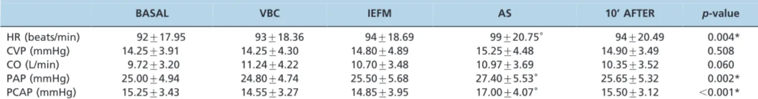
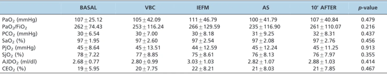
Table 2 presents the data on the behavior of the cardiovascular hemodynamic variables at baseline; after performing the VBC, IEFM and AS maneuvers; and 10 minutes after the end of the protocol. Statistically significant changes were specifically identified for the HR, PAP and PCAP variables when comparing the AS, VBC and IEFM values to baseline, and the variables returned to baseline 10 minutes after the end of AS. No significant differences were observed for arterial or jugular bulb blood gas variables at any time during the protocol (see Table 3).
& DISCUSSION
The results indicate that physiotherapeutic maneuvers do not alter cardiovascular hemodynamics. Rib cage compres-sion can increase intrathoracic pressure, which increases the pressure in the right ventricle, reducing the venous return
(7). However, no increases were noted in pressure (CVP, PAP and PCAP) in this study, suggesting that venous return and cardiac preload and afterload were maintained throughout the VBC and IEFM maneuvers. Discrepancies between studies could be related to differences in the rate of vibration and the force applied during the maneuvers, among other features (8,6).
In addition, in our study, all of the patients were sedated and paralyzed, which might explain the absence of hemodynamic effects. Another possible difference might be related to the lack of change in decubitus, the evaluation of the maneuvers in isolation and the prior analysis of the variables before suctioning, which did not occur in the previous studies.
Regarding blood gas variables, several studies have shown that VBC and IEFM maneuvers, alone and in combination with endotracheal suctioning, did not alter the oxygenation or ventilation of patients (9–11), corrobor-ating our data.
Regarding cerebral variables following VBC and IEFM maneuvers alone, studies have shown that percussion and vibration do not adversely affect ICP, and manual rib-cage vibration and manual expiratory pressure maneuvers do not alter the MAP, ICP or CPP of patients with severe craniocerebral trauma (7,12). Thus, our study confirms the safety of applying physiotherapeutic respiratory maneuvers in patients with severe craniocerebral trauma, with no significant effects on MAP, ICP, CPP, PjO2, SvjO2, AJDO2or
CEO2, thereby demonstrating that there were no signs of
cerebral ischemia or hyperemia during these procedures. The significant increases in the ICP and MAP during AS could be explained by two factors. First, carinal stimulation by the AS catheter promotes vagal stimulation, which results in cerebral vasodilation and a consequent increase in ICP. Second, given the absence of analgesics, the likely presence of pain during the procedure might have con-tributed to the ICP elevation in this study due to an increase in sympathetic activity with a reduction in cerebral blood flow, resulting in changes in CPP (13). A third, less likely, factor is related to hyperinflation and hyperoxygenation
Table 1 -The results of the cerebral hemodynamic variables, reported as means and standard deviations.
VBC IEFM AS
Before After p Before After p Before After p-value
MAP (mmHg) 94.00¡18.78 93.00¡17.54 0.451 94.70¡18.17 96.65¡21.61 0.209 94.25¡18.40 100.65¡22.19 0.011* ICP (mmHg) 16.95¡7.25 16.80¡7.68 0.877 17.75¡7.78 18.80¡7.35 0.107 19.65¡8.24 26.35¡12.82 ,0.001* CPP (mmHg) 76.90¡22.95 76.15¡22.38 0.475 76.90¡22.95 77.65¡25.80 0.634 74.60¡21.97 75.15¡28.77 0.833
Before = pre-maneuver; After = post-maneuver; MAP = mean arterial pressure; ICP = intracranial pressure; CPP = cerebral perfusion pressure; VBC = vibrocompression; IEFM = increased expiratory flow manual; AS = aspiration; Student’s t test, * =p,0.05.
Table 2 -The results of the cardiovascular hemodynamic variables, reported as means and standard deviations.
BASAL VBC IEFM AS 10’ AFTER p-value
HR (beats/min) 92¡17.95 93¡18.36 94¡18.69 99¡20.75˚ 94¡20.49 0.004*
CVP (mmHg) 14.25¡3.91 14.25¡4.30 14.80¡4.89 15.25¡4.48 14.90¡3.49 0.508
CO (L/min) 9.72¡3.20 11.24¡4.22 10.70¡3.48 10.97¡3.69 10.35¡3.52 0.060
PAP (mmHg) 25.00¡4.94 24.80¡4.74 25.50¡5.68 27.40¡5.53˚ 25.65¡5.32 0.002*
PCAP (mmHg) 15.25¡3.43 14.55¡3.27 14.85¡3.95 17.00¡4.07˚ 15.50¡3.12 ,0.001*
prior to AS, which induce increases in intrathoracic pressure, impair cerebral venous return and thus elevate ICP. However, the CVP and CO values were maintained, suggesting that the intrathoracic pressure did not generate hemodynamic effects (13).
Several studies have attested that sedation combined with neuromuscular blocking (NMB) is inversely correlated with increases in ICP (14–16). Neuromuscular blocking paralyzes the intercostal muscles and diaphragm and could be respon-sible for attenuating the expected increases in ICP that occur during endotracheal AS (17). Moreover, stabilization occurred, and ICP returned to baseline 10 to 15 minutes after the maneuvers, in agreement with the findings of this study (13). Other studies have shown that increases in MAP were not prevented by neuromuscular blocking and that increased ICP can occur in the absence of cerebral self-regulation (18,19). These observations support the results of this study, in which increases in MAP occurred during and immedi-ately after endotracheal AS. The cerebral perfusion pressure was not significantly affected, given the concomitant increases in MAP and ICP.
When assessing PjO2, SvjO2, AJDO2 and CEO2 in this
study, no signs of cerebral ischemia or hyperemia were observed. Nevertheless, we cannot confirm the absence of cerebral vasodilation and increased cerebral blood flow, which would constitute reasons for increased ICP during endotracheal AS. A transcranial Doppler evaluation would be required to confirm this hypothesis (20).
Previous studies have observed changes in the behavior of PjO2and SvjO2, combined with increased MAP and ICP,
either with or without changes in CPP and cerebral blood flow (20–22). These results might be explained by the various AS protocols used by the authors.
Although AS-induced hypoxemia has been suggested to explain the increase in MAP, there is clear evidence that hypoxemia did not occur in patients with increased MAP; hyperoxygenation prior to AS assured cerebral oxygenation and provided beneficial effects. However, even in the absence of hypoxemia, increases in ICP remain, combined with synchronous elevations in MAP, as observed in previous studies (10,12,20,23).
The increase in HR observed in this study had no clinical significance, and HR returned to baseline after 10 minutes. These findings agree with other studies in which a slight increase in HR was observed during AS in sedated and paralyzed patients that immediately resolved 2 to 5 minutes after the end of AS (10,24,25).
Regarding the transient increases in PAP and PCAP during AS, three facts should be taken into account (26). First, the manual hyperinflation performed prior to AS increases pulmonary vascular resistance and can generate significant pulmonary hypertension (27,28). Conversely, hyperinflation compresses the heart between the expanded lungs, increasing the right ventricular pressure (reflected by PAP) and left atrial pressure (reflected by PCAP). The second factor is related to hypoxemia. However, due to hyperoxia being performed prior to AS, all of the patients stably maintained their oxygen levels during the protocol (29). The third and most likely factor to have changed PAP and PCAP was the presence of pain and subsequent sympathetic activation and vasoconstriction because of the absence of an analgesic, unlike in previous studies (30).
Therefore, we suggest that in patients with severe craniocerebral trauma, AS should be performed in combina-tion with sedacombina-tion, analgesia, and neuromuscular paralysis; additionally, AS should be carefully applied via a clinical evaluation and ventilatory mechanics (impedance) to avoid accumulating additional hemodynamic changes (even tran-sient changes) in these patients.
The physiotherapeutic respiratory maneuvers of VBC and IEFM did not result in acute changes in cerebral hemody-namics, cardiovascular variables or blood gas variables. Therefore, these maneuvers can be safely performed on sedated or paralyzed patients with CET who are receiving MV.
& ACKNOWLEDGMENTS
The authors would like to thank the staff members of the Intensive Care Unit of the Hospital do Trabalhador (Curitiba PR, Brazil) for their invaluable collaboration in this study.
& AUTHOR CONTRIBUTIONS
Neto ML conceived and designed the research, acquired the data, analyzed and interpreted the data and drafted the manuscript. Cerqueira TC analyzed and interpreted the data and provided critical revisions of the manuscript for important intellectual content. Aquim EE and Moura AV conceived and designed the research and analyzed and interpreted the data. Oliveira MC conceived and designed the research and acquired the data. Rea-Neto A, Silva Ju´nior VM and Santana-Filho VJ provided critical revisions of the manuscript for important intellectual content. Scola RH conceived and designed the research, analyzed and interpreted the data and provided critical revisions of the manuscript for important intellectual content.
Table 3 -The results of the blood gas variables, reported as means and standard deviations.
BASAL VBC IEFM AS 10’ AFTER p-value
PaO2(mmHg) 107¡25.12 105¡42.09 111¡46.79 100¡41.79 107¡40.84 0.479
PaO2/FiO2 262¡74.43 253¡116.24 266¡129.59 235¡116.90 261¡110.07 0.216
PCO2(mmHg) 30¡6.54 30¡7.00 30¡8.18 31¡9.25 32¡8.31 0.437
SaO2(%) 97¡1.95 97¡2.60 97¡2.54 97¡2.08 97¡2.76 0.456
PjO2(mmHg) 45¡8.64 45¡13.51 44¡12.59 45¡12.24 45¡11.25 0.913
SjO2(%) 78¡7.22 77¡8.85 75¡8.61 76¡8.13 76¡7.97 0.355
AJDO2(ml/dl) 2.68¡0.77 2.80¡0.99 3.03¡1.03 2.82¡1.07 2.88¡1.03 0.414
CEO2(%) 19¡5.95 20¡7.75 22¡8.21 21¡8.03 21¡7.85 0.467
Arterial oxygen tension (PaO2), the ratio of the arterial oxygen tension to the fractional inspired oxygen (PaO2/FiO2), arterial carbon dioxide tension
(PaCO2), arterial saturation (SaO2), jugular oxygen tension (PjO2), jugular bulb venous oxygen saturation (SvjO2), the arterio-jugular difference in the
oxygen content (AJDO2), and cerebral extraction of oxygen (CEO2); BASAL = before physiotherapy maneuvers; VBC = vibrocompression; IEFM =
& REFERENCES
1. Haas C, Loik P, Gay S. Airway clearance applications in the elderly and in patients with neurologic or neuromuscular compromise. Respir Care. 2007;52(10):1362-81.
2. Hess D. Airway clearance: physiology, pharmacology, techniques, and practice. Respiratory Care. 2007;52(10):1392-6.
3. Stiller K. Physiotherapy in intensive care. Chest. 2000;118(6):1801-13, http://dx.doi.org/10.1378/chest.118.6.1801.
4. Paratz J, Lipman J, McAuliffe M. Effect of manual hyperinflation on hemodynamics, gas exchange, and respiratory mechanics in ventilated patients. J Intensive Care Med. 2002;17:317-24, http://dx.doi.org/10. 1177/0885066602238034.
5. Gosselink R, Bott J, Johnson M, Dean E, Nava S, Norrenberg M, et al. Physiotherapy for adult patients with critical illness: recommendations of the European Respiratory Society and European Society of Intensive Care Medicine Task Force on Physiotherapy for Critically Ill Patients. Intensive Care Med. 2008;34(7):1188-99, http://dx.doi.org/10.1007/ s00134-008-1026-7.
6. Zeppos L, Patman S, Berney S, Adsett J, Bridson J, Paratz J. Physiotherapy intervention in intensive care is safe: an observational study. Aust J Physiother. 2007;53(4):279-83.
7. Thiesen RA, Dragosavac D, Roquejani AC, Falca˜o ALE, Araujo S, Dantas-Filho VP, et al. [Influence of the respiratory physiotherapy on intracranial pressure in severe head trauma patients]. Arq Neuropsiquiatr. 2005;63(1):110-3, http://dx.doi.org/10.1590/S0004-282X2005000100020. 8. Wong W, Paratz J, Wilson K, Burns Y. Hemodynamic and ventilatory
effects of manual respiratory physiotherapy techniques of chest clapping, vibration, and shaking in an animal model. J Appl Physiol. 2003;95(3):991-8.
9. Avena K, Duarte A, Cravo S, Sologuren M, Gastaldi A. Efeitos da tosse manualmente assistida sobre a mecaˆnica do sistema respirato´rio de pacientes em suporte ventilato´rio total. J Bras Pneumol. 2008;34(6):380-6. 10. Seymour C, Cross B, Cooke C, Gallop R, Fuchs B. Physiologic impact of closed-system endotracheal suctioning in spontaneously breathing patients receiving mechanical ventilation. Respir Care. 2009;54(3):367-74. 11. Unoki T, Kawasaki Y, Mizutani T, Fujino Y, Yanagisawa Y, Ishimatsu S et al. Effects of expiratory rib-cage compression on oxygenation, ventilation, and airway-secretion removal in patients receiving mechan-ical ventilation. Respir Care. 2005;50(11):1430-7.
12. Cerqueira-Neto ML, Moura AV, Scola RH, Aquim EE, Rea´-Neto A, Oliveira MC et al. The effect of breath physiotherapeutic maneuvers on cerebral hemodynamics: a clinical trial. Arq. Neuro-Psiquiatr. 2010;68(4):567-72, http://dx.doi.org/10.1590/S0004-282X2010000400017. 13. Ersson U, Carlson H, Mellstro¨m A, Ponte´n U, Hedstrand U, Jakobsson S. Observations on intracranial dynamics during respiratory physiotherapy in unconscious neurosurgical patients. Acta Anaesthesiol Scand. 1990;34(2):99-103, http://dx.doi.org/10.1111/j.1399-6576.1990.tb03051.x. 14. Oertel M, Kelly D, Lee J, McArthur D, Glenn T, Vespa P, et al. Efficacy of hyperventilation, blood pressure elevation, and metabolic suppression therapy in controlling intracranial pressure after head injury. J Neurosurg. 2002;97(5):1045-53, http://dx.doi.org/10.3171/jns.2002.97.5.1045. 15. Pinaud M, Lelausque J, Chetanneau A, Fauchoux N, Menegalli D,
Souron R. Effects of propofol on cerebral blood flow, intracranial pressure and cerebral metabolism in head injured patients. Ann Fr Anesth Reanim. 1991;10(1):2-9.
16. Gemma M, Tommasino C, Cerri M, Giannotti A, Piazzi B, Borghi T. Intracranial effects of endotracheal suctioning in the acute phase of head injury. J Neurosurg Anesthesiol. 2002;14(1):50-4, http://dx.doi.org/10. 1097/00008506-200201000-00010.
17. Kerr M, Sereika S, Orndoff P, Weber B, Rudy E, Marion D. Effect of neuromuscular blockers and opiates on the cerebrovascular response to endotracheal suctioning in adults with severe head injuries. Am J Crit Care. 1998;7(3):205-17.
18. Bilotta F, Branca G, Lam A, Cuzzone V, Doronzio A, Rosa G. Endotracheal lidocaine in preventing endotracheal suctioning-induced changes in cerebral hemodynamics in patients with severe head trauma. Neurocrit Care. 2008;8(2):241-6, http://dx.doi.org/10.1007/s12028-007-9012-4.
19. White P, Schlobohm R, Pitts L, Lindauer J. A randomized study of drugs for preventing increase in intracranial pressure during endotracheal suctioning. Anesthesiology. 1982;57(3):242-4, http://dx.doi.org/10. 1097/00000542-198209000-00019.
20. Kerr M, Weber B, Sereika S, Darby J, Marion D, Orndoff P. Effect of endotracheal suctioning on cerebral oxygenation in traumatic brain-injured patients. Crit Care Med. 1999;27(12):2776-81, http://dx.doi.org/ 10.1097/00003246-199912000-00028.
21. Cruz J. Combined continuous monitoring or systemic and cerebral oxygenation in acute brain injury: preliminary observations. Crit Care Med. 1993;21(8):1225-32, http://dx.doi.org/10.1097/00003246-199308000-00025.
22. Fortune J, Feustel P, Weigle C, Popp A. Continuous measurement of jugular venous oxygen saturation in response to transient elevations of blood pressure in the head-injured patients. J Neurosurg. 1994;80(3):461-8, http://dx.doi.org/10.3171/jns.1994.80.3.0461.
23. Rudy E, Turner B, Baun M, Stone K, Brucia J. Endotracheal suctioning in adults with head injury. Heart Lung. 1991;20(6):667-74.
24. Cereda M, Villa F, Colombo E, Greco G, Nacoti M, Pesenti A. Closed system endotracheal suctioning maintains lung volume during volume-controlled ventilation. Intensive Care Med. 2001;27(4):648-54, http://dx. doi.org/10.1007/s001340100897.
25. Fernandez M, Piacentini E, Blanch L, Fernandez R. Changes in lung volume with three systems of endotracheal suctioning with and without pre-oxygenation in patients with mild-to-moderate lung failure. Intensive Care Med. 2004;30(12):2210-5, http://dx.doi.org/10.1007/ s00134-004-2458-3.
26. Lookinland S, Appel P. Hemodynamic and oxygen transport changes following endotracheal suctioning in trauma patients. Nurs Res. 1991;40(6):377, 381.
27. Pinsky MR. Cardiovascular issues in respiratory care. Chest. 2005;128(5 Suppl 2):592S-597S, http://dx.doi.org/10.1378/chest.128.5_suppl_2.592S. 28. Stone K, Preusser B, Groch K, Karl J, Gonyon D. The effect of lung
hyperinflation and endotracheal suctioning on cardiopulmonary hemo-dynamics. Nurs Res. 1991;40(2):76-80.
29. Lu Q, Capderou A, Cluzel P, Mourgeon E, Abdennour L, Law-Koune J, et al. A computed tomographic scan assessment of endotracheal suctioning-induced bronchoconstriction in ventilated sheep. Am J Respir Crit Care Med. 2000;162(5):1898-904, http://dx.doi.org/10.1164/ajrccm. 162.5.2003105.