Case Report
Abdominal Mycotic Aneurysm and Cerebral Embolic Event
Associated with Infective Endocarditis in a Patient with Chronic
Rheumatic Valvar Disease
Bernadete Lourdes Liphaus, Cláudia Goldenstein-Schainberg, Letícia Monteiro Kitamura, Clóvis Artur Almeida Silva
Universidade de São Paulo – USP - São Paulo, SP, Brazil
Mailing Address: Bernadete Lourdes Liphaus •
Rua Alves Guimarães, 642/60 – 05410-001 – São Paulo, SP, Brazil E-mail: [email protected]
Manuscript received February 09, 2006; revised manuscript received May 17, 2006; accepted June 1, 2006.
Key words
Endocarditis; rheumatic heart disease; aneurysm, infected; intracranial embolism.
prophylaxis had been prescribed to this patient, its use was irregular. On admission, physical examination revealed right hemiparesis and aphasia of expression; axillary temperature was 38.0°C and heart auscultation revealed no change in°C and heart auscultation revealed no change inC and heart auscultation revealed no change in the systolic heart murmur previously observed at the initial diagnosis. Laboratory tests were as follows: white blood cell count 1.2x10³/µl; hemoglobin 10.8mg/dl; hematocrit 29.6%;µl; hemoglobin 10.8mg/dl; hematocrit 29.6%;l; hemoglobin 10.8mg/dl; hematocrit 29.6%; platelet count 347x103/µl; erythrocyte sedimentation rateµl; erythrocyte sedimentation ratel; erythrocyte sedimentation rate 60mm/h; C-reactive protein 39.9mg/l; antistreptolysin O (ASO) 184 IU/ml; D-dimer 800 ng/ml, PT 81,7%; APTT 41.0 seconds (control 27.2 seconds); APTT ratio 1.5; negative anticardiolipin and lupus anticoagulant antibodies. The diagnosis of cerebral infarction was suggested and confirmed by central nervous system (CNS) computed tomography scan (CT) demonstrating a hypodense area in the region supplied by the left middle cerebral artery and by magnetic resonance angiography, which also showed lesions suggestive of subacute infarctions in the same area, with flow reduction. Transthoracic echocardiography displayed moderate mitral valve failure and a 13-mm echogenic mass in the left atrium, consistent with sterile thrombus or infected vegetation. Blood cultures confirmed the presence of a highly penicillin-susceptible
Viridans Group Streptococci (VGS) and the diagnosis of IE was established1,2,8. Intravenous crystalline penicillin G was started with improvement of the right hemiparesis and aphasia. Subsequently, blood cultures were repeated and cessation of bacteremia was documented.
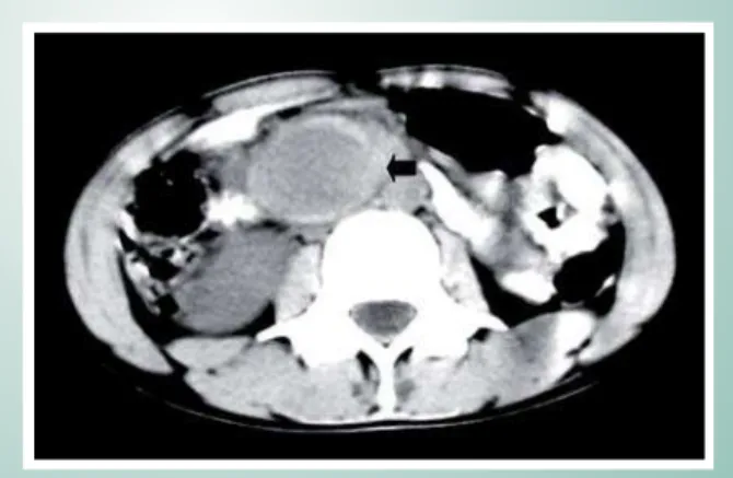
After ten days of treatment she developed persistent abdominal pain associated with vomiting. The abdomen was soft and non-distended, but with epigastric tenderness. Gastroduodenal endoscopy revealed an extrinsic compression of the posterior wall of the gastric body. The diagnosis of pancreatic pseudocyst was suggested, but laboratory tests including aspartate aminotransferase, alanine aminotransferase and amylase were all normal. Abdominal CT disclosed a hypodense image in the superior mesenteric artery suggestive of an aneurysm and abdominal color Doppler ultrasound confirmed the presence of a turbulent flow inside the image (Fig. 1). These findings suggested the diagnosis of a mycotic aneurysm secondary to IE and prompt surgical intervention Infective endocarditis (IE) is rare in children, but its
incidence has increased in the presence of predisposing conditions such as congenital heart disease, previous corrective cardiac surgery, rheumatic valvar disease and catheter use1. Additionally, IE complications are associated with increased morbidity and mortality rates and comprise a broad spectrum that includes acute congestive heart failure, embolic events, periannular extension of infection, arrhythmia, prosthetic device dysfunction and mycotic aneurysm1,2.
The prevalence of rheumatic fever has decreased in developed countries3. On the other hand, two recent reports revised the Brazilian studies regarding rheumatic fever and presented their own experience, confirming the high prevalence of this disease4,5. According to Brazilian Health Ministry data, the prevalence of rheumatic fever is 3% to 5% in children and adolescents6. An increased frequency of carditis and valvar lesions has also been observed5. Consequently, rheumatic fever and its related complications such as valvar disease and IE are still a challenge.
Case Report
On February-11-2003 a 10-year-old girl with chronic rheumatic heart disease was admitted with a 1-day history of gait and speech disturbances associated with fever of a week’s duration. This patient was referred to our center when she was 7 years old with a history of systolic cardiac murmur in the mitral region with axillary irradiation auscultated by the pediatrician. She did not present arthritis, skin lesions or chorea. The echocardiography showed a mild mitral regurgitation with decreased mobility of the posterior leaflet. The diagnosis of rheumatic heart disease was made based on the modified Jone’s criteria7. Although benzathine penicillin We report the case of a girl with chronic rheumatic valvar heart disease who developed infective endocarditis and two complications, ischemic stroke due to a cerebral embolic event and mycotic aneurysm of the superior mesenteric artery.
Case Report
Liphaus et al
ABDOMINAL MYCOTIC ANEURYSM AND CEREBRAL EMBOLIC EVENT ASSOCIATED WITH INFECTIVE ENDOCARDITIS IN A PATIENT WITH CHRONIC RHEUMATIC HEART DISEASE
Arq Bras Cardiol 2007; 88(1) : e6-e8
was performed. Histological specimens demonstrated acute vascular inflammation with the presence of gram-positive bacteria and lesion of the artery wall, confirming the pseudoaneurysm nature of the lesion. Intravenous crystalline penicillin G associated with ampicillin and ceftriaxone therapy was maintained to complete six weeks of treatment. The symptoms resolved and patient was discharged fifteen days after surgery with no ischemic or neurological sequelae and moderate mitral valve failure with no mass inside the left atrium at echocardiography.
According to Brazilian Health Ministry6 and recent
reports4,5, rheumatic fever and rheumatic valvar disease stillvalvar disease stilldisease still represent a public health problem in Brazil. Rheumatic fever is the most common acquired cause of valvar heart diseasevalvar heart diseaseheart disease and consequently of its complications, such as IE1,2,4,5,6. Factors that may additionally predispose children to develop IE complications are: type of organism, location and size of vegetation and the severity of the comorbid cardiac disease1. In adults, mitral lesions have been associated with higher rates of embolization than aortic vegetations (25% versus 10%, respectively); the highest rate of embolization (37%) occurs when vegetations are attached to the anterior rather than the posterior mitral leaflet and patients with large vegetations on echocardiography (> 10 mm) have significantly higher incidence of embolic events than those with small ones (10
mm)1,2. Common organisms related to IE in children are
Streptococcus viridans type, gram-positive cocci, staphylococci and enterococci1. In contrast to what was observed in this
case, Staphylococcus aureaus is the most common organism
associated with neurological manifestations in IE, whereas mycotic aneurysms usually occur secondarily to infections caused by nonhemolytic Streptococcus2,9,10.
Long-term antibiotic therapy is recommended in pediatric endocarditis and generally a 4-week regimen of intravenous aqueous crystalline penicillin G or ampicillin attains a high cure rate1. The majority of studies have reported low frequencies of complications related to IE after the introduction of an adequate antibiotic therapy1,2,9. In contrast, although our patient was undergoing adequate treatment, she developed abdominal pain and surprisingly the abdominal CT revealed an aneurysm in the superior mesenteric artery.
Superior mesenteric artery aneurysms (SMAAs) are the third preferred site (5.5%) for all visceral aneurysms and they most frequently have an infectious etiology10. Although uncommon, SMAAs must be promptly treated to avoid mortality and complications such as expansion and eventual rupture, thrombosis and distal embolization causing intestinal ischemia10. Differently from all other visceral aneurysms, 70% to 90% of SMAAs are symptomatic at presentation and most patients have significant and progressive abdominal pain, as observed in the case here described10. SMAAs presenting signs and symptoms can be misdiagnosed as much more common conditions such as pancreatitis, perforated viscus, ulcer disease and appendicitis.10Duplex ultrasound imaging represents a valuable, quick and reliable tool that allows an appropriate diagnosis, but similarly to what occurred with our patient, abdominal CT and/or angiography may be necessary to confirm the correct diagnosis10. Mycotic aneurysms are uncommon complications in children with IE and can develop in any systemic artery, but the aorta itself and SMAAs are the most common sites2,10.
Currently, opinions differ as to whether early valve surgery can prevent the development of new embolic events in patients with cardiac vegetations, but most authorities agree that one episode of embolization is not an indication for valve replacement, considering the uncommon nature of recurrent emboli during controlled infection1,2,9. However, in the case reported herein, despite improvement of neurological symptoms and sterile blood cultures, a mycotic aneurysm,
Discussion
Focal neurological deficit is rare in children and generally results from a cerebral infarction that most commonly occur as a result of embolization from endocardic vegetation9. Several series have reported incidence rates of 20% to 40% and mortality rates as high as 58% for neurological complications among patients with IE and these complications are more common when both aortic and mitral valves are involved2,9. Early diagnosis and prompt initiation of antimicrobial therapy are of major importance in preventing neurological complications associated with IE1,2,9. Nevertheless, Heiro et al9, studying 218 episodes of IE, showed that in most IE episodes (76%) a neurological manifestation was evident before the start of antimicrobial treatment, being the first sign in 47% of episodes and also that 9 of 13 embolic brain infarctions were located in the region supplied by the middle cerebral artery, as observed in the case described herein.
Our patient presented a chronic rheumatic heart disease, which is a well-known predisposing risk for IE. Patients with prolapsed and regurgitating mitral valves, as it occurs in subjects with congenital heart disease, previous corrective cardiac surgery and rheumatic valvar disease are classified byvalvar disease are classified bydisease are classified by the American Heart Association as presenting moderate risk of developing endocarditis1,2. IE occurs less often in children and accounts for approximately one in 280 pediatric admissions per year in developed countries, where the prevalence of rheumatic fever has declined1,3.
Fig. 1 -Abdominal CT showing a hypodense image in the superior mesenteric artery consistent with aneurysm. (arrow)
Case Report
Liphaus et al ABDOMINAL MYCOTIC ANEURYSM AND CEREBRAL EMBOLIC EVENT ASSOCIATED WITH INFECTIVE ENDOCARDITIS IN A PATIENT WITH CHRONIC RHEUMATIC HEART DISEASE
Arq Bras Cardiol 2007; 88(1) : e6-e8
1. Ferrieri P, Gewitz MH, Gerber MA, Newburger JW, Dajani AS, Shulman ST, et al. Unique features of infective endocarditis in childhood. Circulation. 2002; 105: 2115-7.
2. Baddour LM, Wilson WE, Bayer AS, Fowler VG Jr, Bolger AF, Levison ME, et al. Infective endocarditis diagnosis, antimicrobial therapy, and management of complications: a statement for healthcare professionals from the Committee on Rheumatic Fever, Endocarditis, and Kawasaki Disease, Council on Cardiovascular Disease in the Young, and the Councils on Clinical Cardiology, Stroke, and Cardiovascular Surgery and Anesthesia, American Heart Association. Circulation. 2005;111: e394-e433.
3. Dajani AS. Current status of nonsuppurative complications of group A streptococci. Pediatr Infect Dis J. 1991;10 (Suppl): 525-7.
4. Silva CHM and Pediatric Committee – São Paulo Pediatric Rheumatology Society. Rheumatic Fever: a multicenter study in the state of São Paulo. Rev Hosp Clin Fac Med S Paulo. 1999; 54: 85-90.
5. Borges F, Barbosa MLA, Borges RB, Pinheiro OC, Cardoso C, Bastos C, et al. Clinical and demographic characteristics of 99 episodes of rheumatic fever
in Acre, the Brazilian Amazon. Arq Bras Cardiol. 2005; 84: 111-4.
6. Ministério da Saúde. Coordenação de Doenças Crônicas-Degenerativas. Incidência da febre reumática no Brasil. Brasília; 2003.
7. Dajani AS, Ayoub E, Bierman FZ. Special writing group of the Committee on Rheumatic Fever, Endocarditis, and Kawasaki Disease of the Council ob Cardiovascular Disease in the Young of the American Heart Association. Guideline for the diagnosis of rheumatic fever. Jones criteria,1992 update. JAMA. 268: 2069-73.
8. Li JS, Sexton DJ, Mick N, Nettles R, Fowler VG Jr, Ryan T, et al. Proposed modifications to the Duke criteria for the diagnosis of infective endocarditis. Clin Infect Dis. 2000; 30: 633-8.
9. Heiro M, Nikoskelainen J, Engblom E, Kotilainen E, Marttila R, Kotilainen P. Neurologic manifestations of infective endocarditis: a 17-year experience in a teaching hospital in Finland. Arch Int Med. 2000; 160: 2781-7.
10.Lorelli DR, Caimbra RA, Seabrook GR, Towne JB. Diagnosis and management of aneurysms involving the superior mesenteric artery and its branches: a report of four cases. Vasc Endovascular Surg. 2003; 37: 59-66.
References
which is a second complication of IE, occurred.
Finally, the purpose of this report was to warn pediatricians and clinicians regarding the need of a high level of suspicion when diagnosing IE in patients with rheumatic valvar disease.valvar disease.disease. The early start of adequate antibiotic therapy is mandatory in order to avoid complications associated with high levels of morbidity and mortality. In addition, a wide variety of cerebral and abdominal manifestations may constitute the
first signs and symptoms of an IE and all physicians must be aware of the possibility of a second complication in the same patient, despite the onset of prompt and adequate antibiotic therapy.
Potential Conflict of Interest
No potential conflict of interest relevant to this article was reported.