einstein. 2012;10(2):239-41 CASE REPORT
Imaging findings and cerebral perfusion in arterial ischemic
stroke due to transient cerebral arteriopathy in children
Achados de imagem e perfusão arterial cerebral em acidente vascular cerebral isquêmico
devido à arteriopatia transitória em criança
Alcino Alves Barbosa Junior1, Saada Resende de Souza Ellovitch2, Rita de Cassia Maciel Pincerato3
1 Department of Diagnostic Imaging, Hospital Israelita Albert Einstein – HIAE, São Paulo (SP), Brazil. 2 Neuropediatrics, Hospital Israelita Albert Einstein – HIAE, São Paulo (SP), Brazil.
3 Hospital Samaritano – São Paulo (SP), Brazil.
Corresponding author: Alcino Alves Barbosa Junior – Avenida Albert Einstein, 617, bloco D, 4° andar – Morumbi – Zip code: 05651-901 – São Paulo (SP), Brazil – Phone: (55 11) 2151-2487 E-mail: [email protected]
Received on: Feb 28, 2012 – Accepted on: Apr 3, 2012
ABSTRACT
We report the case of a 4-year-old female child who developed an arterial ischemic stroke in the left middle cerebral artery territory, due to a proximal stenosis of the supraclinoid internal carotid artery, most probably related to transient cerebral arteriopathy of childhood. Computed tomography scan, magnetic resonance imaging, perfusion magnetic resonance and magnetic resonance angiography are presented, as well as follow-up by magnetic resonance and magnetic resonance angiography exams. Changes in cerebral perfusion and diffusion-perfusion mismatch call attention. As far as we know, this is the first report of magnetic resonance perfusion findings in transient cerebral arteriopathy.
Keywords: Stroke; Brain ischemia; Perfusion imaging; Magnetic resonance angiography; Diffusion magnetic resonance imaging; Child; Case reports
RESUMO
Paciente de 4 anos de idade teve um acidente vascular cerebral isquêmico no território da artéria cerebral média esquerda, devido a uma estenose proximal da artéria carótida interna supraclinóidea, provavelmente relacionada à arteriopatia cerebral transitória da infância. Apresentamos os exames de tomografia computadorizada, ressonância magnética, perfusão por ressonância magnética e angiorressonância, e o seguimento por ressonância magnética e angiorressonância. Chamam atenção as alterações na perfusão cerebral e o desacoplamento na difusão-perfusão. Até onde temos conhecimento, este é o primeiro relato de achados de perfusão por ressonância em arteriopatia cerebral transitória.
Descritores: Acidente vascular cerebral; Isquemia encefálica; Imagem de perfusão; Angiografia por ressonância magnética; Imagem de difusão por ressonância magnética; Criança; Relatos de casos
INTRODUCTION
Arterial ischemic stroke (AIS) in childhood is uncommon, and its incidence is estimated as about
3.3/100,000(1). Arteriopathies are the most frequent
risk factor in children(2) and they can be divided into
progressive (systemic vasculitis, Moyamoya disease, sickle cell disease, primary angiitis of the central nervous system etc.) and non-progressive (transient arteriopathy and dissection). Repeated vascular imaging in children with AIS demonstrated the existence of a “transient cerebral arteriopathy” (TCA), characterized by non-progressive unilateral arterial disease, affecting the supraclinoid internal carotid artery and its proximal branches. Since 1998 TCA is known to be an important
cause of AIS in childhood(3).
About 31% of all cases of AIS may be related to previous infection by varicela-zoster virus (VZV) 12
months before the onset of neurological symptoms(4).
The vast majority of children with AIS and anterior circulation intracranial arteriopathy suffer from TCA, presumably of inflammatory origin, that is associated
with preceding VZV in 44% of cases(3).
The course of stenosis related to TCA is monophasic in the majority of cases, with subsequent regression
(partial or complete) and rarely progression(5). Although
the inflammatory phase of TCA is supposedly transient, 59% of children present permanent neurological deficits, and 77% have residual arterial lesions at
repeated angiography(3).
einstein. 2012;10(2):239-41
240 Barbosa Junior AA, Ellovitch SR, Pincerato RC
CASE REPORT
We present the case of a 4-year-old female child, referred to the emergency service of our institution complaining about visual blurring and drowsiness of acute onset. She underwent computed tomography (CT) scan of the brain without and with intravenous iodine contrast (Figure 1), which revealed a hypoattenuating area with no contrast enhancement in centrum semiovale and subcortical parietal white matter in the left cerebral hemisphere, suggesting a recent AIS.
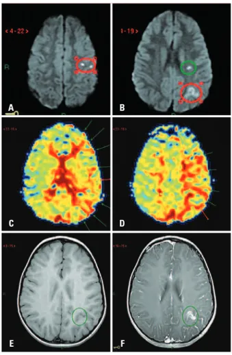
Five days later, magnetic resonance (MR) of the brain, MR cerebral perfusion with intravenous paramagnetic contrast and MR angiography (MRA) of intracranial arteries were performed. MR (Figure 2) confirmed the presence of an acute/subacute AIS, since we could observe low apparent diffusion coefficients in diffusion weighted images (DWI), as well as some areas of contrast enhancement (disrupted blood brain barrier), affecting discontinuous cortical segments of the precentral gyrus, inferior parietal lobule, subcortical parietal white matter and centrum semiovale on the left, all within the superior division of middle cerebral artery branches.
MR perfusion was performed with echo-planar gradient-echo technique, after the first bolus of intravenous contrast passing through the microvasculature, followed by analysis of parametric color maps, which revealed a delay in mean time to transit (MTT) in regions around the infarcts in the left hemisphere, most likely representing hypoperfusion in vascular border zones, mismatched with diffusion. Relative cerebral blood volume and flow (rCBV and rCBF, respectively) were not altered in comparison with the opposite hemisphere.
MRA with time-of-flight (TOF) technique without intravenous contrast (Figure 3) showed a moderate stenosis of the left supraclinoid internal carotid artery, extending to segment A1 of anterior cerebral artery and to segment M1 of middle cerebral artery, with some irregular contours, with no evidence of collateral vessels or neoangiogenesis in the territory of lenticulo-striate arteries or leptomeningeal arteries, except for slightly prominent lateral lenticule arteries, supporting the hypothesis of recent onset of an arteriopathy, rather than an insidious chronic arteriopathy. There was no sign of mural clot or intimal flap, such as
T1-hyperintensity on vessel walls(6), which could favor
the diagnosis of dissection.
These results allowed us to consider TCA of childhood as the main presumptive diagnosis, although the possibility of initial unilateral Moyamoya disease, systemic vasculitis or primary angiitis of central nervous
Figure 2. Magnetic resonance of the brain. Diffusion weighted images (A and B) reveal areas of diffusion restriction in the centrum semiovale (green circle), precentral gyrus and inferior parietal lobule (red circles). Parametric color map based on mean time to transit (C and D) shows areas of hypoperfusion mismatched with diffusion (arrows) in vascular border zones of the left hemisphere. T1-weighted images before (E) and after (F) IV paramagnetic contrast demonstrate areas of disrupted blood brain barrier in the inferior parietal lobule (green circles)
A
C
E
B
D
F
Figure 1. Computed tomography scan of the head without (A) and with (B) IV iodine contrast shows a non-enhanced hypoattenuating area in the centrum semiovale and parietal subcortical white matter in the left (red circles)
241
Imaging findings and cerebral perfusion in arterial ischemic stroke
einstein. 2012;10(2):239-41
system could not be entirely ruled out based only upon the first exam.
A detailed investigation of past diseases was conducted, and the parents finally recalled a recent infection by VZV 4 months before the onset of neurological symptoms, which is an important risk
factor for AIS in childhood(4), supporting the hypothesis
of TCA.
The patient underwent antiplatelet therapy with acetylsalicylic acid (50 mg/day) and oxcarbamazepine (15 mg/kg/day) was also prescribed. She recovered from symptoms within 48 hours, was discharged from hospital with no perceptible neurological deficits, and has not presented any other episodes 6 months after the first event.
Four months after the beginning of the disease, she was re-evaluated through MR and MRA exams (Figure 3). As expected, the ischemic lesions were detected as areas of gliotic sequelae. An increase in the caliber of supraclinoid internal carotid artery, segment A1 of anterior cerebral artery and segment M1 of middle cerebral artery was also noticed, persisting some irregular contours, less prominent than in the previous
exam, leading to the conclusion of a partial regression of stenosis.
Considering the clinical evolution and follow-up exams, the possibility of progressive arteriopathy was very unlikely and TCA, on the other hand, became the most probable cause of stenosis.
DISCUSSION
In this case, imaging exams were very important in the investigation, as they led to a diagnosis that was not considered in the first clinical assessment. Particularly MRA – which is essential in evaluation of patients with ischemic stroke – showed determinant data to the final conclusion, revealing the point of stenosis. Follow-up exams also played a crucial role by excluding progressive disorders.
Despite the favorable clinical course of this case, it was speculated whether cerebral perfusion impairments, mismatched with diffusion observed in the first exam, suggesting some degree of ischemic injury surrounding the infarct, could somehow be implicated in the fluctuation or worsening of symptoms that some patients with TCA experience in the beginning of the
disease(3). We believe that systematic cerebral perfusion
studies in patients with TCA in childhood may improve our understanding of the pathophysiology of this condition.
ACKNOWLEDGEMENT
The authors would like to acknowledge the contribution of Dr. Cristiane Wosni, radiology resident at Hospital Israelita Albert Einstein.
REFERENCES
1. Lynch JK, Hirtz DG, DeVeber G, Nelson KB. Report of the National Institute of Neurological Disorders and Stroke workshop on perinatal and childhood stroke. Pediatrics. 2002;109(1):116-23.
2. Mackay MT, Wiznitzer M, Benedict SL, Lee KJ, Deveber GA, Ganesan V; International Pediatric Stroke Study Group. Arterial ischaemic stroke risk factors: the International Pediatric Stroke Study. Ann Neurol. 2011;69(1):130-40. 3. Braun KP, Bulder MM, Chabrier S, Kirkham FJ, Uiterwaal CS, Tardieu M, et al.
The course and outcome of unilateral intracranial arteriopathy in 79 children with ischaemic stroke. Brain. 2009;132(Pt 2):544-57.
4. Askalan R, Laughlin S, Mayank S, Chan A, MacGregor D, Andrew M, et al. Chickenpox and stroke in childhood: a study of frequency and causation. Stroke. 2001;32(6):1257-62.
5. Lanthier S, Armstrong D, Domi T, de Veber G. Post-varicella arteriopathy of childhood: natural history of vascular stenosis. Neurology. 2005;64(4):660-3. 6. Dlamini N, Freeman JL, Mackay MT, Hawkins C, Shroff M, Fullerton HJ, et al.
Intracranial dissection mimicking transient cerebral arteriopathy in childhood arterial ischaemic stroke. J Child Neurol. 2011;26(9):1203-6.
Figure 3. Magnetic resonance angiography of intracranial arteries, maximum intensity projection (A) and volume rendering image (B) revealed moderate stenosis of supraclinoid internal carotid artery, A1 segment of anterior cerebral artery and M1 segment of middle cerebral artery (red circles). Four months later, magnetic resonance angiography for follow-up (C and D) shows increase in arterial caliber, persisting some irregular contours (red circles)
A
C
B