AneurysmAl bone cyst At c2
Imaging evaluation after intralesional injection
of calcitonin and methylprednisolone
Elise T. Tonomura¹, Paulo Ramos², Paulo Miguel Hemais³,
Edson Marchiori
4, Emerson L. Gasparetto
5Abstract – Objective: To demonstrate imaging findings during the follow-up of patients with aneurysmal bone cyst (ABC) of C2 treated with intralesional injection of calcitonin and methylprednisolone. Method:
Three patients with ABC of C2 were treated percutaneously with intralesional injection of calcitonin and methylprednisolone. All the patients were females, with ages varying from 6 to 13 years. All of them presented with cervical masses, but without neurological symptoms. Results: Imaging follow-up with CT and plain films showed progressive ossification and reduction of the blood-filled cavities with volume reduction of all lesions. No recurrence was noted during the follow-up. Conclusion: Intralesional injection of calcitonin and methylprednisolone is a safe treatment option for cervical ABC. The CT scan is especially valuable as a guide of injection site and for the follow-up of ossification of the treated ABCs.
KEY WORDS: aneurysmal bone cyst, calcitonin, methylprednisolone.
cisto ósseo aneurismático em c2: avaliação por imagem após injeção intralesional de calcitonina e metilprednisolona
Resumo – Objetivo: Demonstrar os aspectos de imagem no acompanhamento de pacientes submetidos ao tratamento de cisto ósseo aneurismático com injeção intralesional de calcitonina e metilprednisolona. Método:
Três pacientes com cisto ósseo aneurismático em C2 foram tratados com injeção intralesional percutânea de calcitonina e metilprednisolona. Os três pacientes eram do sexo feminino com idades variando de 6 a 13 anos. Os três pacientes apresentavam massa cervical sem sintomas neurológicos. Resultados: O acompanhamento por imagem com tomografia computadorizada e radiografia simples demonstrou ossificação progressiva e redução das cavidades preenchidas por sangue, com redução do volume das lesões. Não foi percebida recidiva durante o acompanhamento. Conclusão: A injeção intralesional de calcitonina e metilprednisolona é uma opção de tratamento segura para o cisto ósseo aneurismático cervical. A tomografia computadorizada é especialmente útil para orientar o sítio da punção e para o acompanhamento da ossificação dos cistos ósseos aneurismáticos tratados.
PALAVRAS-CHAVE: cisto ósseo aneurismático, calcitonina, metilprednisolona.
1Médica Radiologista do Instituto Nacional de Traumato-Ortopedia (INTO) Rio de Janeiro RJ, Brasil e Professora Assistente do Departamento de
Ra-diologia da Faculdade de Medicina da Universidade Federal do Rio de Janeiro, Rio de Janeiro RJ, Brasil (UFRJ); 2Médico Ortopedista do INTO; 3Médico
Radiologista do INTO; 4Professor Titular do Departamento de Radiologia da Universidade Federal Fluminense e Professor Associado do Departamento
de Radiologia da Faculdade de Medicina da UFRJ; 5Professor Adjunto do Departamento de Radiologia da Faculdade de Medicina da UFRJ.
Received 8 May 2008, received in inal form 29 July 2008. Accepted 9 August 2008.
Dra. Elise Tonomura – Avenida Prefeito Dulcídio Cardoso 2500 / bloco 5 / apto 2502 - 22631-051 Rio de Janeiro RJ - Brasil. E-mail: etonomura@gmail.com
Aneurysmal bone cysts (ABC) are relatively rare lesions that occur more commonly among patients in the irst and second decades, especially in females. Characteris-tically, the ABC has many cavities full of blood. Most of the lesions occur at long bones, vertebrae and lat bones. The cervical vertebrae involvement is uncommon1,2. When seen in this location, the lesion involves initially the pos-terior arch and pedicles, with bone destruction and
cor-tical expansion. Occasionally, the ABCs compress the spi-nal cord and nerve roots.
We present the imaging follow-up of three patients with ABC in the posterior arch of C2, who were treat-ed with percutaneous intralesional injection of calcito-nin and methylprednisolone.
method
This study evaluated a small series of patients with ABCs in the posterior arch of C2, who were treated with intralesional in-jection of calcitonin and methylprednisolone. All the patients signed informed consent and the Institutional Review Board of the Hospital approved the study. The patients underwent clini-cal and imaging investigation, including plain ilms, CT scans and also magnetic resonance imaging (MRI) in one case. The histolog-ical diagnosis of ABC was deined after surghistolog-ical biopsy. The in-tralesional injection of calcitonin and methylprednisolone was performed between 2 and 6 times (mean of 4 times), with dos-es of 200 UI and 120–160 mg, rdos-espectively. The follow-up was
done with clinical and imaging examinations, ranging from 2 to 18 months (mean of 7. 7 months).
results
Case 1
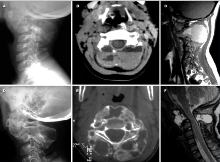
A six-year-old female patient was referred to our insti-tution with diagnosis of ABC deined in another hospital. She had the irst medical consultation 3 years before, and the main complain was reduced cervical mobility, which was treated initially as torticollis, without any radiologi-cal study. The symptoms did not improve, and when she was 4-years old a cervical spine CT scan showed a lyt-ic expansible lesion involving the body and the posteri-or arch of C2 (Fig 1A), with luid-luid level content. This inding was also demonstrated in the MRI as hyperintense and isointense luid-luid levels. The plain ilm showed the “bubble” aspect of the lesion involving the vertebral
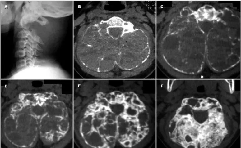
Fig 3 (Case 3). Plain ilm (A) of the cervical spine shows a soft-tissue mass at the topography of the C2 posterior arch, which is not seen. The CT scan (B) shows the egg-shell-thin cortex and the luid-luid levels. Notice the sparing of the cervical canal. The CT scans during the one year long follow-up (C–F) show the progressive thickening of the outer cortex and concentric occupation of the cavities with bone until almost com-plete ossiication of the lesion.
Fig 2 (Case 2). (A) Plain ilm of the cervical spine showing the needle inside the lesion that is involving the posterior arch of C2. (B) The CT scan demonstrates the expansive lytic lesion involving the body and posterior arch of C2, which presents luid-luid levels. The plain ilm (C) and sagittal CT scan (D) performed one year later show the shrinkage of the lesion and the ossiication of its walls. Notice the reduction of size of the cavities.
body and the posterior arch of C2 (Fig 1B). The plain ilm and the MRI performed at our institution one year later showed signiicant reduction of the lesion size, although remaining with areas of high-signal on T2-weighted im-ages (Fig 1C). Subsequently, a percutaneous iniltration of 200 IU of calcitonin and 120 mg of methylprednisolone were infused in the lesion. The injection was performed inside the operation room and under general anesthesia to prevent patient movements during the procedure. She completed 4 sessions within 6 months and underwent the follow-up CT that showed intense ossiication of the
le-sion with lesser cavities illed with blood. She abandoned the treatment for 18 months, returning two years later, when she received another iniltration. The follow-up CT showed that the lesion was completely ossiied (Fig 1D). MRI showed shrinking hyperintense lesion and sparing of the vertebral canal (Fig 1F).
Case 2
an expansible lytic bone lesion at the posterior arch of C2. The CT scan corroborated the indings and demonstrated the characteristic luid-luid levels inside the lesion. She underwent a surgical biopsy that conirmed the diagnosis of aneurysmal bone cyst. At the same time, the irst in-tralesional iniltration of 200 IU of calcitonin and 160 mg of methylprednisolone was performed. Two months lat-er anothlat-er iniltration was done, and the plain ilms and the CT showed that the cyst ossiied intensely, with vol-ume reduction (Fig 2).
Case 3
An 11-year-old girl present with a 5-month history of cervical pain, associated with growing volume in the pos-terior region of the neck. The cervical radiographs showed a lytic expansible lesion involving the body and especial-ly the posterior arch of C2, which presented multiple luid-luid levels at the CT scan (Fig 3A). The surgical bi-opsy deined the diagnosis of aneurysmal bone cyst. At the same procedure, 200 IU of calcitonin and 120 mg of methylprednisolone were injected inside the lesion. This treatment was repeated twice with one-week interval, and then repeated monthly three times. Three months after the last injection, the patient referred signiicant pain re-duction, and the CT scan showed the lesion intensely os-siied and volume reduction (Fig 3).
discussion
Aneurysmal bone cysts are lesions containing thin-walled cystic cavities illed with blood. The ABC occurs more frequently among female patients of the irst and second decades. Most of the lesions involve long bones, vertebrae and flat bones1. In the spine the ABCs occur more frequently at the thoracic and lumbar segments, be-ing rare in the cervical spine. In this study, we evaluated three cases of ABCs at the C2 level, showing the satisfac-tory imaging evaluation after treatment with intralesional injection of calcitonin and methylprednisolone.
The ABC affects initially the posterior arch and pedi-cles of the vertebrae, with bone destruction and cortical expansion. In some cases, it may compress the spinal cord and neural roots. The clinical presentation of vertebral ABCs is varied, and patients may present local deformity, movement limitations, muscular stiffness, torticollis, local heat, and occasionally, murmur over the affected region4,7-9. In our study, the lesion affected mainly the posterior arch, but also the vertebral body. In addition, none of our cases presented signiicant neurological symptoms, even when there was reduction of the vertebral canal size and change of the vertebral axis.
The main pathological characteristic of the ABC is the presence of many blood cavities separated by thin
ibrous septa. This inding is demonstrated with CT scans and MR imaging, which show separation of the blood con-tent forming levels of different luid densities. However, the luid-luid signal is not exclusive to ABCs, and can be seen in other bone lesions, such as chondroblastoma, gi-ant cell tumor and osteoblastoma. The septations of the lesion are hypervascularized and the venous injection of contrast media can help to differentiate the thin and smooth septa of ABC from other tumors, which would have nodularities4,10-13.
The vertebral ABCs may present on plain radiographs a characteristic “ballooning” of the posterior elements with a thin rim, although it may be difficult to see in some cases14. Pathological fractures are common and oc-casionally may progress to vertebra plana15. Involvement of multiple vertebrae may occur4,16 and the expansion of bone into the spinal canal can compress the dural sac. The hypervascular complex, with pathological circula-tion and even arteriovenous shunts, can be shown in se-lective angiography11,12. The CT scan is highly sensitive to detect calcium and demonstrates the extent of the bone destruction, showing the many cavities illed with luid-luid levels. On MRI the ABCs have heterogeneous signal, forming multiloculated cysts with luid-luid levels bet-ter seen on T2-weighted images. On T1-weighted images, the luid-luid levels are hyperintense, presumably due to methemoglobin deposits4,11,12,17.
In our study, the three cases of ABC at C2 appeared as expansible lytic lesions of the posterior arch, showing an eggshell thin cortex barely seen on plain ilms. The CT scans demonstrated better the thin outer cortex of the lesion with the cavities showing hypo/isodense luid-luid levels. MRI performed in case 1 corroborated the pres-ence of blood inside the lesion, showing hyperintense and isointense luid-luid levels on T1-weighted images.
The treatment of aneurysmal bone cyst with less re-currence index was during many years the complete sur-gical resection. However, the resection of cervical verte-brae ABC is risky, and can cause instability, which is even more undesirable for growing child3,4,8,18-20. The treatment approach of our cases, based in the iniltration of the le-sion with calcitonin and methylprednisolone, was recently described7. Another less aggressive surgical treatment is curettage of the lesion with bone grafting. However, in most of the cases the curettage is incomplete, increasing the risk of recurrence20. Radiation therapy for ABCs was already performed, but no more accepted due to the risk of posterior malignization and development of scoliosis caused by asymmetric growing of the irradiated site1.
when compared to ibrosing agent. The calcitonin can in-duce the formation of cancellous bone and inhibit the os-teoclastic activity. The corticoid can reduce the ibroblas-tic action and the angiogenesis7. This kind of treatment can take years to cure, however, previous studies showed good results, without the common recurrence and the side effects of the ibrosing agents4,5.
In our cases, the doses of calcitonin and methylpred-nisolone were decided empirically, following the doses injected in a previous study7. In each section we injected under general anesthesia 200 IU of calcitonin and 120–160 mg of methylprednisolone. Although a CT-guided iniltra-tion could be performed, we avoided this method due to the risk of patients’ movements during the procedure, since all of them were younger than 15 years. However, we deined the site of puncture and injection based on the CT scans, which were also used as the imaging method of choice for the follow-up. In all cases the follow-up CT scans showed that the osteoblastic activity was persistent, with progressive ossiication of the cavities in a concentric way, thickening the walls and reducing the blood content inside them. At the end of the treatment, the ABCs were almost all ossiied, with shrinkage of the lesions. After being considered out of risk of fracture or bleeding of the lesion, the children had functional radiographs of the cervical spine performed with lexion and extension, as-suring the stability of C2.
In conclusion, the direct injection of methylpredniso-lone and calcitonin inside the ABC cavities seems to be a safe procedure, with no side effects that are usually seem in cases of iniltration using ibrosing agents. The CT scan is the imaging method of choice for the follow-up of the ABCs treatment, as it shows the progressive ossiication of the cavities in a concentric way. We believe that three iniltration sessions with two month intervals result in sig-niicant bone thickening, being enough to prevent patho-logic fractures or bleeding. Further studies could consider a longer follow-up, as the children are still growing and it
is expected that the ossiied lesions of C2 will be remod-eling together with the other growing vertebrae.
references
1. Mankin HJ, Hornicek FJ, Ortiz-Cruz E, Villafuerte J, Gebhardt MC. Aneu-rysmal bone cyst: a review of 150 patients. J Clin Oncol 2005;23:6756-6762. 2. Senac MO, Isaacs H, Gwinn JL. Primary lesions of bone in the 1st decade of
life: retrospective survey of biopsy results. Radiology 1986; 160:491-495. 3. Garg S, Mehta S, Dormans J. Modern surgical treatment of primary an-eurysmal bone cyst of the spine in children and adolescents. J Pediat Orthop 2005;25:387-392.
4. Liu JK, Brockmeyer DL, Dailey AT, Schmidt MH. Surgical management of aneurysmal bone cysts of the spine. Neurosurg Focus 2003;15:E4. 5. Topouchian V, Mazda K, Hamze B, Laredo JD, Penneçot GF.
Aneurys-mal bone cysts in children: complications of ibrosing agent injection. Radiology 2004;232:522-526.
6. Vale BP, Alencar FJ, Aguiar GB, Almeida BR. Vertebral aneurysmatic bone cyst: study of three cases. Arq Neuropsiquiatr 2005;63:1079-1083. 7. Gladden ML Jr, Gillingham BL, Hennrikus W, Vaughan LM.
Aneurys-mal bone cyst of the irst vertebrae in a child treated with percutane
-ous intralesional Injection of calcitonin and methylprednisolone: a case report. Spine 2000;25:527-530.
8. Eastwood B, Biggs HK. Aneurysmal bone cyst. E-medicine January 23, 2006.
9. Rai AT, Collins JJ. Percutaneous treatment of pediatric aneurysmal bone cyst at C1: a minimally invasive alternative: a case report. AJNR 2005;26:30-33.
10. Hudson TM. Fluid levels in aneurysmal bone cysts: a CT feature. AJR 1984;142:1001-1004.
11. Kransdorf MJ, Sweet DE. Aneurysmal bone cyst: concept, controversy, clinical presentation, and imaging. AJR 1995;164:573-580.
12. Beltran J, Simon DC, Levy M, Herman L, Weis L, Mueller CF. Aneurys-mal bone cysts: MR imaging at 1.5 T. Radiology 1986;158:689-690. 13. Tsai JC, Dalinka MK, Fallon MD, Zlatkin MB, Kressel HY. Fluid-luid
level: a non-speciic inding in tumors of bone and soft tissue. Radiol
-ogy 1990;175:779-782.
14. Sampaio PM, Pellizzaro B. Aneurysmal bone cysts of the spine: report of 2 cases with spinal cord compression. Arq Neuropsiquiatr 1972;30:245-249. 15. Papagelopoulos PJ, Currier BL, Galanis EC, Sim FH. Vertebra plana of
the lumbar spine caused by an aneurysmal bone cyst: a case report. Am J Orthop 1999;28:119-124.
16. Tillman PP, Dahlin DC, Lipscomb PR, Stewart JR. Aneurysmal bone cyst: an analysis of ninety-ive cases. Mayo Clin Proc 1968;43:478- 495. 17. Cory DA, Fritsch SA, Cohen MD, et al. Aneurysmal bone cysts:
imag-ing indimag-ings and embolotherapy. AJR 1989;153:369-373.
18. Docquier PL, Delloye C. Treatment of aneurysmal bone cysts by intro-duction of demineralized bone and autogenous bone marrow. J Bone Joint Surg Amer 2005;87:2253-2258.
19. Levin DA, Hensinger RN, Graziano GP. Aneurysmal bone cyst of the second cervical vertebrae causing multilevel upper cervical instability. J Spinal Disord Tech 2006;19:73-75.