Congenital musCular dystrophy
Part II: a review of pathogenesis and therapeutic perspectives
Umbertina Conti Reed
1abstract – The congenital muscular dystrophies (CMDs) are a group of genetically and clinically heterogeneous hereditary myopathies with preferentially autosomal recessive inheritance, that are characterized by congenital hypotonia, delayed motor development and early onset of progressive muscle weakness associated with dystrophic pattern on muscle biopsy. The clinical course is broadly variable and can comprise the involvement of the brain and eyes. From 1994, a great development in the knowledge of the molecular basis has occurred and the classification of CMDs has to be continuously up dated. In the last number of this journal, we presented the main clinical and diagnostic data concerning the different subtypes of CMD. In this second part of the review, we analyse the main reports from the literature concerning the pathogenesis and the therapeutic perspectives of the most common subtypes of CMD: MDC1A with merosin deficiency, collagen VI related CMDs (Ullrich and Bethlem), CMDs with abnormal glycosylation of alpha-dystroglycan (Fukuyama CMD, Muscle-eye-brain disease, Walker Warburg syndrome, MDC1C, MDC1D), and rigid spine syndrome, another much rare subtype of CMDs not related with the dystrophin/glycoproteins/extracellular matrix complex.
Key WorDs: congenital muscular dystrophy, MDC1A, collagen VI related disorders, glycosylation of alpha-dystroglycan, Fukuyama DMC, muscle-eye-brain (MeB) disease, Walker-Warburg syndrome, rigid spine syndrome.
distrofia muscular congênita. parte ii: revisão da patogênese e perspectivas terapêuticas
resumo – As distrofias musculares congênitas (DMCs) são miopatias hereditárias geralmente, porém não exclusivamente, de herança autossômica recessiva, que apresentam grande heterogeneidade genética e clínica. são caracterizadas por hipotonia muscular congênita, atraso do desenvolvimento motor e fraqueza muscular de início precoce associada a padrão distrófico na biópsia muscular. o quadro clínico, de gravidade variável, pode também incluir anormalidades oculares e do sistema nervoso central. A partir de 1994, os conhecimentos sobre genética e biologia molecular das DMCs progrediram rapidamente, sendo a classificação continuamente atualizada. os aspectos clínicos e diagnósticos dos principais subtipos de DMC foram apresentados no número anterior deste periódico, como primeira parte desta revisão. Nesta segunda parte apresentaremos os principais mecanismos patogênicos e as perspectivas terapêuticas dos subtipos mais comuns de DMC: DMC tipo 1A com deficiência de merosina, DMCs relacionadas com alterações do colágeno VI (Ullrich e Bethlem), e DMCs com anormalidades de glicosilação da alfa-distroglicana (DMC Fukuyama, DMC “Muscle-eye-brain” ou MeB, síndrome de Walker Warburg, DMC tipo 1C, DMC tipo 1D). A DMC com espinha rígida, mais rara e não relacionada com alterações do complexo distrofina-glicoproteínas associadas-matriz extracelular também será abordada quanto aos mesmos aspectos patogênicos e terapêuticos.
PAlAVrAs-ChAVes: distrofia muscular congênita, merosina, colágeno VI, glicosilação da alfa-distroglicana, DMC Fukuyama, DMC “muscle-eye-brain”-MeB, síndrome de Walker-Warburg, espinha rígida.
Departamento de Neurologia, Faculdade de Medicina da Universidade de são Paulo, são Paulo sP, Brazil: 1Professora Titular da Disciplina de Neuro-logia Infantil.
received 31 october 2008. Accepted 14 March 2009.
The congenital muscular dystrophies (CMDs) are ge-netically and clinically heterogeneous hereditary myop-athies with a predominant autosomal recessive mode of inheritance that are characterized by congenital hypoto-nia, delayed motor development and early onset of pro-gressive muscle weakness, as well as dystrophic pattern on muscle biopsy. The clinical course is broadly variable and can comprise the involvement of the brain and eyes1-7.
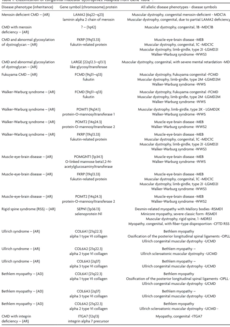
From 1994 and mostly in the first years of the cur-rent century a great input in the knowledge of the mo-lecular basis has occurred, and the classification of CMDs has to be continuously up dated. The oficial jour-nal of the World Muscular society, Neuromuscular Dis-orders, periodically publishes the revised classiication (Table 1)8. A computerized version of the classiication
is accessible at http://www.musclegenetable.org and http://194.167.35.195/. Most of the different genes in-volved with the pathogenesis of the CMD subtypes are related to the function of the dystrophin-glycoproteins associated complex (DGC) in the sarcolemma and extra-cellular matrix and their mutations lead either to defects in the glycosylation of alpha-dystroglycan (alpha-dystro-glycanopathies) or to abnormalities of extracellular ma-trix proteins (MDC1A and collagen VI related disorders)1-7.
A fourth subtype, rigid spine CMD, is related to a defect of an endoplasmic reticulum protein, selenoprotein N9,
and recently a new subtype10 was associated to a defect
of a nuclear protein, lamin A/C.
last month, the irst part of this review focused on the clinical and diagnostic aspects of the different sub-types of CMD. Presently, the second part emphasizes the main data on pathogenesis and therapeutic perspectives for the most common subtypes of CMD, i.e. MDC1A, col-lagen VI related disorders, CMDs caused by defects of gly-cosylation of alpha-DG, and inally the much rarer rig-id spine CMD.
Before reviewing the essential pathogenic data about the most common subtypes of CMD, we summarize the general aspects of the organization of the DGC and ex-tracellular matrix.
dystrophin-glyCoproteins assoCiated Complex and extraCellular matrix: general remarks on struCture and funCtion
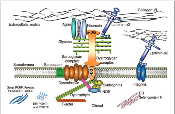
The DGC is an assembly of proteins spanning the sar-colemma of skeletal muscle ibers that forms a chain of links between the contractile actin in the cytoskeleton and the extracellular matrix11,12 (Fig 1). Defects in the DGC
can disrupt these links and alter the basement membrane organization, resulting in sarcolemmal instability and mus-cle cells apoptose. In addition to the function of stabiliz-ing the sarcollema, the DCG is also essential in organizstabiliz-ing molecules involved in cellular signaling12.
The irst component of the chain of links in the inner cytoskeleton is dystrophin that through the amino-termi-nus domain binds to actin and through the carboxyl
Table 1. Classiication of congenital muscular dystrophies. Adapted from Gene Table58.
Disease phenotype (inheritance) Gene symbol (chromosome) protein All allelic disease phenotypes - disease symbols Merosin deicient CMD – (Ar) lAMA2 (6q22–q23)
laminin alpha 2 chain of merosin
Muscular dystrophy, congenital merosin-deicient - MDC1A Muscular dystrophy, congenital, due to partial lAMA2 deiciency CMD with merosin
deiciency – (Ar)
? – (1q42) Muscular dystrophy, congenital, 1B -MDC1B
CMD and abnormal glycosylation of dystroglycan – (Ar)
FKrP (19q13.33) fukutin-related protein
Muscle-eye-brain disease -MeB Muscular dystrophy, congenital, 1C -MDC1C Muscular dystrophy, limb-girdle, type 2I -lGMD2I
Walker-Warburg syndrome -WWs3 CMD and abnormal glycosylation
of dystroglycan – (Ar)
lArGe (22q12.3–q13.1) like-glycosyltransferase
Muscular dystrophy, congenital, with severe mental retardation -MDC1D
Fukuyama CMD – (Ar) FCMD (9q31–q33) fukutin
Muscular dystrophy, Fukuyama congenital -FCMD Muscular dystrophy, limb-girdle, type 2M -lGMD2M
Walker-Warburg syndrome -WWs Walker-Warburg syndrome – (Ar) FCMD (9q31–q33)
fukutin
Muscular dystrophy, Fukuyama congenital -FCMD Muscular dystrophy, limb-girdle, type 2M -lGMD2M
Walker-Warburg syndrome -WWs Walker-Warburg syndrome – (Ar) PoMT1 (9q34.1)
protein-o-mannosyltransferase 1
Muscular dystrophy, limb-girdle, type 2K - lGMD2K Walker-Warburg syndrome -WWs Walker-Warburg syndrome – (Ar) PoMT2 (14q24.3)
protein-o-mannosyltransferase 2
Muscle-eye-brain disease -MeB Walker-Warburg syndrome -WWs2 Walker-Warburg syndrome – (Ar) FKrP (19q13.33)
fukutin-related protein
Muscle-eye-brain disease -MeB Muscular dystrophy, congenital, 1C -MDC1C Muscular dystrophy, limb-girdle, type 2I -lGMD2I
Walker-Warburg syndrome -WWs3 Muscle-eye-brain disease – (Ar) PoMGNT1 (1p34.1)
o-linked mannose beta1,2-N-acetylglucosaminyltransferase
Muscle-eye-brain disease -MeB Walker-Warburg syndrome -WWs
Muscle-eye-brain disease – (Ar) FKrP (19q13.33) fukutin-related protein
Muscle-eye-brain disease -MeB Muscular dystrophy, congenital, 1C -MDC1C Muscular dystrophy, limb-girdle, type 2I -lGMD2I
Walker-Warburg syndrome -WWs3 Muscle-eye-brain disease – (Ar) PoMT2 (14q24.3)
protein-o-mannosyltransferase 2
Muscle-eye-brain disease -MeB Walker-Warburg syndrome -WWs2 rigid spine syndrome (rss) – (Ar) sePN1 (1p36.13)
selenoprotein N1
Desmin-related myopathy with Mallory bodies -rsMD1 Minicore myopathy, severe classic form -rsMD1
Muscular dystrophy, rigid spine, 1 -MDrs1
Myopathy, congenital, with iber-type disproportion -CFTD rss Ullrich syndrome – (Ar) Col6A1 (21q22.3)
alpha 1 type VI collagen
Bethlem myopathy
ossiication of the posterior longitudinal spinal ligaments -oPll Ullrich congenital muscular dystrophy -UCMD Ullrich syndrome – (Ar) Col6A2 (21q22.3)
alpha 2 type VI collagen
Bethlem myopathy –
Ullrich scleroatonic muscular dystrophy -UCMD Ullrich syndrome – (Ar) Col6A3 (2q37)
alpha 3 type VI collagen
Bethlem myopathy –
Ullrich congenital muscular dystrophy -UCMD Bethlem myopathy – (AD) Col6A1 (21q22.3)
alpha 1 type VI collagen
Bethlem myopathy
ossiication of the posterior longitudinal spinal ligaments -oPll – Ullrich congenital muscular dystrophy -UCMD Bethlem myopathy – (AD) Col6A3 (2q37)
alpha 3 type VI collagen
Bethlem myopathy –
Ullrich congenital muscular dystrophy -UCMD Bethlem myopathy – (AD) Col6A2 (21q22.3)
alpha 2 type VI collagen
Bethlem myopathy
Ullrich scleroatonic muscular dystrophy UCMD -CMD with integrin
deiciency – (Ar)
ITGA7 (12q13) integrin alpha 7 precursor
Myopathy, congenital -ITGA7
minus binds to dystroglycan (DG)11,12. Dystrophin changes
lead to the X-linked Duchenne and Becker muscular dys-trophies, and are not involved in any type of CMD. The following link is dystroglycan (DG) that has two compo-nents: beta and alpha-DG. Beta-DG is a transmembrane glycoprotein and alpha-DG is extracellular but with close contact with the peripheral membrane11-13. Both are
en-coded by the same gene and then cleaved into two pro-teins, alpha and beta11-13. Primary mutations in the gene
encodingDG have not been reported and the knockout mouse for DGis embryonically lethal14.
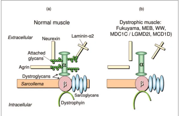
Alpha-DG is involved in the next step of the chain of links and connects the sarcolemma to the basement mem-brane. however, for performing this step, alpha-DG needs to be glycosylated11-15 (Fig 2). The glycosylation depends on
the biosynthesis of glycans that occurs by the enzymat-ic activity of glycosyltransferases, and it is necessary for the correct function of many animal proteins, the glyco-proteins. Dystroglycan glycosylation is highly conserved during evolution. The covalently addiction of sugar chains (glycans), forming a glycoprotein, induces a modiication that increases the availability of the protein for ligand in-teraction with laminin, agrinand perlecan in skeletal mus-cle, as well as with laminin and neurexin in the brain11-18.
The glycoproteins act as biosignals for cell-cell communi-cation, intracellular signaling, protein folding, and target-ing of proteins within cells11-15. each glycosyltransferase has
speciic expression and localization in tissues or cell type
as well as along the different stages of development, and its speciicity is determined by the type of glycan compo-nent. The most common type of glycosylation that occurs in mammals’ proteins is by N-glycan linkage. however, in a limited number of glycoproteins of brain, nerve, and skel-etal muscle, including alpha-DG, the glycosylation is by o-glycan linkage and is named o-mannosyl-glycosylation because mannosylglycans are the speciic sugars that pro-mote the interaction between alpha-DG and extracellu-lar matrix ligands. The glycosylation pattern of alpha-DG is speciic not only for different tissues but also for dif-ferent regions of the muscle iber, such as the sarcolem-ma and the neuromuscular junction15. Protein o-mannose
beta-1,2-N-acetylglucosaminyltransferase (PoMGnT1) that was isolated in 200116, is the irst human
glycosyltrans-ferase which was found to participate in o-mannosyl gly-can synthesis in muscle and brain by adding N- acetylg-lucosamine to O-linked mannose16. soon after, Michele
et al.17 and Moore et al.18 suggested that, in addition to
PoMGnT1, fukutin and acetylglucosaminyltransferase-like protein (lArGe) may participate in a similar pathway “that ultimately results in transfers of sugar to dystroglycan”. In patients with FCMD or MeB disease and in dystrophic mice they demonstrated that the o-glycosylation of al-pha-DG, mediated by different glycosyltransferases, is es-sential for muscle and brain development and functions. During the o-linked mannose glycosilation the glycosyl-transferases add different glycans directly to the
yl groups of alpha-DG on either serine or threonine resi-dues, following successive steps19. In this way, alpha-DG is
sequentially modiied by glycosyltransferases during the transportation from the rough endoplasmic reticulum to the trans-Golgi network20. Protein o-mannosyl tranferase
1 (PoMT1) and protein o-mannosyl tranferase 2 (PoMT2) act in endoplasmic reticulum during the irst step of the o-mannosyl glycan synthesis21, and PoMGnT1 acts during
the second step in the Golgi apparatus16. PoMT1 transfers
a mannosyl residue from dolichyl phosphate mannose to a serine or threonine residue in alpha-DG, and PoMGnT1 adds an N-acetylglucosamine residue16. Co-expression of
PoMT1 and PoMT2 is necessary for O -mannosyltrans-ferase activity within the er of mammalian cells21.
how-ever, it is still unclear whether these glycans are directly involved in ligand binding or play some other role. Fuku-tin and FKrP are putative glycosyltransferases and their exact function and localization are controversial. Initial-ly, it seemed that fukutin acted in the cis-Golgi compart-ment, while FKrP probably acted in the rough endoplas-mic reticulum. Matsumoto et al.22 analysed the precise
localization of fukutin and FKrP in muscle cells and sug-gested that FKrP localizes in the rough endoplasmic re-ticulum and with PoMT1 may play a role in the irst O -mannosylation step of α-DG. others reported that FKrP and fukutin are localized subcellularly in the medial-Gol-gi apparatus23,24 but that the mutations in the FKrP gene
would lead to endoplasmic reticulum retention and con-sequent diminution in the Golgi apparatus24. According
to yamamoto et al.25, although the functions of fukutin
continue unclear, it seems to interact with alpha-DG dur-ing glycosylation but binddur-ing to the core area of alpha-DG instead of its sugar chain. Also concerning FKrP, it has recently been supposed that in normal and mutant mice it is detected in the sarcollema and coexists with DG in the native dystrophin-glycoprotein complex, perhaps hav-ing its localization mediated by DG26. It has not yet been
demonstrated that lArGe is a glycosyltransferase itself but the Kanagawa et al.27 found that alpha-DG interacts
with lArGe at two different domains, one involved with the glycosilation process itself and the other at intrac-ellular level (N-terminal domain of α-DG) for deining a recognition motif necessary to initiate functional glyco-sylation. Therefore, in order to stimulate alpha-DG hy-perglycosylation lArGe needs to both physically inter-act with alpha-DG intracellularly and function as a glyco-syltransferase28. The two interactions are essential for the
occurrence of the link between alpha-DG and laminin as well as other extracellular matrix ligands. lArGe also in-teracts with a homologous glycosyltransferase (glycosyl-transferase-like 1B-GylTl1B) named lArGe2 that is highly similar and also localizes to the Golgi apparatus but has lesser tissue expression and has not been detected in
mus-cle and brain29. It is supposed that also lArGe2 is involved
in alpha-DG glycosylation and that both show functional integration29. Although it has not yet been demonstrated
that lArGe and lArGe 2 are glycosyltransferases them-selves, their overexpression in cultured cells induce al-pha-DG hyperglycosilation and laminin-binding; therefore, this biological activity in vivo suggests that both act as glycosyltransferases30. Indeed, lArGe2 was found to
sup-port the maturation of alpha-dystroglycan more effec-tively than lArGe31.
The structure, biosynthesis, and pathology of o-man-nosyl glycans have been described in details by endo13,15,32,33,
Barresi and Campbell34 and Martin35. It is still under
analy-ses whether these glycans are directly involved in ligand binding or play some other role30.
Molecular changes in genes that codified glycosyl-transferases affecting this step of the DAG chain of links are responsible for the pathogenesis of ive CMDs named alpha-dystroglycanopathies or CMDs by defects of the o-mannosyl-glycosylation of alpha-DG1-7,15,30,33,36-50: Fukuyama
CMD (FCMD), “muscle-eye-brain” (MeB) disease, Walker-Warburg syndrome (WWs), MDC1C and MCD1D.
After being glycosylated alpha-DG links to the follow-ing component of the DAG chain, laminin alpha-2, a gly-coprotein that together with collagen IV is a major con-stituent of the basement membrane of the extracellular matrix51 (Fig. 1). Its linkage to alpha-DG was irstly
demon-strated by Ibraghimov-Beskrovnaya51 All the components
of the laminin family are essential for the assembly and architectural integrity of the mammals’ basement mem-branes52. The molecule of laminin is a heterotimer
com-posed of three chains, alpha, beta e gamma, whose spa-tial arrangement has the form of a cross. The long arm of the cross corresponds to the long arms of the three chains closely assembled and each one of the three short arms of the cross comes from a different chain. At the moment 16 laminin isoforms are known, each one formed by a speciic combination of alpha, beta, and gamma chain that allows a broad capacity of interaction with cellular receptors such as integrins and other extracellular ligands. Differ-ent laminins have their own spatial and temporal expres-sion. In a recent review on the role of laminins in physio-logical and pathogenic conditions, schéele et al.53
report-ed that one of the speciic basement membrane functions is “to govern cell fate by inducing intracellular signalling cascades”. In fact the broad spatial distribution of laminin genes products offers tissue speciicity as each tissue has its proper ligands. The temporal expression of laminins inluences the processes of proliferation, differentiation, adhesion, and migration in different stages of normal life or in pathologic states52. The role of laminin in the
in peripheral nerve regeneration, inluencing neurite out-growth, neural differentiation, and synapse formation54.
laminin-2 or laminin alpha-2, also named merosin, is a heterotrimer composed of alpha-2, beta-1, and gamma-1 chains, that is speciically found in the basement mem-branes of striated muscle, schwann cells and placental trophoblasts55. Vuolteenaho et al.56 described the
com-plete primary structure of the human laminin M chain (merosin), assigned it to 6q22-23 and reported its tecid-ual distribution, including in human fetal tissues. The term merosin was changed to muscle laminin alpha-2 by Burgeson et al.57 in 1994. In muscle, laminin alpha-2 acts
as the most important ligand for the surface receptors of the muscular iber, and is essential for controlling the transmission of force from the interior of the cytoskel-eton. During the myogenesis, laminin alpha-2 is also in-volved in the stability and survival of the myotubes that develop normally in the irst stages but collapsed and de-generated when the mature, contractile function of the muscular cell begins58. The biological functions of the rest
of the chains of laminin complex (alpha-1, alpha-4 and al-pha-5 chains) include the formation of basement mem-branes during muscle myogenesis and development, and probably a role in signal transmission events during mus-cle formation and musmus-cle regeneration59.
Molecular changes in the gene responsible by codify-ing laminin alpha-2 are the source of the most common form of CMD, termed MDC1A or merosin-deicient CMD60.
In addition to the connection with alpha-DG, laminin al-pha-2 also connects with the sarcolemma through the gly-coprotein named integrin, and to the rest of the extracel-lular matrix network mainly through collagen IV unit (Fig 1). Integrins are heterodimeric cell adhesion receptors or-ganized in a transmembrane arrangement and extracellu-lar domains that organize the cytoskeletand links to the extracellular matrix. They constitute the major cell sur-face receptors allowing cell-extracellular matrix adhe-sion61. In humans at least 18 alpha and eight beta subunits
have been identiied that compose 24 heterodimers with a great diversity of functions and tecidual speciicity61,62.
Playing as adhesion receptors, integrins act in the trans-duction of signals to the cell interior and receive intrac-ellular signals that control their own ligand-binding af-inity. similarly to laminin, through these signaling path-ways, integrins participate in proliferation, differentiation, apoptosis, and cell migration at different developmen-tal stages61,62.
The speciic integrin for the muscular ibre is integrin alpha-7/beta-1 that represents another link between the muscle iber and the extracellular matrix that is indepen-dent of DG. Mutations in the integrin alpha7 gene are re-sponsible by a very rare form of CMD63.
The link with laminin is the last step of the DAG chain
but in the basement membrane, laminin alpha 2 binds to other laminins, nidogen (that binds to collagen IV and per-lecan), agrin, ibronectin and different macromolecules64.
In addition to laminin, type IV collagen is the most abun-dant glycoprotein in the basement membrane and all to-gether the components of the basement membrane form a thin pericellular matrix that is involved in many cellu-lar functions (gene expression, adhesion, migration, dif-ferentiation, and apoptosis)64,65. Through its
carboxyl-ter-minal globular domain collagen IV interacts with the ami-no-terminal domain of the collagen VI alpha-1 chain65 and
this connection is essential for anchoring the basement membrane of the muscular iber to the other components of the extracellular matrix and maintaining the structural muscle integrity65-68. Collagen VI provides a
microilamen-tous network in the extracellular matrix of the muscular tissue, as well as in other organs66-68. lampe and Bushby69
revised the literature concerning the main roles of colla-gen VI: it also interacts with several extracellular matrix components such as other ibrillar collagens, perlecan, glycoproteins associated to elastin, proteoglycans and i-bronectin. In addition to these mechanical functions it also acts in cycle signaling, homeostasis control, tissue de-velopment and architecture, as well as in repair process-es such as wound healing69.
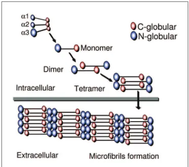
Collagen VI is formed by three chains: alpha-1, alpha-2, and alpha-3, each one encoded by a different gene. The structure and macromolecular organization of type VI col-lagen was described by engel et al.66, and later by Baldock
et al.70 and Ball et al.71 reported in details the biochemical
basis of the tetrameric and microibrillar organization. The three chains contribute to the formation of a triple heli-cal monomer: irst they associate through the C-terminal globular domain and then the triple helix folds from the C to N terminus to form the monomer that is the basic col-lagen VI structure66. The globular amino and
carboxi-ter-minal regions of each monomer are assembled in an an-tiparallel manner by disulide bonds into dimers that are aligned laterally to form tetramers. each tetramer is thre-fore made by eight smaller subunits corresponding to the globular amino and carboxi-terminal regions of the mo-nomeric components of the microibril. The tetramers are then secreted and associated end-to-end to form a char-acteristic microibrils repeat that constructs a network of microibrils (Fig 3). The formation of monomers, dimers and tetramers occurs intracellularly, while the microibril assembly occurs in the extracellular space66,69-71.
Mutations in the genes that codify the three colla-gen VI subunits lead to the second most common form of CMD, Ullrich atonic-sclerotic CMD72-75 and to Bethlem
myopathy76,77 that now is included into CMD classiication8.
the main pathogenic aspects and therapeutic perspectives of the most common forms of CMD that are caused by changes in this complex: CMD with merosin (laminin al-pha-2) deiciency (MDC1A); collagen type VI related CMDs (Ullrich CMD and Bethlem myopathy); CMDs with abnor-mal glycosylation of alpha-DG (Fukuyama CMD, MeB dis-ease, WWs, MDC1C, MDC1D).
Finally, we present the main pathogenic data and ther-apeutic researches about the much rare rigid spine CMD caused by a defect of an endoplasmic reticulum protein, selenoprotein N.
merosin-defiCient Congenital musCular dystrophy: mdC1a
Pathogenesis
MDC1A is caused by mutations in the laminin alpha-2 gene (lAMA2) and was irst described by Tomé et al. in 199460. In the same year, hillaire et al.78 demonstrated that
this speciic CMD is linked to 6q2, and soon after heib-ling-leclerc et al.79 found the irst homozygous mutations
in lAMA2 gene in two patients who had been reported by Tomé et al.60. The disease has congenital onset and
progressive course and patients manifest severe muscu-lar weakness and atrophy, diffuse contractures, inability to walk and facial dysmorphism61,2,60. Diffuse low-density
areas in the cerebral white matter are the hallmark of this condition1,2,60. In a few patients who have partial laminin
alpha-2 deiciency, a later onset and a milder course can be observed1,2. on muscle biopsies the dystrophic
pat-tern is marked with iber size variation, huge interstitial
proliferation and fatty iniltration1,2,60. Necrosis and
re-generation features are less prominent. Taniguchi et al.80
showed that regenerative features are less prominent in MDC1A and FCMD than in Duchenne muscular dystrophy. FCMD and MDC1A muscular samples of patients at differ-ent stages of life showed increased expression of extracel-lular matrix genes and downregulation of genes that cod-ify structural components of mature muscle. This active interstitial ibrosis with less active regeneration of muscle cell components suggested that FCMD and MDC1A could be primary ibrotic diseases80.
Due to the fact that the alpha-2 subunit of laminin is also expressed in the basal lamina of schwann cell-axon unit, a peripheral demyelinating neuropathy affecting pre-dominantly motor ibers81, but also the sensitive ones82,
is a feature of laminin alpha-2 -deicient CMD. In the dy
mouse model laminin alpha-2 is expressed in the en-doneurium surrounding the schwann cell/myelin sheath and interacts with dystroglycan and/or alpha 6 beta 4 in-tegrin located at the outer membrane of schwann cell/ myelin sheath. Its expression inluences the peripheral myelinogenesis83. however, peripheral motor nerves
in-volvement has not been found in Brazilian children with MDC 1A84,85 and this fact might be due to the type and
lo-cation of the mutation84.
The characteristic pattern of white matter abnormal-ity associated to MDC1A has been extensively analysed. opposite to the peripheral nerve, in which laminin alpha 2 is associated only with myelinated and not with unmy-elinated nerve ibers and is involved in the myelin stabil-ity86, a role for laminin alpha-2 in central myelination has
not been conirmed. Villanova et al.87 found that
lamin-in alpha-2 chalamin-in is localized to the basal lamlamin-ina of all ce-rebral blood vessels and supposed that it may be impor-tant for the selective iltration capability of the blood-brain barrier. In patients with MDC1A the lack of lamin-in alpha-2 may lead to an abnormality of the blood-bralamin-in barrier causing impaired selective iltration. Caro et al.88
postulated that disruption of the blood-brain barrier as-sociated with laminin alpha-2 leads to increased water content, resulting in abnormal white matter signal inten-sity. Using magnetic resonance spectroscopy and appar-ent diffusion coeficiappar-ent mapping, leite et al.89 detected
abnormally high free-water concentrations in the white matter of our Brazilian patients with MDC1A and more prominent changes in the parietal, frontal, and tempo-ral white matter. They also found no correlation between the extent of white matter abnormality on MrI and the clinical status as well as the degree of laminin alpha-2 de-iciency (partial or total). sijens et al.90 in a patient with
MDC1A observed that the concentrations of all metabo-lites detected by brain magnetic resonance spectroscopy were decreased only in the white matter. This observation Fig 3. Partial reproduction of Fig 1 from Baldock et al.114: “a diagram
combined with fractional anisotropy decrease and appar-ent diffusion coeficiappar-ent increase in the white matter indi-cated a presence of vasogenic edema in the white matter. The same was concluded by Brockmann et al.91 that
ob-served a metabolite proile suggesting white matter ema which supports the hypothesis that laminin alpha2 de-iciency results in leakage of luids across the blood-brain barrier. Anyway, the pathophysiology of the white matter changes is not completely elucidated.
lAMA 2 mutations are markedly variable spanning all protein domains92-94 and, in general, the molecular
diag-nosis is not considered a priority in children with MDC1A, due to the homogeneous phenotype, the easy immunohis-tochemical analysis of laminin alpha-2 chain in muscle1,2,60
and skin95, and the characteristic white matter
abnormali-ties on neuroimaging. however, siala et al.96,97 have
empha-sized the utility of mrNA analysis in cases of MDC 1A to understand the mechanism of the mutation and the gen-otype-phenotype correlation. recently, also oliveira et al.98 emphasized the utility of analyzing
genotype/phe-notype correlations in MDC1A. In 26 patients they report-ed 96% of mutation detection rate using full genomic se-quencing and complementary DNA analysis. They detect-ed new polymorphisms and novel nonsense and truncat-ing mutations, as well as the irst fully characterized gross deletion. Interestingly, in the two patients with the mild-er clinical presentation and a partial reduction in muscle laminin-alpha2 one of the alleles showed a missense mu-tation that was not observed in any of the severe cases98.
Therapeutic perspectives
From 1994, knock-out mouse or spontaneousmutant mouse strains have been identiied as animal models for MDC1A with total and partial deiciency99-105, and
experi-mental therapeutic strategies have been attempted 102,103,106-116. In mice, Kuang et al.102,103 were successful in obtaining
the expression of a human laminin alpha 2 chain trans-gene under the regulation of a muscle-speciic creatine kinase promoter. They detected the synthesis of laminin alpha-2 in muscle and a marked improvement of the clin-ical and histopathologclin-ical changes; however, there was no improvement of the peripheral neuropathy and they con-cluded that potential new therapies have to be also di-rected to restore laminin alpha-2 in other tissue as CNs and peripheral nerves. Vilkin et al.106 studying both human
and mouse myogenic cells veriied that pure muscle cells are able to synthesize laminin alpha-2 chain. Using primary muscle cell culture transplantation, they found that lami-nin alpha-2 is secreted and expressed in both non-phic muscles of an immunodeicient mouse and dystro-phic muscles of a CMD animal model. however, not all myoblasts or muscle ibers that they form are capable of secretion and deposition in vivo as cell spreading and
dif-fusion of the protein remain restricted to the injection site. In addition, the success of myoblast transplantation depends on future advances for controlling the immune response of the receptor and his biochemical adequacy with the donor. In dyW/dyW mice a “replacement therapy” using a mini-agrin (miniaturized form of agrin) resulted in a lesser muscle degeneration and in a decrease of mortality that is mediated through the linking of muscle basement membrane to the DGC, and not to integrin107. The irst
at-tempt of somatic gene therapy to treat mice with laminin alpha-2 deiciency was performed by Qiao et al.108. They
used adeno-associated viral vector for delivering mini-agrin gene into dyW/dyW mice and obtained an improve-ment of the dystrophic pattern. Mini-agrin not only ame-liorates the phenotype in those mice but also decelerates disease progression when applied at late stages. Meinen et al.109 demonstrated that full-length agrin is also capable
of promoting this improvement This result indicates the role of DG not only in the pathogenesis of CMDs caused by hypoglycosilation of alpha-DG, but also in MDC1A. The improvement of the phenotype may be due to the link between ‘non-muscle’ laminins and DG through miniagrin, therefore maintaining the sarcollema stability. These au-thors109 also supposed that since the genetic manipulation
in dyW/dyW mice can prevent apoptosis, it might be pos-sible that antitiapoptotic agents act synergistically with mini-agrin. Therefore, a future therapeutic attempt for patients with MDC1A could be the combination of anti-apoptotic drugs with the expression of mini-agrin in mus-cle, and/or the up-regulation of endogenous agrin. Gaw-lik et al.110-112 generatedlaminin alpha-2 deicient mice
ex-pressing laminin alpha-1 inperipheral nerves and muscles.
Dy3K/dy3K mice harboring the laminin alpha-1 transgene in these tissues presented reducedmuscular dystrophy and show peripheral myelination. The inding that trans-genically expressed laminin alpha-1compensates the lack of laminin alpha-2 in muscles and peripheral nerves indi-cates that laminin alpha-1 can be an ideal candidate for replacing laminin alpha-2 chain in MDC1A112. In addition,
the authors considered that laminin alpha-1 overexpres-sion induced by gene therapywould be paralogous, there-fore minimizing adverse immunological responses. They also generated generated dy3k/dy3k laminin-a2 deicient
mice in which the expression of alpha-DG in muscle is increased112. Using transgenic expression of laminin-a1,
they obtained a restoration of the basement membrane and a normalization of alpha-DG hyperexpression, there-fore demonstrating a probable interaction between lami-nin-a1 chain and alpha-DG112. laminin-a1 also restored the
link with the integrin at the sarcollema, what represents an additional way for increasing the muscle cell stabili-ty. hagiwara et al.113 tested bone marrow transplantation
in-crease in lifespan, growth rate, muscle strength, and re-spiratory function. Xu et al.114 demonstrated that
embry-onic or postnatal overexpression of cytotoxic T cell Gal-NAc transferase (Galgt2) (the latter using adeno-associat-ed virus) is effective in inhibiting the development of the muscular dystrophy or altering the disease progression in skeletal muscles of dy(w) by increasing the expression
of agrin. Fukada et al.115 reported another possible
ther-apeutic strategy for MDC1A. In normal animals, laminin alpha-2 is produced in CD90(+) cell fractions (from mes-enchimal cells) not depending on its fusion with myogen-ic cells. They demonstrated that the number of C90+cells increases during muscle regeneration process in vivo and as those cells can be transplanted to the animal model of MDC 1A, perhaps they represent a new source of cellular theraphy. Allamand et al.116 using drugs that force
read-ing through premature termination codons (PTCs) dem-onstrated that the mutant mrNAs were strongly stabi-lized in myotubes derived from MDC1 A after administra-tion of negamycin but were not able to allow re-expres-sion of laminin alpha-2, perhaps consequently to transla-tional or post-translatransla-tional troubles. They concluded that this novel form of therapy requires more studies to de-ine the nucleotide context of PTCs, the mechanism of mrNA stability and its translation into a functional pro-tein. Finally, the future treatment of lamininopathies may include stem-cell approaches as well as gene therapy53.
Collagen Vi related musCle disorders
Pathogenesis
Mutations in each one of the three collagen VI genes, Col6A1 (21 q22.3), Col6A2 (21 q22. 3), and Col6A3 (2 q37) that encode respectively the alpha-1, alpha-2 and alpha-3 chains of collagen VI, cause two types of muscle disor-ders: Bethlem myopathy, with mild or moderate pheno-type, and Ullrich CMD, with severe phenotype1-4,6,7,69,117.
Bethlem myopathy is a slowly progressive disorder with variable age of onset, congenital or within the irst or sec-ond decade of life, and marked lexion contractures of several joints7,69,76,77,117-119. Ullrich CMD is an early onset
con-dition with severe weakness and progressive course that manifests proximal joint contractures and marked distal hyperlaxity. Protruding calcanei, follicular hyperkeratosis, and abnormal scarring are other frequent clinical signs1-4, 6,7,69,72,73,118,119.
Until few years ago, Bethlem myopathy and Ullrich CMD were separate entities with distinct modes of in-heritance; after the recognition of heterozygous in-frame deletions acting in a dominantly-negative way in patients with Ullrich phenotype the boundaries between Bethlem myopathy and Ullrich CMD became narrow69. According
to lampe and Bushby69 it is becoming clear that BM and
UCMD represent opposite end points of a clinical
con-tinuum in which individuals presenting with intermedi-ate phenotypes could be considered to have either ‘‘mild UCMD’’ or ‘‘severe BM’’. The concept of a spectrum of col-lagen VI-related disorders with marked clinical and genet-ic heterogeneity has emerged from the recent advances on the molecular mechanism of both diseases69. The
com-plex genotype/phenotype correlations that have been broadly analysed in these two conditions clearly indicate that in both collagen VI-related disorders the main path-omechanism is due to the disruption of collagen VI an-chorage to the basal lamina of the muscular ibers74,75,120-138.
Presently more than 60% of patients with collagen VI related disorders have autosomal dominant or recessive mutations identiied121; however, the highly polymorphic
nature of the three genes suggests the need of speciic methods of mutations analysis for performing an adequate genetic counseling121. since the triple helical deletions are
readily detectable by rT-PCr analysis, lampe et al.122
rec-ommend that mutation analysis checks these regions irst by rT-PCr prior to screen all the 107 coding exons.
The data from molecular analysis in Bethlem myopa-thy revealed that Col6A1 is the most involved gene and a splice site mutation seems to be the most common, not only in Col6A1 as also in Col6A2 and Col6A3 genes123,124.
The α1exon 14 skipping mutation is the most commonly reported in Bethlem patients75,121,123-125, leading to reduced
amounts of collagen VI protein, some of which is struc-turally abnormal with an impaired ability to form micro-ibrils69. As different kinds of mutations have been found,
the pathogenic mechanism of Bethlem miopathy and the degree of clinical severity depend on the effect of the mutation on the structure, biosynthesis, secretion and as-sembly of collagen VI. In patients with different types of mutations, many have attempted to interpret the way by which the mutation alters the collagen VI organization and deines genotype/phenotype correlations. For ex-ample, a heterozygous nonsense mutation causing an al-pha-1 premature stop codon and a decrease of available alpha-1 chains consequently reduces production and se-cretion of collagen VI126. otherwise, a heterozygous
mis-sense mutation interrupting the repetitive amino acid se-quence (glycine mutation) that forms the characteristic collagen triple helix either in the alpha-1or alpha-2 chains does not affect collagen VI intracellular monomer, dimer, and tetramer assembly or secretion, but the mutant te-tramers are anomalous (kinked) and the extracellular mi-croibrils are functionally deicient127. Patients reported
with a heterozygous in-frame deletion in the triple-heli-cal domain resulting in exon skipping in the Col6A1 gene have either a typical clinical picture with inger contrac-tures125 or a course more severe than that commonly
re-ported128. some data indicate that large deletions and
he-lix formed by alpha-1, 2 and 3 polypeptides are associat-ed with a phenotype more severe than those mutations occurring in the amino-terminal globular region118. Pepe et
al.129 found two large and highly similar heterozygous
Co-l6A1 genomic deletions, spanning from intron 8 to exon 13 or intron 13, in two patients, one with Bethlem myopa-thy and the other with moderate Ullrich CMD. They not-ed that as a similar deletion had been previously report-ed in the severe Brazilian case with Ullrich CMD, the pres-ence of a deletion-prone region involving the minisatellite in intron 8 of Col6A1 could be suggested129.
lampe and Bushby69 summarized the most frequent
pathogenic mechanisms in Bethlem myopathy: single amino acid substitutions disrupting the Gly-Xaa-yaa mo-tif of the triple helical domain in Col6A1, Col6A2, or Col6A377,121,127,130,and splice site mutations75,121,125,128 which
cause skipping of Col6A1 exon 14 during pre-mrNA splic-ing and consequently in-frame deletion of 18 amino acids from the triple helical domain of the a1 chain. In the irst case, mutations towards the N terminal region of the tri-ple helix may cause kinking of the tetramers in the nor-mally straight supercoiled triple helical region, thus re-ducing their ability to form microibrils127 and exerting a
dominant negative effect. Baker et al.124 conirmed that
the majority of the Bethlem myopathy mutations occur toward the N-terminal end of the triple helix, that is a critical region of the collagen VI monomer (α1:α2:α3) is critical for eficient dimer, tetramer, and microibril as-sembly. however, in the second case, the interpretation of the inluence of missplicing mutations on collagen VI organization is dificult and needs caution69.
recently, in one patient with Bethlem myopathy it was described a novel mutation that apparently did not affect the assembly, and it was suggested that its effect could be inluencing collagen VI interactions in the extracellu-lar matrix124.
In relation to Ullrich phenotype, the initial molecu-lar studies in Col6A2 and Col6A3 genes pointed only to recessive mutations73,74,118. lampe and Bushby69 revised the
literature about the type of mutations in Ullrich CMD and reported a large number of the mutations that introduce premature termination codons with consequent nonsense mediated mrNA decay and loss of the mutated chain; an-other common type of mutation is by splicing that leads to in-frame exonic deletions as well as in-frame genom-ic deletions. Different missense changes can also occur within the triple helical and C terminal domains of Co-l6A2 and the N-terminal domains of Col6A3 their inter-pretation is dificult.
In 2003, the irst heterozygous in-frame deletions act-ing in a dominantly-negative way was found in the Co-l6A1 gene75 in one Brazilian patient with severe Ullrich
phenotype131; soon, more patients with a dominantly
act-ing mutation in the Col6A1 were reported132-134, and inally
this same type of mutation has also been found in Ullrich patients with mutations in Col6A2 and Col6A3 genes132.
heterozygously occurring N-terminal triple helical in-frame deletions allow mutant monomers to form dimers but secreted tetramers are abnormal with a consequent dominant negative effect on microibrillar assembly132. In
general, the complete absence of collagen VI in the extra-cellular matrix depends on mutations that exert a strong dominant-negative effect and compromise intracellular assembly of dimers, tetramers, and extracellular micro-ibrils132. however, the pathogenic effect of the in-frame
heterozygous deletions is still poorly understood and the genetic counselling of patients and their families is a difi-cult task, as it has been also demonstrated by Peat et al.135.
Pace et al.136 recently considered that in spite of the
great number of different types of mutations that have been already identiied detailed analyses of the effects of the mutations on assembly of the protein have been conducted only in a small number of patients75,124,125,127,132.
Pace et al.136 investigated in eight patients the mechanism
by which different dominant glycine mutations in the col-lagen VI triple helix produce a spectrum of clinical phe-notypes from mild Bethlem myopathy to severe UCMD, therefore pointing to the fact that the collagen VI relat-ed disorders form a continuum of clinical severity. Their study136 identiied that in both, Bethlem and Ullrich CMD,
heterozygous glycine mutations toward the N-terminal end of the triple helix impair the collagen VI assembly in two different ways, therefore producing different pheno-types: in the irst that correspond to less compromised patients, collagen VI dimers accumulated in the cell but microfibril formation in the medium is moderately re-duced, with no signiicant collagen VI decrease in the ex-tracellular matrix; in the second, that corresponds to se-verely affected Ullrich patients, the assembly defects are more pronounced, some secreted collagen VI tetramers are not disulide bonded, microibril formation in the me-dium is altered, and collagen VI is decreased in the extra-cellular matrix136. Pace et al.136 supposed that the relative
positions of the mutations contribute to understand why some glycine mutations in this region have no detectable effect on disulide bonding whereas others result in se-cretion of nondisulide bonded tetramers. In conclusion, they demonstrated that in patients with different domi-nant glycine mutations a mild phenotype is associated to a mild assembly defect whereas severe assembly defects result in moderate-to-severe phenotype.
okada et al.138 found that in Japanese population
26 Japanese patients with primary collagen VI deiciency that in Japan accounts for 7.2% of CMD cases. By immu-nohistochemical analysis they found that most patients had sarcolemma-speciic collagen VI deiciency and ive had complete collagen VI deiciency, i.e. sarcollema plus interstitium. In the former group all mutations were spo-radic dominant; however, in spite of the occurrence of this apparently speciic localization of mutated collagen VI, they could not deine any genotype/phenotype corre-lation. Kawahara et al.139,140 also found that in Ullrich CMD
due to heterozygous mutations in Col6 genes collagen VI is preserved in the interstitium but lost in the sarco-lemma. As in normal conditions collagen VI is particu-larly abundant close to the cells and in intimate contact with basement membranes surrounding myocytes, its lack caused by heterozygous mutations in Col6 genes dimin-ish the anchorage of collagen VI microibrils to the extra-cellular matrix.
Finally, lampe et al.122 recently concluded that in
col-lagen VI related disorders the genotype-phenotype cor-relation does exist and that the type and location of exon skipping mutation is predictive for severity and inheri-tance. They compared the molecular data of patients with Ullrich CMD with de novo dominant negative heterozy-gous splice mutations in Col6A1, Col6A2, and Col6A3, Ullrich CMD with recessively acting splice mutations, and Bethlem myopathy with heterozygous splice mutations. For the three types of mutations, i.e. dominant and re-cessive Ullrich CMD as well as dominant Bethlem myo-pathy, they discussed the different mechanisms by which the location of the skipped exon in relation to the mo-lecular structure of the collagen chain and to the degree of interference on the collagen VI microibrils assembly, strongly correlated with the clinical phenotype. There-fore, they add new evidence to the knowledge that in last instance the clinical severity depends on the ability of mutant chains to be incorporated in the inal multimeric structure of collagen VI microibrillar network122.
Although rarely, Ullrich phenotype can be detected even in patients who do not harbor mutations in the col-lagen VI genes and show a normal amount of colcol-lagen VI in the interstitium. Ishikawa et al.141 considered that these
patients could have a primary abnormality of other not yet identiied protein interacting with collagen VI in the sarcolemma causing a failure of collagen VI to anchor the basal lamina to the interstitium. however, according to okada et al.138 the possibility of mutations affecting the
promoter regions or introns, or of overlooked mutations must be considered in such cases.
In conclusion, it is clear that the complex genotype-phenotype correlations of collagen VI- related disorders are next to be elucidated. recently137, three novel
colla-gen VI chains, alpha-4, alpha-5, and alpha-6 were found
in mice in or close to basement membrane. These chains structurally resemble the collagen VI alpha-3 chain, appar-ently having the ability of substitute it in the constitution of the heterotrimers. This identiication could implicate in new indings related to the molecular biology of Beth-lem myopathy and Ullrich CMD.
Therapeutic perspectives
As for other types of muscular dystrophies, many re-searches focusing on possible effective therapies have occurred. An animal model of human Bethlem myopa-thy has already been described and the details about the composition and the role of collagen VI have been wide-ly discussed117,142.
For at least some types of mutations, Kawahara et al.139
suggested a therapeutic strategy based on a replacement therapy: in ibroblasts culture from patients with Ullrich CMD and Col6A1 glycine mutation, they veriied that i-broblasts adhesion was markedly reduced but could be re-covered by the addition of a medium with normal colla-gen VI139. recently, Merlini et al.143 reported the results of
an open pilot trial with oral cyclosporin A in ive patients with collagen VI myopathies. After a month of treatment a new muscular biopsy showed a decrease of the mito-chondrial and apoptotic changes in the myoibers as well as an increase of regenerative signs143. According to
An-gelin et al.144,145, an anomalous mitochondrial
depolariza-tion has been demonstrated in myoblasts from patients with Ullrich CMD, and cyclosporin A as well as intracel-lular Ca (2+) chelators, probably, prevent such anomalous mitochondrial depolarization and a consequent ATP de-pletion. Also Zou et al.146 recently reported another
pos-sible implication for future therapies. By means of immu-noluorescence staining and Western blot analysis in vit-ro they found that the secretion and deposition of colla-gen VI in the extracellular matrix depends on interstitial ibroblasts but not on myogenic cells146. studies on animal
models for collagen VI related disorders may further offer good perspectives for therapeutic researches147,148.
disorders of glyCosylation of alpha-dg (alpha-dystroglyCanopathies)
Pathogenesis
The alpha-dystroglycanopathies include different forms of dystrophies whose clinical severity varies from WWs that is associated with ocular abnormalities and CNs malformations and is not compatible with survival beyond the irst years of life, to forms of lGMD with lat-er onset latlat-er and pure muscular involvement. Five forms of CMDs are alpha-dystroglycanopathies1-7: FCMD, MeB
and MCD1D, with only one report, is also associated to se-vere muscular and CNs involvement. MDC1C can manifest pure, although severe, muscular involvement or presents associated clinical and radiological CNs involvement, sug-gested by mental retardation, cerebellar cysts and white matter abnormalities on neuroimaging149-151. The lack of
CNs involvement in some patients can suggest that skel-etal muscle is more sensitive to perturbation of dystro-glycan30. The clinical and diagnostic particularities of each
dystroglycanopathy were discussed in Part I of this review. The genetic and clinical heterogeneity of dystroglycano-pathies are remarkable and, except for FCMD is impossi-ble to correlate a particular gene with a speciic pheno-type (Table 2).
The pathogenic mechanism that explains the se-vere forms of CMDs associated with CNs manifestations emerged in 2001 from different works16, 152-154.
Brocking-ton et al.152 identified a gene that codifies a protein of
the fukutin family termed fukutin related protein (FKrP) assigning it to 19q13.3. Mutations in the FKrP gene were found in children with a severe form of CMD who man-ifested inability to walk and muscle hypertrophy but no CNs changes and whose muscle samples showed a sec-ondary deiciency of laminin alpha-2 immunostaining and a decreased expression of alpha-dystroglycan (DG). They
named this new genotype/phenotype association as MD-C1C and suggested that the mutations in the FKrP gene causing a defective glycosilation (hypoglycosilation) of al-pha-DG would explain the basic pathologic mechanism in MDC1C152. hayashi et al.153 found that the highly
glyco-sylated epitope of alpha-DG was markedly deicient in skeletal and cardiac muscle and reduced in brain tissue obtained from patients with FCMD. These indings sug-gested that also fukutin could be involved in the glyco-sylation of alpha-DG that, when altered, could induce a disruption in the extracellular surface membrane of the muscle iber, also inluencing CNs development153.
yoshi-da et al.16 found that the MeB gene codiies a
glycosyl-transferase named PoMGnT1 that is involved in o-man-nosyl glycosylation and, therefore, suggested that an al-tered glycosylation could be a new pathomechanism for CMDs with neuronal migration disorder16. Kano et al.154
re-ported a selective deiciency of alpha-DG in MeB patients; therefore, they suggested that PoMGNT1 acts on alpha-DG and that altered glycosylation of alpha-alpha-DG is involved in the pathogenesis of MeB.
At last, in 2002 two works from Campbells’ research group at the Iowa University17,18 demonstrated that
pa-tients with MeB and FCMD, who carry mutant PoMGnT1 and fukutin genes, respectively, had an abnormal
hypo-Table 2. Genotype/Phenotype correlations in CMDs caused by alpha-dystroglycanopathy (the most common phenotype associated to each gene is underlined, and the bibliography refers to the irst molecular identiication).
FKrP: fukutin
related protein FKTN: fukutin PoMGnT1
PoMT1: protein o-mannosyl transferase 1
PoMT2: protein o-mannosyl transferase 2
glycosyiltransferase “like”: lArGe
lGMD2I197 FCMD199 MeB16 WWs205 WWs156 two patients
with severe CNs involvement: MDC1D208 and WW209 MDC 1C152 WWs200 WWs50 MDC with mental retardation,
microcephaly, structural brain changes, muscle hypertrophy
and eventual myopia206
MeB171
WWs198 MeB50 lGMD2? with severe myopia and normal
intelligence50,203
MeB50 MDC with microcephaly, severe mental deiciency, possible ocular changes171,172 MeB198 Myocardiopathy,
minimal or absent muscle weakness, normal ntelligence201
MDC with brain malformations without ocular abnormalities204
MDC and cerebellar involvement50
MDC with
cerebellar cysts151 lGMD2M
202 lGMD2? mild, with normal
intelligence and inlammatory changes on muscle biopsy207 MDC with variable
cortical, cerebellar and pontine dysplasic changes149
lGMD2? with mental retardation50
glycosilation of alpha-DG that abolished its binding with the ligands, laminin alpha-2, agrin, and neurexin of the ex-tracellular matrix. The same occured in the myodystro-phy (myd) mouse that carried a lArGe mutation and in the knock-out for brain-selective alpha-DG mouse. In the knock-out mouse they found aberrant cerebral and cere-bellar migration probably derived from a discontinuos pial surface basal lamina (glia limitans); they considered that the aberrant migration, similar to that seen in patients with MeB and FCMD, was due to the lack of DG preventing its high-afinity binding to the extracellular matrix lamin-in. These results identify alpha-DG as having an essential role in both muscle and brain development and function and suggested that PoMGnT1, fukutin, and lArGe may have a role in transferring sugars to alpha-DG, i.e. act as glycosyltransferases. The recognition of the involvement of other two glycosyltransferases, PoMT1 and PoMT2, in the pathogenesis of CMDs came soon after155-156.
During the last seven years, many have discussed and elucidated the pathogenesis of the defective glycosyla-tion of alpha-DG, and the particular cortical involvement that is observed in many of them5,711,13,15,17,18,30,33,34,36-47,157-161. It
is supposed that other subtypes of CMD will prove to de-pend on mutations in genes as yet unidentiied but also involved in alpha-DG glycosylation39,45,46,48. In addition,
from the irst reports on congenital disorders of glyco-sylation, it seemed unlikely that the glycosyltransferases could have a role only in glycosylation of alpha-DG and the importance of looking for other protein targets has been emphasized37,38.
The role of alpha-DG in developing brain and in neu-ronal migration has been studied in animals models11,18,158, 159,162,163. Zaccaria et al.162 found that in adult mice alpha-DG
is expressed in neurons of the cerebral cortex, hippocam-pus, olfactory bulb, basal ganglia, thalamus, hypothala-mus, brainstem and cerebellum; in addition the astrocytes and their endfeet around blood vessels and the endotheli-al cells at the blood-brain barrier, endotheli-also expressed DG. Dur-ing CNs development, alpha-DG is expressedin the ven-tricular zone and in basement membranes, therefore par-ticipating in neuronal proliferation, in the constitution of the meningeal layer and in migration process as the radi-al glia is attached to the piradi-al basement membrane158. In
DG-null mice, then abnormal glycosylation of alpha-DG leads to different defects of neuronal migration that at the maximum of rupture of the pial-glial limitans, closely resemble the cobblestone aspect of cortex in human lis-sencephaly type II18.
The number of glycosiltransferases involved in the gly-cosylation of alpha-DG and their different possibilities of regional and developmentalexpression explain the spec-trum of malformations variability and clinical severity that are observed in the different CMDs caused by
alpha-dys-troglycanopathies. yamamoto et al.164 analysed the
expres-sion and localization of fukutin, PoMGnT1, and PoMT1 in the CNs and discussed the mechanism of lesions linked to these three glycosyltransferase whose changes are rep-resented by migration disorders in FCMD, MeB, and WWs. They observed that fukutin, PoMGnT1, and PoMT1 are ex-pressed especially in astrocytes that compose the astro-cytic glia limitans and therefore is involved in the forma-tion of the basement membrane. As the latter was found disrupted in fetal cases of FCMD, MeB, and WWs the de-fect in any of the three glycosyltransferases can be relat-ed to the pathogenesis of CNs lesions. These are also ex-pressed in immature neurons what suggests their proba-ble involvement in neuronal migration itself. In mature neurons PoMGnT1 and PoMT1 are expressed but fukutin is rarely positive indicating that at late stages of develop-ment PoMGnT1 and PoMT1 have a more crucial role than fukutin163. In addition, fukutin is expressed in pancreas,
kidneys, bronchi, salivary gland, alimentary tract and skin in both fetal and adult mice, and in autopsied FCMD cas-es it was found decreased in kidneys, lung, skin and intcas-es- intes-tine163. These indings demonstrate the role of alpha-DG
glycosylation also in non-muscle systems both during de-velopment and in the adult; studies with the aim of clari-fy why muscle and brain symptoms predominate in FCMD would be useful for better understanding the complex pathogenic mechanism of alpha-dystroglycanopathies163.
The wide phenotypical variation of the alpha-dystro-glycanopathies, in particular the FKrP-related myopathies, has not a clear pathogenic basis. A study analysed patients with FKrP mutations who had also CNs alterations and concluded that thedistribution of FKRP gene mutations did not allow any particular genotype/phenotype corre-lation concerning the amount of CNs malformations149.
Keramaris et al.24 considered that the type of missense
point mutation could in part explain the phenotypical variation of FKrP gene mutations that alter the normal subcellular localization of FKrP protein in the Golgi ap-paratus: the protein is kept in the endoplasmic reticulum and diminishes in the Golgi apparatus. Therefore, each mutation could act in two ways, directly altering the pre-sumed glycosyltransferase activity and further altering the protein function by the mislocalization24. In patients with
FKrP mutations, the residual expression of alpha-DG de-tected by immunolabeling and Western blot can be corre-lated with the clinical severity and with the type of muta-tion168. Patients with MDC 1C who exhibit a severe
are compound heterozygotes (a missense mutation and either another missense or a nonsense mutation) while patients at the milder end (later onset) generally present variable immunohistochemical pattern, sometimes only minimally changed, and in general are homozygous for the common C826A/leu279Ile FKrP mutation168.
PoMT1 and PoMT2 mutations are not associated to the same clinical heterogeneity that is observed in the FKrP and in general cause classical WW or MeB pheno-types. however, out of these classical phenotypes, dif-ferent types and degrees of cortical and posterior fossa malformations have been described156, 169-174, with or
with-out eye involvement; mental retardation, and calf as well as thigh enlargement has also been related170,171.
yanagisa-wa et col172 hypothesized that patients with PoMT1 and
PoMT2 mutations could share the same phenotype be-cause, according to Manya et al.21, both
glycosyltransferas-es form a heterodimeric complex that is rglycosyltransferas-esponsible for the catalysis of the irst step in O-mannosyl glycan synthe-sis. Messina et al.175 emphasized that PoMT1 and PoMT2
mutations have a wider clinical spectrum than initially thought and that, excluding MeB and WWs phenotypes, mutations causing frameshifts and stop codons were asso-ciated to the most severe phenotypes with predominant cerebellar hypoplasia commonly found. Godfrey et al.50
analysed eight patients with PoMT1 mutations and nine with PoMT2 mutations and suggested that POMT1 and
POMT2 mutations do not seem to manifest a hierarchical involvement ofmuscle and brain, being associated with signiicant central nervoussystem involvement even in pa-tients with relatively mild weaknessand who remain am-bulant (lGMD2K). In this large revision about alpha-dys-troglycanopathies not related to FKrP mutations Godfrey et al.50 found a broad correlation betweenthe amount of
depleted glycosylated epitope and phenotypic severity. recently another study165 demonstrated a good
cor-relation between the clinical phenotype and the amount of glycosylated alpha-DG with the most severe cases be-ing associated to a lack or marked reduction of glycosylat-ed alpha-DG. however, this correlation was strong in pa-tients with PoMT1, PoMT2 and PoMGnT1 mutations, but not consistent in patients whose mutations had occurred in fukutin and FKrP genes.
The amount of dystroglycan hypoglycosylation is de-tected utilizing two monoclonal antibodies (IIh6 and VIA41) that recognize only the functional glycosylated epitope of alpha-DG30; further immunohistochemical studies will
be useful for identifying all the glycan structures induced by glycosyltransferases, in particular lArGe activity30.
Considering all the alpha-dystroglycanopathies that have already been supposed by means of hypoglycosila-tion on the immunohistochemical analysis of muscle bi-opsies, Muntoni et al.5 pointed to a rate of conirmed
mo-lecular diagnosis around 65% of the cases, suggesting that more genes have yet to be identiied. The same is clearly demonstrated by the work from Godfrey et al.50 that
de-scribed many patients who had a phenotype suffesting al-pha-dystroglycanopathy but no mutations in any of the six glycosyltransferases already identiied. Therefore, they concluded that undeined, genes are likely to beinvolved in the pathogenesis of the dystroglycanopathies and that theidentiication of these genes may provide additional informationon the pathway of glycosylation of alpha-DG.
other great evidence pointing to the probability of the existence of more genes codifying glycosyltransferas-es not yet identiied comglycosyltransferas-es from the molecular studiglycosyltransferas-es from patients with WWs. In spite of its worldwide dis-tribution and therefore the good availability of patients’ DNA for genomic analysis, only one third of the cases has been associated to mutations in one of the six genes in-volved in the o-manosylation pathways50,166,167. It is
sup-posed then that additional loci are going to be identi-ied soon30,167.
Finally, Moore and hewitt30 recently hypothesized
that genetic variation between patients for other genes that might have compensatory functions could contrib-ute to this clinical heterogeneity.
Therapeutic perspectives
Glycobiology of o-mannosyl glycans is a broad and open ield of research that can favor the understanding of the pathogenesis and consequently the advent of a possible thearapeutic strategy for the alpha-dystrogly-canopathies. The researches about the long-term poten-tial of glycotherapies for CMD have begun with the irst reports about the defective alpha-DG glycosylation as a new pathogenic mechanism for some subtypes of 7,11,13,15,32-34,36-48,157,176,177. The central point of these researches is that
the overexpression of lArGe promotes the attachement of glycans to alpha-DG, therefore restoring its ligand-binding function178.
Barresi et al.178 demonstrated that in large(myd)mice
overexpression of lArGe leads to a recovery of alpha-DG function as a receptor and ameliorates the dystrophic phenotype; in normal mice induces the synthesis of gly-can-enriched alpha-DG with high afinity for extracellular ligands. They also demonstrated that in cell lines derived from patients with WWs and MeB (PoMT1, PoMGnT1 or fukutin mutations) as well as in transfected cell cultures from all cell lines, gene transfer of lArGe restores the laminin-binding activity of alpha-DG and results in hy-perglycosylation of alpha-DG; these data indicate that, at least in this experimental over-expression system, lArGe may act in a different glycosylation pathway178. Therefore,