Disciplina de Neurologia Infantil - Departamento de Neurologia - Faculdade de Ciências Médicas da Universidade Estadual de Campinas (FCM/UNICAMP) Campinas SP, Brasil: 1 Mestre - Ciências Biomédicas; 2Livre Docente; 3Professor Titular.
Received 21 June 2005, received in final form 24 August 2006. Accepted 19 October 2006. Dra. Sylvia Maria Ciasca - Praça XV de Novembro 40/41 - 13024-180 Campinas SP - Brasil.
CEREBROVASCULAR DISEASE IN CHILDHOOD
Neuropsychological investigation of 14 cases
Inês Elcione Guimarães
1, Sylvia Maria Ciasca
2, M. Valeriana L. Moura-Ribeiro
3ABSTRACT - There are few studies about the development of a child after a cerebrovascular accident (CVA), and they usually describe problems such as diminishing in intellectual capacities, difficulties in linguistic and visual-motor skills, as well as in spatial organization and integration. In this study, there were 28 chil-dren participating, being 14 placed in the experimental group (EG) after clinical diagnosis and ischemic CVA imaging, and other 14 children without past history of CVA, who formed the control group (CG). The neuropsychological research protocol included an intelligence test, a visual-motor coordination test, human figure drawing, a cortical functions battery and the medical records of the children from the EG. The analy-sis of the results of this study revealed that the best performances after the CVA are related to the short-est time of functional recovery; CG presented better performances than EG in all the instruments used, in cognitive, perceptual and motor skills. It has been noticed that CVA may lead to intellectual reduction in case of a recurrence of the vascular insult.
KEY WORDS: cerebrovascular disease, childhood, neuropsycological investigation.
Doença cerebrovascular na infância: investigação neuropsicológica em 14 casos
RESUMO - Nos poucos estudos acerca do desenvolvimento da criança após o acidente vascular cerebral (AVC) são descritos problemas como rebaixamento intelectual, dificuldades na capacidade lingüística, viso-motora, na organização e integração espacial. Neste estudo fizeram parte 28 crianças, sendo 14 perten-centes ao grupo experimental (GE) com diagnóstico clínico e de imagem de AVC isquêmico. Outras 14 cri-anças sem história de AVC que formaram o grupo controle (GC). O protocolo de investigação neuropsi-cológica constou de teste de inteligência, teste de coordenação viso-motor, desenho da figura humana, bateria de funções corticais e prontuário médico das crianças do GE. A análise dos resultados deste estu-do mostrou que os melhores desempenhos após o AVC, estão relacionaestu-dos ao menor tempo de recupe-ração funcional; houve desempenho superior do GC sobre o GE em todos os instrumentos utilizados, nas áreas cognitiva, perceptual e motora. Constatou-se ainda que o AVC pode levar a rebaixamento intelec-tual, quando ocorre um quadro de repetição do insulto vascular.
PALAVRAS-CHAVE: doença cerebrovascular, infância, avaliação neuropsicológica.
Literature shows that studies in the area of neu-ropsychological research with children who had cere-brovascular accident (CVA) are scarce; however, over the last years there has been a significant increase in studies which take into account neuroradiological and neurosurgerical aspects1.
In the few studies on children development after CVA, neuropsychological deficits are described2-6. Armstrong et al.7assessed 194 children with sickle cell anemia, amongst whom there were 9 with past history of CVA. These children presented difficulties in linguistic and visual-motor skills, as well as in spa-tial organization and integration. De Schryver et al.5 found impaired capacity for solving problems in post
CVA children, when compared to the general popu-lation. Rodrigues et al.8described children with CVA assed evolutionally by Piaget´s clinical method had present a performance significantly inferior when compared to the control group. The sequelae of a CVA during childhood are not yet as widely known as of one during adulthood; however, the more stud-ies there are in the area, the more details shall be discovered, favoring professionals involved in the rehabilitation of such children9.
METHOD
Twenty-eight children participated in this study; 14 of them belonged to the experimental group (EG) with both clinical and imaging diagnostic of ischemic CVA. Children with vascular problems related to brain-skull trauma and associated to genetic syndromes were excluded. The oth-er 14 children woth-ere placed in the control group (CG), paired according to gender and age to the EG, and besides not having a past history of CVA or another neurological prob-lem, they presented appropriate academic performances. Two public schools of the region of Campinas (Brazil) were chosen by simple random draw, the children were select-ed by the pselect-edagogue and the teacher, whose criterion was a good performance according to the grade.
The participation of each child in both groups was authorized by parents who agreed with the research by signing a Free Consent Agreement Form approved by the Ethical Committee on research of the Faculdade de Ciências Médicas (FCM) of the Universidade Estadual de Campinas (UNICAMP).
Based on the protocol for neuropsychological research on cerebrovascular disease, the following instruments were used: the medical records of each child from the EG belong-ing to the Clinical Hospital (CH) of UNICAMP; the Wechsler Intelligence Scale for Children - WISC10; the Visual-Motor
Gestalt Test11, the Luria-Nebraska Battery (LNB)12and the
Test of human figure drawing.
RESULTS
In order to form the EG a total of 35 children were assessed – who according to inclusion and exclusion criteria were or were not selected for the study. The EG consisted of 14 children, being 7 males and 7
fe-males; the age ranged from 7 years and 1 month to 14 years and 1 month, with an average of 9 years and 10 months. As for schooling, it ranged from 1stto 6th grades, besides special education and no schooling.
As the CG was paired according to age and gen-der to the EG, it was also formed by 14 children, 7 males and 7 females, and with age range during the assessment period of 7 years and 5 months to 14 years and 2 months, with average age of 9 years and 11 months. Schooling ranged from 2ndto 8thgrades.
The results of the study with the total 28 children were organized in a way that relations among the instruments used as procedure of this work and also between both groups (EG and CG) could be demon-strated.
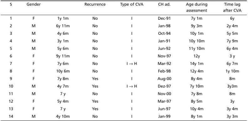
The data from the EG children (Table 1) refer to the information obtained from the medical records at the Clinical Hospital - UNICAMP, showing that the first CVA occurred between the ages of 13 months and 10 years and 6 months, being 2 children during the interval from 0 to 3 years of age; 8 children bet-ween 3 and 6 years of age; and 4 in the period from 9 to 10 years of age. There has been recurrence in 4 children. All the children presented ischemic type CVA and 2 of them had a CVA with hemorrhagic transformation. The admission to UNICAMP’s Clinical Hospital happened from January of 1991 to No-vember of 2000. Children’s age during the assessment ranged from 7 years and 1 month old to 14 years and 1 month old, with average of 9 years and 10 months.
Table 1. Cerebrovascular disease: identification.
S Gender Recurrence Type of CVA CH ad. Age during
assessment
Time lag after CVA
1 F 1y 1m No I Dec-91 7y 1m 6y
2 M 6y 11m No I Jan-98 9y 3m 2y 4m
3 M 4y 6m No I Oct-94 10y 1m 5y 5m
4 M 3y 1m No I Jan-91 10y 10m 7y 9m
5 M 5y 6m No I Jun-92 11y 10m 6y 4m
6 F 9y 11m No I Nov-97 12y 3 y
7 F 7y 6m No I →H Mar-92 14y 1m 6y 7m
8 F 10y 6m No I Feb-98 12y 4m 1y 10m
9 F 7y 8m Yes I Aug-00 8y 4m 8m
10 M 4y 7m Yes I →H Dez-97 7y 10m 3y3m
11 M 7 y No I Nov-00 7y 8m 8m
12 F 5y 4m Yes I Mar-97 8y 5m 3y
13 F 7 y Yes I Jun-97 10y 4m 3y 4m
14 M 4y 10m No I Jan-99 8y 1m 3y 3m
The time lag between ages when vascular insult and assessment occurred ranged from 8 months to 7 years and 9 months, revealing an approximate average of 4 years and 4 months, being 9 of the children aged under the average in the period of assessment and 5 above.
At the time of the vascular insult occurrence, the clinical symptoms presented were: 14 (100%) motor symptoms; 10 (71%) changes in speech; 8 (57%) head-ache; 5 (36%) vomiting; 4 (48%) generalized convul-sive seizure; 3 (21%) fever; 3 (21%) consciousness changes; 3 (21%) somnolence; 4 (48%) visual
diffi-culty (impairment); 2 (14%) sialorrhea; 2 (14%) abdo-minal pain; and 1 (7%) had other symptoms such as dizziness, diarrhea and hypertension.
Table 2 presents information from the imaging exam and neurological evolution after vascular insult. Brain injury in the left hemisphere was found in 7 (50%) of the cases; in the right hemisphere in 5 (36%) and in both hemispheres in 2 (14%) of the cases. In 6 of these cases, the subcortical area was affected, being 2 cortical, 5 cortical-subcortical and 3 in the brain stem. The neurological examination present-ed alterations in 11 subjects and was normal in the
Table 2. Results of the imaging and neurological exams - experimental group.
S Hemisphere Localization Neurological exam Neurological evolution
1 L Subcortical – –
2 R Subcortical and brain stem X Hemicorrhea L
3 R Cortical and brain stem X Hemiparesis L
4 R Subcortical X Hemiparesis L
5 L Subcortical X Hemiparesis R
6 L Subcortical X Hemiparesis R
7 R Cortical-subcortical X Hemiparesis L
8 L Subcortical – –
9 B Cortical-subcortical X Hemiparesis R
10 L Brain stem X Hemiparesis R
11 R Cortical-subcortical – –
12 B Cortical-subcortical X Hemiparesis L
13 L Cortical X Hemiparesis R
14 L Cortical-subcortical X Hemiparesis R
R, right; L, left; B, bilateral; ( x ) presence of symptoms; ( – ) absence of symptoms.
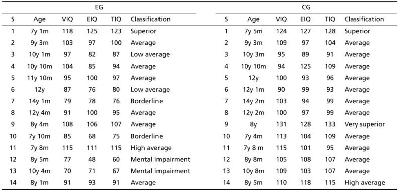
Table 3. Results of the WISC - experimental group and control group.
EG CG
S Age VIQ EIQ TIQ Classification S Age VIQ EIQ TIQ Classification
1 7y 1m 118 125 123 Superior 1 7y 5m 124 127 128 Superior
2 9y 3m 103 97 100 Average 2 9y 3m 109 97 104 Average
3 10y 1m 97 82 87 Low average 3 10y 3m 95 89 91 Average
4 10y 10m 104 85 94 Average 4 10y 10m 94 125 109 Average
5 11y 10m 95 100 97 Average 5 12y 100 93 96 Average
6 12y 87 76 80 Low average 6 12y 1m 90 99 93 Average
7 14y 1m 79 78 76 Borderline 7 14y 2m 103 94 99 Average
8 12y 4m 91 100 95 Average 8 12y 2m 100 97 99 Average
9 8y 4m 108 106 107 Average 9 8y 131 128 133 Very superior
10 7y 10m 85 68 75 Borderline 10 7y 4m 113 104 109 Average
11 7y 8m 115 111 115 High average 11 7y 8 m 115 101 95 Average
12 8y 5m 77 48 60 Mental impairment 12 8y 8m 105 108 107 Average 13 10y 4m 70 71 67 Mental impairment 13 10y 8m 109 103 107 Average
14 8y 1m 91 93 91 Average 14 8y 5m 110 118 115 High average
other 3 children, and it was also found right hemi-paresis in 6 children, left hemihemi-paresis in 4 subjects and left hemicorrhea in only one child.
As for the cognitive assessment (WISC) of the EG, the following classification was obtained for lectual quotient (IQ): one child at the superior intel-ligence range; one child at the high average intelli-gence range, 6 at the average intelliintelli-gence range, 2 children at the low average range, 2 children at the borderline deficiency range and 2 children with re-sults which were compatible to mental impairment.
The following is the CG classification for IQ: 1 child at the very superior intelligence range, 1 child at the superior range, 1 child at the high average range and 11 children at the average intelligence range. These results are organized in Table 3.
Figure 1 shows a comparison of the WISC results from both groups, which reveals an inferiority of the responses of EG over CG. This datum was expected since in the first group there are children with intel-lectual reduction.
The results of the perceptive motor development of the EG in the Gestaltic visual-motor test (Bender) were obtained under two correction systems, known as Santucci and Koppitz. Based on the results, the data were grouped together according to the cor-rection system as follows.
The scores obtained through the Santucci system13 were: 8 (57.1%) children presented average percep-tive motor development, 5 (37.7%) had inferior per-formances and 1 (7.1%) child had a score above the average for his chronological age. While in the Kop-pitz system14– 11 (78.5%) children reached the aver-age and 3 (21.4%) presented perceptive motor matu-rity below average. There was predominance of dif-ficulty in the shape inversion item, followed by rota-tion, integration and perseverance.
The CG presented the following results, which also refer to gross scores from the two correction sys-tems of the Bender test. The perceptive-motor devel-opment of this group shows the following pattern: in the Santucci system – 8 (57.1%) children with aver-age development; 4 (28.5%) above the averaver-age and 2 (14.2%) with perceptive development below the
Fig 1. Comparison of the WISC results from - experimental group and control group.
expected for the age. And in the Koppitz, 14 (100%) of the children presented average pattern for per-ceptive-motor development. Rotation was the most difficult item, followed by shape inversion, integra-tion and perseverance.
In both groups, there has been a concurrence of 85.7% in the correction systems used.
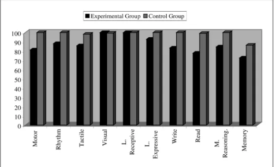
The correct marks obtained in each function of the LNB from EG and CG were transformed into per-centage of correct marks, as it may be noticed in fig-ure 2, comparing both groups. As for the statistical analysis, significative results from CG over EG were found in the following functions: motor skill, tactile skill, writing, reading and memory.
The following results refer to conceptual maturi-ty of the human figure drawing for both EG and CG population. For the EG the percentile variation rang-ed from 1 to 100 while in the CG it rangrang-ed from 22 to 100, increasing in this way the classification of the CG over the EG.
The following classificatory distribution was found in the EG: 2 (14.2%) much superior performances; 1 (7.1%) superior score; 3 (21.4%) above average; 4 (28.5%) average performances, 2 (1.2%) borderline performances and 2 (1.2%) with impaired perform-ances.
While in the CG the following data were found: 3 (21.4%) much superior scores; 5 (35.7%) superior scores; 1 (7.1%) above the average performance; 4 (28.5%) average performances; and 1 (7.1%) border-line performance regarding the maturity of the human figure drawing.
DISCUSSION
CG’s superior performance in the LNB was signif-icant in the following functions: motor and tactile skills, writing, reading and memory. The motor seque-lae in the EG altered the capacity for both global and fine motor coordination, what hindered successful performances in such activities. There was no direct relation between the worst performances and cog-nitive capacity but with motor difficulty, more specif-ically with the hemiparesis condition.
As for the tactile skill both groups presented sig-nificant differences. The left-right domain, mediat-ed in this test, showmediat-ed the maturity function to be delayed, and according to Le Bouch15the stabiliza-tion of the lateral funcstabiliza-tion occurs between 6 and 8 years of age. The statistical significance of CG over EG for writing and reading skills can be justified by factors such as: difficulty in management of the learn-ing-teaching process by both parents and teachers;
longer schooling time of the CG and intellectual reduction of the EG.
There was difference between EG and CG in rela-tion to memory ability, mainly in the exercise of im-mediate auditory memory (word), which made it pos-sible to correlate the results of this test to the digit span subtest, and EG presented low scores in both tests. Regarding neuropsychological mechanisms of memory organization, Barbizet and Duizabo16reveal that the verbal information quantity (verbal or visu-al span) which the individuvisu-al is able to memorize af-ter a single exposition is limited. This capacity de-mands the integrity of functional areas; immediate memory is decreased constantly in cases of damaged cortical areas.
In the works of Lefèvre17and Tabaquim18with chil-dren, the first related to Moyamoya disease and the second about hemiparethic cerebral palsy and learn-ing disorders, the similarity subtest was the one that presented the best performances. The same thing may be observed in both groups of this study; this subtest demands conceptual thought capacity and it is less influenced by social or formal education than other verbal subtests19.
The intellectual deficit found in the EG of this study is superior when compared to the general pop-ulation; however, these indexes may not be consid-ered permanent, once the capacity of cerebral reor-ganization was clearly observed, and adding to this conclusion, there is the fact that remarkable changes in IQ are common in pediatric age groups20, as well as in all age groups.
Vargha-Khadem21reported that cognitive reduc-tion is more common when the CVA occurs between the ages of 2 and 4 years old, but at this present study this finding was not confirmed. Cognitive reduction occurred in children who had CVA between the ages of 5 and 7 years old, which can have been influenced by repetition of the vascular condition.
hemi-sphere) who had not acquired yet or were still in process of acquisition of these functions.
According to Wood et al. in Oliveira22between the ages of 5 and 8 years old, the left hemisphere be-comes more specialized for language, regarding the development of reading and writing functions. Pitch-ford also cited by Oliveira22presented a five children study with CVD in the left hemisphere, being that two of them were in pre-school age. Damage in ver-bal performance with signs of reading and writing difficulty was observed. For Luria23the damages are greater when there are lesions in the primary area, yet lesions of secondary areas imply participation of other functional circuits for functional recovery.
Bender’s constructive visual task, through percep-tion and responses to stimuli, does not constitute a simple process, but involves neuropsychological as-pects of cerebral functioning15. The performance (EG) in the gestaltic visual-motor test, in both correction systems, revealed that reception, integration and expression of information were being processed appropriately.
Antunha24explains that the development of inter-nal image of one’s own body results from the somes-thetic projection (somessomes-thetic and parietal-occipital cortices). In this test, human figure drawing, a good performance was observed in the EG, but it was noticed that the CG has a better group performance, suggesting that both groups present an appropriate representation of body image or scheme.
One aspect that deserves attention is the cogni-tive performance related to gender. Studies have shown that gender differences change cerebral pro-cessing, that is, men and women have different cog-nitive styles25,26.
It may be observed that in this study it was pos-sible to form EG with seven girls and seven boys. Through their performances as a subgroup, the fe-males are found with two individuals presenting cog-nitive performances that are compatible to border-line deficiency and mental impairment. While the males present only one borderline deficiency individ-ual. Another important point is the fact that the female subgroup presents more individuals with CVA recurrence in relation to the male one, a 3:1 ratio.
A valuable analysis of the relation between gen-der and abilities in the population studied here is considered to be impracticable due to cognitive re-duction observed in the female subgroup of the EG. Such finding affects the performances of this sub-group incisively. Nevertheless, it may also be noticed
that in this study the females with a past history of vascular insult present greater possibilities of intel-lectual reduction and CVA recurrence.
In this study, 4 years and 4 months was the aver-age time span between vascular insult and assess-ment. It was not possible to establish stronger strict-ness for the control of this variable due to the low number of casuistic. Out of five children who pre-sented above the average performances only one showed borderline cognitive performance, while the others had normal variation of intelligence. As for the other nine children evaluated who were under this average, two presented evolution with intellec-tual performance compatible to mental impairment, borderline deficiency and the others with normal variation of the cognitive capacity. Moreover, four children had a recurrence of the vascular insult.
In summary, it may be suggested that the children who were assessed above the average of 4 years and 4 months after the CVA had better cerebral reorgan-ization in response to the lesion, revealing more adaptative cognitive and behavioral resources. How-ever, it must be observed that for the children who were assessed under this average the recurrence fac-tor denotes the severity of cerebral damage, once the repercussion of the lesion impact seems to dimin-ish possibilities of plasticity, which may be noticed from behavioral expressions that are or are not in accordance with the age group to which the child belongs. Mello and Muszakat27reveal that a devel-oping brain is less vulnerable to the effect of a lesion. The present study shows the importance of specific evaluations in groups of post-CVA children, because neuropsychological deficits which hinder the individ-ual’s satisfactory development were found. The parameters of a child development after a CVA are still being grounded, yet it is believed that some con-tributions in order to reach them could be substan-tiated in this work.
REFERENCES
1. Bowen M, Marks MP, Steinberg GK. Case report: neuropsychological recovery from childhood moyamoya disease. Brain Dev 1998;20: 119-123.
2. Koelfen W, Freund M, Konig S, Varnholt V, Rohr H, Schultze CH. Results of parenchymal and angiographic magnetic resonance imag-ing and neuropsychoological testimag-ing of children after stroke as neonates. Eur J Pediatr 1993;152:1030-1035.
3. Ganesan V, Isaac E, Kirham FJ. Variable presentation of cerebrovascular disease in monovular twins. Dev Med Child Neurol 1997;39:628-631. 4. Ciasca SM, Alves HL, Guimarães IE,et al. Comparação das avaliações
neuropsicológicas em menina com doença cerebrovascular bilateral (Moyamoya) antes e após a intervenção cirúrgica. Arq Neuropsiquiatr 1999;57:1036-1040.
6. Guimaraães IE, Ciasca SM, Moura-Ribeiro MLL. Neuropshychological evaluation of children after ischemic cerebrovascular disease. Arq Neuropsiquiatr 2002;60:386-389.
7. Armstrong FD, Thompson RJ, Wang W, et al. Cognitive function and brain magnetic resonance imaging in children with sickle cell disease. Pediatrics 1996;97:864-870.
8. Rodrigues SM, Ciasca SM, Moura-Ribeiro MVL. Ischemic cerebrovas-cular disease in chilhood: cognitive assessment of 15 patients.Arq Neuropsiquiatr 2004;62:802-807.
9. Ganesan V, Hogan A, Shack N, Gordon A, Isaacs E, Kirkham FJ. Out-come after stroke in childhood. Dev Med Child Neurol 2000;42:455-456. 10. Wechsler D. Manual for the Wechsler intelligence scale for children.
New York: Psychological Corporation, 1974.
11. Bender L. Teste guestáltico visomotor. Buenos Aires: Paidós, 1964. 12. Golden CY. The nebraska neuropsychological children`s battery. In:
Reynolds CR, Fletcher-Jansen E (eds). Handbook of clinical child neu-ropsychology. New York: Plenum Press, 1989:193-204.
13. Zazzo R. Manual para exame psicológico da criança. São Paulo: Editora Mestre Jou, 1968.
14. Cunha JA. Psciodiagnóstico-R. 4ed. Porto Alegre: Artes Médicas, 1993. 15. Le Bouch J. O desenvolvimento psicomotor: do nascimento até 6 anos.
6.Ed. Porto Alegre: Artes Médicas, 1992.
16. Barbizer J, Duizabo Ph. Manual de neuropsicologia. Porto Alegre: Artes Médicas; São Paulo: Masson, 1985.
17. Lefèvre BH. Neuropsicologia infantil. São Paulo: Savier, 1989. 18. Tabaquim MLM. Avaliação neuropsicológica: estudo comparativo de
crianças com paralisia cerebral hemiparética e distúrbios de aprendiza-gem. Tese, Campinas, 2002.
19. Lezak MD. Neuropsychological assessment. New York: Oxford Universit Press, 1983.
20. Levin HS, Soukup VM, Benton AL, Fletcher JM, Satz P. Avaliação neu-ropsicológica e intelectual de adultos. In: Kaplan H, Sadock BJ (eds). Tratado de psiquiatria. 6.Ed. Porto Alegre: Artmed, 1999:613-633. 21. Varga-Khaderm F. Neuropsychological outcome (Abstr). In 8th
Inter-national Child Neurology Congress. Slovenia, 1998:282-283. 22. Oliveira KT. Distúrbio adquirido em crianças com doença
cerebrovas-cular: aquisição de linguagem em lactentes e pré-escolares. Dissertação. Campinas, 2002.
23. Luria AR. Fundamentos de neuropsicologia. São Paulo:Edusp, 1981. 24. Antunha ELG. Avaliação neuropsicológica na infância. In Oliveira VB,
Bossa ANA. (org). Avaliação psicopedagógica da criança de zero a seis anos. 2.Ed. Petrópolis: Editora Vozes, 1994:89-122.
25. Kolb B, Whishaw IQ. Neurociência do comportamento. Barueri:Manole,2002. 26. Goldberg E. O cerebro executivo: lobos frontais e a mente civilizada.
Rio de Janeiro: Imago, 2002.