Echocardiographic Features of Non-Compaction Cardiomyopathy.
Missed and Misdiagnosed Disease
Francisco Martínez-Baca López
1, Rosa Marisol Alonso Bravo
1, Domingo Arturo Rodríguez Huerta
2Department of Echocardiography1 , Pathologist, Department of Pathology2, Hospital de Cardiología, Centro Médico Nacional SXXI, IMSS, México
D.F., México.
Mailing Address: Francisco Martínez Baca López •
Floresta No. 146-2, Colonia Clavería 02080 México, D.F, México Email: [email protected]
Manuscript received March 13, 2008; revised manuscript received July 8, 2008; accepted July 8, 2008.
Non-compaction cardiomyopathy is a rare disease, anatomically characterized by a prominent trabecular pattern and deep intertrabecular recesses. Its clinical manifestations include severe left ventricular dysfunction, arrhythmias, systemic embolism, and sudden death. In this report, two cases of patients of different ages with non-compaction cardiomyopathy are described: a male schoolboy whose pathology was associated with mitral stenosis and regurgitation and a 50-year-old female with history of high blood pressure and cardiac failure.
Introduction
Non-compaction cardiomyopathy is a genetically heterogeneous congenital disorder, characterized by a pattern of excessively prominent ventricular trabeculations and deep intertrabecular recesses that are not connected with coronary circulation and are covered by an endocardium layer continuous with ventricular wall, making it susceptible to local thrombus formation.1,2. The cause of non-compaction cardiomyopathy
appear to be a morphogenetic abnormality that arrests compaction of myocardium fibers during embryogenesis3,4.
The prevalence of non-compaction cardiomyopathy is 0.014 in adults3,5. The purpose of this study was to analyze the
clinical and echocardiographic features of non-compaction cardiomyopathy and its diagnostic difficulty in two patients.
Case 1
An 11-year-old male schoolboy had presented dyspnea to great efforts in the last 4 years. Physical examination revealed stable vital signs; auscultatory findings demonstrated systolic murmur III/VI followed by a low tone mid-diastolic murmur; the 2nd sound was normal. The electrocardiogram showed
sinus rhythm with right bundle branch blockade. The chest x-ray revealed an enlarged heart with double outline of the large left atrium. The two-dimensional echocardiogram was compatible with non-compaction cardiomyopathy, revealing
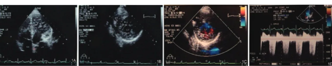
prominent ventricular trabeculations in the left ventricle (Figures 1A, 1B). The color Doppler echocardiogram imaging showed sinusoidal recesses filled with blood (Figure 1C) and an endocardium-epicardium ratio of 2:1. Mitral stenosis with a valvular area of 1.6 cm2 and mild mitral and tricuspid
regurgitation were observed (Figure 1D); systolic pressure of the pulmonary artery was 41 mmHg and ejection fraction was 71%. A remarkable fact in this case was the family history, as the patient’s parents were consanguineous; one of the patient’s brother had died suddenly during the first months of life and another sibling died at the age of 16 years, with a diagnosis of dilated cardiomyopathy; however, when reviewing his autopsy, we discovered that his heart transmural histological section showed compatible features with non-compaction cardiomyopathy (Figures 2A, 2B, 2C).
Case 2
The patient was a 50-year-old female with family history of heart failure and sudden death. She had had hypertension for at least 22 years. Eight years previously, she began to present progressive dyspnea Four years previously, she had suffered a cerebrovascular accident, which resulted in left hemiplegia with complete recovery; at that time, she was diagnosed with dilated cardiomyopathy. Physical examination revealed normal vital signs; cardiac auscultation showed mitral and tricuspid regurgitation murmur II/VI. At rest, the 12-lead electrocardiogram recorded sinus rhythm with left bundle branch blockade image. The chest x-ray showed pulmonary fields with slight hilar congestion and grade III enlarged heart. The two-dimensional echocardiogram was compatible with non-compaction cardiomyopathy. We observed trabeculations and deep sinusoidal recesses in the left ventricle, generalized hypokinesia of the left ventricle with two separated layers, and an endocardium-epicardium ratio of 2.5:1; Doppler color image with sinusoidal recesses filled with blood; dilated left ventricle with a restrictive-congestive filling pattern and mild mitral and tricuspid regurgitation. Systolic pressure of pulmonary artery was 44 mmHg, and ejection fraction, 20%. The cardiac cineangiography revealed normal epicardial coronary arteries; the ventriculogram demonstrated global hypokinesia, ventricular dilatation, and severe left ventricular dysfunction. Apical endomyocardial biopsy disclosed markedly thickened endocardium, interstitial fibrosis, and myocytic degeneration.
Discussion
Left ventricular non-compaction cardiomyopathy currently remains an unclassified cardiomyopathy according
Key Words
Cardiomyopathies; Ventricular Dysfunction, Left; Arrhythmias, Cardiac; Embolism; Death, Sudden, Cardiac.
to the World Health Organization1. It is a genetic disorder
due to mutations in the G4.5 and the alpha-dystrobrevin genes that results in the arrest of the compaction process of the myocardial meshwork during endomyocardial embryogenesis1,3,4. This cardiomyopathy is a disease with
familial recurrence of which clinical manifestations may appear in childhood or early youth6. It has been observed
that the patients in this study had a relevant family history of sudden death, and cardiac failure; therefore, they had high risk to present this cardiomyopathy. Initial symptomatology in both cases was characterized by progressive dyspnea. Case 2 presented the antecedent of stroke. Different series have reported that clinical manifestations of this pathology are characterized by progressive left ventricular dysfunction, severe cardiac failure, and pulmonary and systemic embolism which may result from impaired ventricular function and from thrombus formation within the intertrabecular recesses6,7. Physiopathology of myocardial perfusion may play
a crucial role in non-compaction cardiomyopathy, resulting in abnormalities of the coronary microcirculation producing alterations in global segmental motion, ventricular dilatation and cardiac failure1,8. The two-dimensional echocardiogram
of the two patients described here showed numerous and prominent left ventricular trabeculations prevailing in the mid-ventricular, apical, and mid-inferior regions and a two-layered structure with a endocardium-epicardium ratio >2 at the end of systole. The color Doppler echocardiography in both patients revealed sinusoidal recesses filled with
blood proceeding from ventricular cavity. Non-compaction cardiomyopathy is apt to be missed or misdiagnosed due to the lack of awareness of the disease and because other heart diseases can present similar characteristics; hence, it is necessary to distinguish it from pathologies in which increased thickness of the ventricular wall and prominent trabeculations are observed similarly to hypertrophic cardiomyopathy and hypertensive cardiopathy, as well as in dilated cardiomyopathy, in which generalized hypokinesia, dilatation of cardiac cavities and severe left ventricular dysfunction are found. In children, this cardiomyopathy must be differentiated from pulmonary valve atresia, intact interventricular septum and from pathologies that induce outflow obstruction of the left ventricle9,10. Some cardiac
tumors such as hemangiomas, which are characterized by proliferation of blood vessels, may resemble recesses7. In our
study, a brother of case 1 had been misdiagnosed with dilated cardiomyopathy, but the autopsy study revealed he had non-compaction cardiomyopathy, demonstrated by the presence of the features of this disease. In case 1, this cardiomyopathy could have been missed or misdiagnosed because of the associated mitral stenosis and regurgitation, which made us consider a diagnosis of rheumatic cardiopathy. Case 2 was first diagnosed with dilated cardiomyopathy, due to the observed dilated left ventricular cavity and generalized hypokinesia; however, in both cases, the presence of prominent trabeculations in the left ventricle, deep sinusoidal recesses filled with blood, and an endocardium-epicardium Figure 1 - Two-dimensional apical-four chamber echocardiogram and short axis view demonstrating left ventricle with prominent trabeculations and deep intertrabecular
recesses (A, B). Color Doppler image, short axis view, showing a meshwork with sinusoidal recesses illed with blood (C). Continuous-wave Doppler revealing stenosis mitral regurgitation was associated (D).
Figure 2 - Histological section with Masson´s trichrome stain showing compacted epicardial layer and non-compacted endocardial layer. Endocardium/epicardium rate
was 2.4:1 with mean recesses of 1.3 x 0.7 mm and trabeculation thickness mean of 0.4 mm. (A) Cross-section of the myocardial layer demonstrated markedly thickened endocardium, interstitial ibrosis, and myocytic degeneration (B, C).
Case Report
Contreras et al Prinzmetal’s Angina
e22
López et al
Non-Compaction Cardiomyopathy
ratio >2 led us to establish a diagnosis of non-compaction cardiomyopathy. The two-dimensional echocardiogram and Doppler undoubtedly proved to be reliable methods to carry out the diagnosis of this pathology.
Conclusions
Non-compaction cardiomyopathy is a rare congenital disease that may be missed or misdiagnosed. The classification of this disorder by WHO as a different cardiomyopathy is important to foster awareness of the disease and its early diagnosis. The established criteria for its diagnosis allow us to increase knowledge of the disease and to distinguish it from other pathologies that can present similar characteristics. The two-dimensional and Doppler echocardiograms are
reliable, non-invasive methods of choice to diagnose this cardiomyopathy.
Potential Conflict of Interest
No potential conflict of interest relevant to this article was reported.
Sources of Funding
There were no external funding sources for this study.
Study Association
This study is not associated with any post-graduation program.
Referências
1. Oechslin E, Jenni R. Isolated left ventricular no-compaction: increasing recognition of this distinct, yet unclassified cardiomyopathy. Eur J Echocardiogr. 2002; 3 (4): 250-1.
2. Jenni R, Oechslin E, Schneider J, Jost C, Kaufmann P. Echocardiographic and pathoanatomical characteristics of isolated left ventricular no-compaction: a step towards classification as a distinct cardiomyopathy. Heart. 2001; 86: 666-71.
3. Oechslin R, Attenhofer CH, Rojas J, Kaufmann P, Jenni R. Long-term follow-up of 34 adults with isolated left ventricular noncompaction: a distinct cardiomyopathy with poor prognosis. J Am Coll Cardiol. 2000; 36: 493-500.
4. Ichida F, Tsubata S, Bowles K, Haneda N, Uese K, Miyawaki T, et al. Novel gene mutations in patients with left ventricular noncompaction or barth syndrome. Circulation. 2001; 103: 1256-63.
5. Ritter M, Oechslin E, Sütsch G, Attenhofer CH, Schneider J, Jenni R. Isolated noncompaction of the myocardium in adults. Mayo Clin Proc. 1997; 72 (1): 26-31.
6. Chin T, Perloff J, Williams R, Jue k, Mohrmann R. Isolated nocompaction of left ventricular myocardium: a study of eight cases. Circulation. 1990; 82: 507-13.
7. Ichida F, Hamamichi Y, Miyawaki T, Ono Y, Kamiya T, Akagi T, et al. Clinical features of isolated noncompaction of the ventricular myocardium: long term clinical course, hemodynamic properties, and genetic background. J Am Coll Cardiol. 1999; 34: 233-40.
8. Jenni R, Wyss CH, Oechslin E, Kauffman P. Isolated ventricular noncompaction is associated with coronary microcirculatory dysfunction. J Am Coll Cardiol. 2002; 39: 450-4.
10. Bax J, Lamb H, Poldermans D, Schalij M, de Ross A, Van der Wall E. Non-compaction cardiomyopathy-echocardiographic diagnosis. Eur J Echocardiogr. 2002; 3: 301-2.
11. Engberding R, Bender F. Identification of a rare congenital anomaly of the myocardium by two-dimensional echocardiography: persistence of isolated myocardial sinusoids. Am Heart J. 1984; 53: 1733-4.
Case Report
Contreras et al Prinzmetal’s Angina
e23
López et al Non-Compaction Cardiomyopathy