Case Report
Key Words
Oto-renal-syndrome; heart defects, congenital.
We report the case of a 43-day-old boy with branchio-oculo-facial syndrome (BOFS) and congenital heart defect. On clinical examination, he presented growth retardation, epicanthal folds, small palpebral fissures, telecanthus, broadened nasal bridge, lip pseudocleft, micrognathia, dysplastic and posteriorly-rotated ears, branchial clefts, short and webbed neck, supernumerary nipple, hypotonia and decreased deep tendon reflexes. Echocardiography showed the presence of a type-A complete atrioventricular septal defect and patent ductus arteriosus. This description strengthens the possibility of congenital heart defects being part of the spectrum of anomalies seen in BOFS.
Branchio-Oculo-Facial Syndrome (BOFS) and Congenital Heart
Defects
Rafael Fabiano Machado Rosa, Paulo Ricardo Gazzola Zen, Carla Graziadio, Giorgio Adriano Paskulin
Clinical Genetics, Universidade Federal de Ciências da Saúde de Porto Alegre (UFCSPA) and Santa Casa de Porto Alegre Hospital Complex (CHSCPA), Porto Alegre, RS – Brazil
Mailing address: Giorgio Adriano Paskulin•
Genética Clínica (UFCSPA) Rua Sarmento Leite, 245 / 403, Centro 90050-170, Porto Alegre, RS - Brazil
E-mail: [email protected]
Manuscript received June 16, 2008; revised manuscript received July 10, 2008, accepted July 10, 2008.
Introduction
Branchio-oculo-facial syndrome (BOFS) (MIM 113620)1,
first named in 1987 by Fujimoto et al2, is a rare autosomal
disease with a highly variable expressivity3. Mutations in
genes of the EYA-DACH-SIX-PAX pathway, initially considered candidates for the syndrome because of their relationship with the development of some oro-facial-cervical structures and diseases overlapping with BOFS, such as the branchio-oto-renal syndrome (BOR; MIM 113650) and Townes-Brocks syndrome (TB; MIM 107480), have not been identified in BOFS patients. However, more recently, Milunsky et al4
studied a sample of BOFS patients and verified the presence of mutation in the TFAP2A gene, which is located in chromosome 6 (region 6p24) and previously related to the development of the anterior chamber of the eye1,4. Despite these findings,
the authors concluded that more patients should be studied to exclude possible genetic heterogeneity4. The syndrome is
clinically characterized by the presence of branchial defects covered by a portion of normal skin, associated or not with pre-auricular or auricular pits; microphtalmia; coloboma; nasolacrimal duct obstruction; lip pseudoclefts (abnormal upper lip and filtrum resembling a poorly repaired cleft lip);
partial or total clefts of upper lip, and premature graying of hair2,3.
Although renal malformations are frequent in patients with the syndrome, the involvement of other organs, particularly congenital heart defects (CHD) have been rarely described3,5.
We report a rare case of BOFS associated with CHD.
Case Report
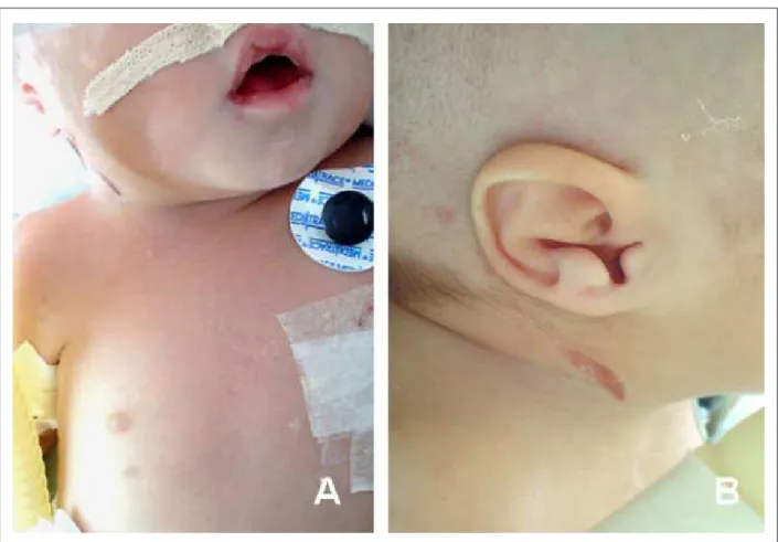
The patient was a white boy, the first child of healthy nonconsanguineous young parents. His family history was negative for any congenital defect or genetic disease. He was born at the 36th week gestation, by cesarean section, in breech presentation. His birth weight was 2870 g (10-50th percentile), his height was 45 cm (10-50th percentile) and the 5-minute Apgar score was 9. On clinical examination at 43 days of age, his weight was 2590 g (< 3rd percentile), his height was 46 cm (< 3rd percentile), head circumference of 34 cm (< 2nd percentile), and he presented epicanthal folds, small palpebral fissures, telecanthus, broadened nasal bridge, lip pseudocleft, migrognathia, dysplastic and posteriorly-rotated ears, branchial clefts (areas of aplasia cutis) in the neck below the ears, short and webbed neck, and supranumerary nipple in right side of the chest (Figure 1).
The neurological examination revealed hypotonia and decreased deep tendon reflexes. Brain ultrasonography showed prominent lateral ventricles. Abdominal ultrasonography, eye examination and high-resolution GTG-banding karyotype were normal.
An echocardiography performed soon after birth showed the presence of an atrioventricular septal defect (AVSD) and patent ductus arteriosus (PDA).
Although cardiac surgery had been performed on the first month of life, the child progressed with hemodynamic and respiratory instability due to the heart condition, and died in the tenth month of life. Autopsy was not performed.
Discussion
The presence of some specific craniofacial anomalies in our patient, particularly telecanthus, broadened nasal bridge, lip pseudocleft, dysplastic and posteriorly-rotated ears associated with branchial clefts, which are considered possibly pathognomonic of BOFS3, supported the diagnosis. In
medical databases such as OMIM1and LMD (London Medical
Database)6, there is no description of the association between
BOFS and congenital heart defects.However, in our review, we found four confirmed cases of BOFS presenting CHD which
Case Report
Rosa et al BOFS and congenital heart defects
Arq Bras Cardiol 2009; 92(2) : e6-e8
Figure 1 -A - Patient at 43 days of life presenting lip pseudocleft, branchial cleft below the right ear, short and webbed neck, and supranumerary nipple on the right side of the chest ; B - Detail of the dysplastic ear and right branchial cleft .
included: ostium secundum atrial septal defect (two cases)3,7,
tetralogy of Fallot (one case)5 and pulmonary valve stenosis
(one case)8 (Table 1). All patients presented facial findings
characteristic of BOFS associated with branchial defects1,3,5,8.
The present report is the first description of a patient with both BOFS and AVSD, one of the CHD more frequently associated with extracardiac anomalies, especially with Down syndrome9. Hing et al10 described a patient presenting multiple
malformations and AVSD resembling BOFS. He presented some atypical findings such as holoprosencephaly and meningoencephalocele. The authors suggested that this patient could present a lethal variant of BOFS, or, alternatively, a syndrome not previously reported10. This case was reevaluated
by Lin et al3, who considered it atypical, probably unaffected3.
Although conotruncal heart defects – malformations related to the abnormal migration of cells from the neural crest into the branchial arches, are frequently found in other syndromes with branchial arch anomalies3, Bennaceur et al5 were the
only authors to describe a patient with BOFS presenting this type of CHD (a patient with tetralogy of Fallot)5. The other
reports included heart defects pathogenetically classified in the group of intracardiac blood flow defects3,7,8 and extracellular
matrix anomalies (the present case) (Table 1). Thus, this new BOFS report strengthens the possibility of congenital heart defects being part of the spectrum of anomalies observed in this syndrome.
Table 1 – Reports of BOFS associated with congenital heart defects described in the literature
Patient Author Year Congenital heart disease
1 Lin et al3 1995 Ostium secundum atrial septal defect (ASD)
2 Bennaceur et al5 1998 Tetralogy of Fallot (TOF)
3 Kapoor et Kapur8 2004 Pulmonary valve stenosis
4 Verret et al7 2005 Ostium secundum atrial septal defect (ASD)
5 Present report Type-A complete atrioventricular septal defect (AVSD) and patent ductus arteriosus (PDA)
Case Report
Rosa et al
BOFS and congenital heart defects
Arq Bras Cardiol 2009; 92(2) : e6-e8
References
1. OMIM (TM). On line Mendelian Inheritance in man (TM). Baltimore: Center for Medical Genetics of the Johns Hopkins University; 2000. [Acessed in 2008 June 12]. Available from: http://www.3.ncbi.nlm.nih.gov:80/htbin-post/omnin/dispmin?229300.
2. Fujimoto A, Lipson M, Lacro RV, Shinno NW, Boelter W, Jones KL, et al. New autosomal dominant branchio-oculo-facial syndrome. Am J Med Genet. 1987; 27: 943-51.
3. Lin AE, Gorlin RJ, Lurie IW, Brunner HG, van der Burgt I, Naumchik IV, et al. Further delineation of the branchio-oculo-facial syndrome. Am J Med Genet. 1995; 56: 42-59.
4. Milunsky JM, Maher TA, Zhao G, Roberts AE, Stalker HJ, Zori RT, et al. TFAP2A mutations result in branchio-oculo-facial syndrome. Am J Hum Genet. 2008; 82: 1171-7.
5. Bennaceur S, Buisson T, Bertolus C, Couly G. Branchio-oculo-facial syndrome with cleft lip and bilateral dermal thymus. Cleft Palate Craniofac J. 1998; 35:
454-9.
6. Winter R, Baraitser M. Winter-Baraitser Dysmorphology Database (WBDD). In: London Medical Database (LMD) Version 1.0. London: Oxford University Press; 2005.
7. Verret DJ, Murray AD, Hobar PC. Branchio-oculo-facial syndrome with ectodermal parathyroid tissue. Otolaryngol Head Neck Surg. 2005; 133: 983-4.
8. Kapoor S, Kapur N. Branchio-oculo-facial syndrome with valvular pulmonic stenosis. Indian Pediatr. 2004; 41: 1180-1.
9. Marino B, Digilio MC. Congenital heart disease and genetic syndromes: specific correlation between cardiac phenotype and genotype. Cardiovasc Pathol. 2000; 9: 303-15.
10. Hing AV, Torack R, Dowton SB. A lethal syndrome resembling branchio-oculo-facial syndrome. Clin Genet. 1992; 41: 74-8.
Potential Conflict of Interest
No potential conflict of interest relevant to this article was reported.
Sources of Funding
There were no external funding sources for this study.
Study Association
This study is not associated with any post-graduation program.