Neurohormonal Profile of Rheumatic Patients with Significant
Chronic Aortic Regurgitation
Guilherme Sobreira Spina, Flávio Tarasoutchi, Roney Orismar Sampaio, Marcelo Luiz Campos Vieira, Célia Strunz,
Francisco Rafael Laurindo, Max Grinberg
Instituto do Coração (InCor), Hospital das Clínicas da Faculdade de Medicina da Universidade de São Paulo, São Paulo, SP - Brasil.
Summary
Background: Neurohormones are involved in the physiopathology of heart failure, but little is known about its behavior in significant chronic aortic regurgitation (AR). We aimed at analyzing the behavior of these mediators in AF.
Objective: We aimed at analyzing the behavior of these mediators in AF.
Metods: We analyzed 89 patients with AF, whose mean age was 33.6±11.5 years and of whom 84.6% were males, 60% asymptomatic, all with rheumatic etiology. After the clinical and echocardiographic assessment, plasma measurements of tumor necrosis factor (TNF), soluble TNF receptor types I and II (sTNFRI e sTNFRII), interleukin-6 (IL-6), its soluble receptor (sIL6R), endothelin-1 and B-type natriuretic peptide (BNP) were carried out; 12 healthy individuals were used
as controls.
Results: The mean values of the left ventricle diastolic diameter (LVDD) were 71.9±8.3mm, whereas the mean values of the LV systolic diameter (LVSD) were 50.4±9.3mm. The neurohormonal levels were elevated in patients with AF (TNF 92.65±110.24 pg/mL vs. 1.67±1.21 pg/ml in controls, p<0.001), (IL-6 7.17±7.78pg/ml vs. 0.81±0.38pg/mL in controls, p=0.0001) and TNFRI (894.75±348.87pg/mL vs. 521.42±395.13pg/ml, p=0.007). Except for the BNP levels, symptomatic
and asymptomatic patients presented a similar neurohormonal profile. There was a correlation between TNFRII and
LVDD (r=-0.329, p=0.038) and LVSD (r=-0.352, p=0.027). BNP levels were significantly higher in symptomatic patients
and only in the latter it was possible to establish a correlation between BNP and ventricular diameters.
Conclusions: Patients with significant chronic AF present high neurohormonal levels, with no correlation with the symptomatic status. The TNFRII and BNP levels could be correlated with ventricular diameters, but only the latter could be correlated with symptoms. (Arq Bras Cardiol 2009;92(2):143-149)
Key words: Aortic valve insufficiency; rheumatic diseases; cardiomegaly; pituitary hormones posterior.
Mailing address: Guilherme Sobreira Spina •
Av. Dr. Eneas de Carvalho Aguiar, 44 - Unidade Clinica de Valvopatia, Cer-queira César, 05.403-000, São Paulo, SP - Brazil.
E-mail: [email protected]
Manuscript received March 12, 2008; revised manuscript received July, 01, 2008; accepted July 01, 2008.
Abbreviations
TNF- Tumor-necrosis factor
sTNFRI – soluble TNF receptor type I
sTNFRII – soluble TNF receptor type II IL-6 – Interleukin-6
sIL6R – soluble Interleukin-6 receptor IL1-Ra – Interleukin-1 receptor antagonist ET-1 – Endothelin-1
BNP – B-type natriuretic peptide LVDD – Left Ventricle Diastolic Diameter LVSD – Left Ventricle Systolic Diameter LVEF- Left Ventricle Ejection Fraction
HF- Heart Failure
RF – Rheumatic Fever
Introduction
The significant chronic aortic regurgitation (AR) causes one of the most important myocardial hypertrophy responses observed in cardiac diseases, being a classic model of ventricular remodeling. The patient remains asymptomatic for a long period and, according to the moment of the natural history and the degree of hypertrophy, can develop chronic heart failure (CHF) or left ventricular dysfunction. However, the mechanisms that direct these evolutions remain unknown1.
During the last two decades1, the clinical studies were mainly based on the left ventricle (LV) dimensions and the systolic function through the Doppler echocardiogram, radioisotope ventriculography and hemodynamics as an indication of the ideal moment for the aortic valve replacement. To date, the ideal moment for the interruption of the natural history is controversial.
cytokines such as the tumor necrosis factor alpha (TNF-α) and interleukin-6 (IL-6) showed to have a central role in the physiopathology of the left ventricular remodeling, in different etiologies of CHF2-4.
Myocardial pressure overload and myocardial fiber distension provide sufficient stimulation to stimulate the secretion of TNF by the cardiac myocytes5, increasing their genic expression5 and triggering a chain reaction that also increases the levels of other cytokines6. The natriuretic peptides, especially the B-type natriuretic peptide (BNP) are other examples of proteins of which secretion is stimulated by mechanical stress to the myocardium.
The increase in the peripheral vascular resistance is a key event in the development of heart failure and endothelin is one of the most potent vasoconstrictors in this disease7. In addition to the vasoconstriction, endothelin also stimulates the cytokine secretion8,9 and has an important role in the development of myocardial hypertrophy.
Thus, the objective of the present study was to determine the behavior of serum levels of these mediators related to the myocardial hypertrophic response in AF and their correlation with left ventricular symptoms and remodeling.
Methods
Patient selectionAF was defined according to the criteria of Spagnuolo et al10, which require a cardiothoracic index >0.5, electrocardiographic evidence of left ventricular hypertrophy, pulse pressure > 80 mmHg and diastolic arterial pressure < 60 mmHg. The exclusion criteria included any valvulopathy except AF, atrial fibrillation, any active inflammatory disease, renal failure or neoplasias. As all of the patients presented rheumatic fever, prior to their inclusion in the protocol they underwent inflammatory activity tests to exclude those in the acute phase of rheumatic fever. Patients younger than 18 years or older than 60 years were also excluded. The rheumatic etiology was defined as a patient with a typical history of rheumatic fever (RF) in childhood or echocardiographic finding compatible with RF. All patients signed the free and informed consent form prior to study enrollment.
Clinical evaluation
All patients were evaluated and examined by the same observer before the collection of the neurohormonal profile. Symptomatic patients were defined as those that presented heart failure, precordial pain or syncope.
Neurohormonal proile
The blood was collected from the antecubital vein in tubes with EDTA. The tubes were immediately immersed in ice water and centrifuged at 1500 rpm at 4oC for 15 minutes. The plasma was separated and frozen at -80oC until the analysis. The measurement of cytokines was carried out by commercially available methods. An automated chemiluminescent assay (Immulite, DPC, USA), was used for TNF, IL-6 and interleukin 1-beta; for the soluble TNF receptors types I and II (TNFRI and TNFRII), soluble IL-6 receptor and IL-1 receptor antagonist,
traditional plate ELISA was used (Quantikine, R & D Systems, USA). Endothelin-1 was measured by a specific enzymatic method (Parameter, R & D Systems, USA). BNP was measured by a specific immunoassay (Advia Centaur BNP, Bayer Diagnostics, Germany).
Echocardiography
Echocardiograms were obtained no later than 2 weeks after the patient’s inclusion in the study. The left ventricular diastolic diameter (LVDD), systolic diameter (LVSD) and ejection fraction (LVEF) were measured. Table 1 shows the general and echocardiographic variables of the study patients. The intensity of the AF was defined semi-quantitatively by determining the jet length and width at the color Doppler.
The cutoff values for the analysis of diameters and function have been previously defined. We used the values defined by the guidelines of the American Heart Association (AHA) such as “significant left ventricular dilation”11: these cutoff values for the analysis were LVDD > 75mm, LVSD > 55mm and LVEF < 0.50.
Statistical analysis
The data are expressed are means ± SD. For the comparison of means between two independent groups, we used the Student’s t test. When the data normality was rejected, the Mann-Whitney test was used. The analysis of three different groups was carried out with the analysis of variance (ANOVA). When the data normality was rejected, the Kruskall-Wallis test was used, with multiple comparisons made by the Dunn’s test.
Pearson’s coefficient of correlation was used to determine the correlations between the neurohormones and the diameter and LV function measurements. The logarithmic transformation of the neurohormone levels was also used to obtain the normalization of the data. The level of significance was set at p<0.05.
Results
Patient GroupEighty-nine consecutive outpatients met the inclusion criteria from January 2004 to December 2005, with 54 of them (60%) being asymptomatic and 35 (40%), symptomatic. The mean age was 33.6±11.5 years and 84.6% of them were males, all of rheumatic etiology. At the clinical assessment, 60% were asymptomatic and were prescribed exclusively Penicillin G benzathine every 21 days for secondary prophylaxis for RF. The symptomatic patients, while waiting for surgical treatment, received digitalis, diuretics and angiotensin-converting enzyme inhibitors (ACEI). As the ACEI can modify the serum levels of cytokines, these were withdrawn for 7 days prior to the collection of blood samples. The control group consisted of 12 healthy volunteers, with a mean age of 30±9.5 years.
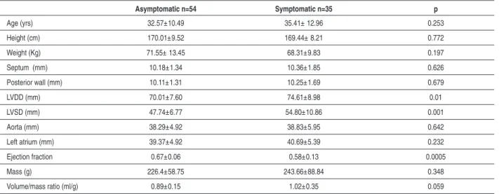
Table 1 – Clinical and echocardiographic data of symptomatic and asymptomatic patients with AR.
Asymptomatic n=54 Symptomatic n=35 p
Age (yrs) 32.57±10.49 35.41± 12.96 0.253
Height (cm) 170.01±9.52 169.44± 8.21 0.772
Weight (Kg) 71.55± 13.45 68.31±9.83 0.197
Septum (mm) 10.18±1.34 10.36±1.85 0.626
Posterior wall (mm) 10.11±1.31 10.25±1.69 0.679
LVDD (mm) 70.01±7.60 74.61±8.98 0.01
LVSD (mm) 47.74±6.77 54.80±10.86 0.001
Aorta (mm) 38.29±4.92 38.83±5.95 0.642
Left atrium (mm) 39.37±4.92 40.69±5.39 0.232
Ejection fraction 0.67±0.06 0.58±0.13 0.0005
Mass (g) 226.4±58.75 243.66±88.84 0.348
Volume/mass ratio (ml/g) 0.89±0.15 1.02±0.35 0.059
LVDD - left ventricular diastolic diameter; LVSD - left ventricular systolic diameter.
the demographic and echocardiographic data for the symptomatic and asymptomatic patients.
Serum levels of neurohormones
Patients with AF presented significantly higher levels of TNF, IL-6 and TNFRI when compared to the control group. The asymptomatic and symptomatic patients had similar levels of neurohormones, except for BNP, which was significantly higher in the symptomatic patients. All 89 patients had undetectable levels of interleukin 1-β (< 5pg/ml).
The serum levels of neurohormones in asymptomatic and symptomatic patients are shown in Table 2.
Table 2 - Neurohormonal proile in symptomatic and asymptomatic
patients with AR.
Aortic Regurgitation
Controls Asymptomatic Symptomatic
TNF-α (pg/ml )1 86.9±85.2 103.5± 141.4 1.7±1.2*
sTNFRI(pg/ml)1 906.8± 299.6 881.4± 404.4 521.4±395.1*
sTNFRII (pg/ml)3 1868.7±530.5 1891.7± 675.9 n/a
IL-6 ( pg/ml)2 6.5±7.4 8.3±8.4 0.9±0.4*
IL-6R ( ng/ml)3 33.5±12.5 34.5±6.8 n/a
IL1-RA ( pg/ml)3 134.1±230.2 19.8±60.1 n/a
ET-1 ( pg/ml )3 7.1± 5.1 7.6±8.3 n/a
BNP (pg.ml) 3 † 31.2±37.4 164.5±274.7 n/a
TNF-� - Tumor-necrosis factor-alpha; sTNFRI – soluble TNF receptor� - Tumor-necrosis factor-alpha; sTNFRI – soluble TNF receptor - Tumor-necrosis factor-alpha; sTNFRI – soluble TNF receptor type I, sTNFRII – soluble TNF receptor type II, IL-6 – Interleukin-6, sIL6R – soluble interleukin-6 receptor; IL1-Ra –interleukin-1 receptor antagonist; ET-1 – Endothelin-1; BNP – B-type natriuretic peptide; n/a - control group
not available; * - p<0.05, patients with AF (symptomatic and asymptomatic)
vs. controls; † - p<0.05 patients with AF, symptomatic vs. asymptomatic; 1
- Kruskal-Wallis test ; 2 - ANOVA; 3 - Mann-Whitney test.
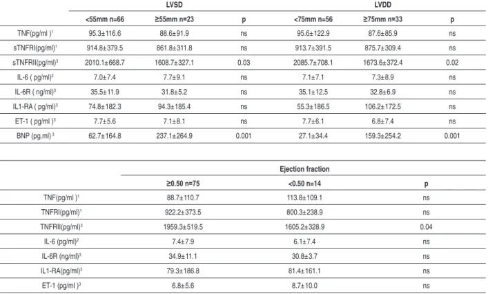
We observed significantly lower levels of TNFRII in patients with “significant ventricular dilation”, according to the criteria of the AHA9 (LVSD > 55 mm and lower EF (LVEF < 0.50) (Table 3).
BNP was higher in patients with higher ventricular diameters and lower ejection fraction (Table 3).
There was a significant correlation between the levels of TNFRII and LVDD 0.329, p=0.038) and LVSD (r=-0.352, p=0.027), with a decrease in the TNFRII levels with the increase in ventricular diameters (Figure 1). There was a correlation of the BNP with ventricular diameters only in symptomatic patients (Figure 2).
There was no correlation of TNF, TNFRI, IL-6, IL6R, IL-1RA and endothelin (Table 3) with diameters or LVEF.
Discussion
General proile of cytokines
The increase in the ventricular diameters is an essential mechanism to adapt the left ventricle to the volume-pressure overload observed in AF, allowing the adjustment of the large regurgitating volume with the maintenance of the left ventricular end-diastolic pressure at normal levels. Mediators such as IL-6 and TNF are essential in the process of myocardial hypertrophy12,13, which can justify, even in the asymptomatic phase of AF, the observed high concentrations of these mediators. Cytokines seem to contribute to the eccentric hypertrophy and ventricular dilation5,6,14-16 that help the myocardium to deal with the high hemodynamic overload present in AF.
Table 3 - Neurohormonal proile of patients with AF, divided according to parameters of diameter and function derived from the AHA
guidelines8
LVSD LVDD
<55mm n=66 ≥55mm n=23 p <75mm n=56 ≥75mm n=33 p
TNF(pg/ml )1 95.3±116.6 88.6±91.9 ns 95.6±122.9 87.6±85.9 ns
sTNFRI(pg/ml)1 914.8±379.5 861.8±311.8 ns 913.7±391.5 875.7±309.4 ns
sTNFRII(pg/ml)3 2010.1±668.7 1608.7±327.1 0.03 2085.7±708.1 1673.6±372.4 0.02
IL-6 ( pg/ml)2 7.0±7.4 7.7±9.1 ns 7.1±7.1 7.3±8.9 ns
IL-6R ( ng/ml)3 35.5±11.9 31.8±5.2 ns 35.1±12.5 32.8±6.9 ns
IL1-RA ( pg/ml)3 74.8±182.3 94.3±185.4 ns 55.3±186.5 106.2±172.5 ns
ET-1 ( pg/ml )3 7.7±5.6 7.1±8.1 ns 7.7±6.1 6.8±7.4 ns
BNP (pg.ml) 3 62.7±164.8 237.1±264.9 0.001 27.1±34.4 159.3±254.2 0.001
Ejection fraction
≥0.50 n=75 <0.50 n=14 p
TNF(pg/ml )1 88.7±110.7 113.8±109.1 ns
TNFRI(pg/ml)1 922.2±373.5 800.3±238.9 ns
TNFRII(pg/ml)3 1959.3±519.5 1605.2±328.9 0.04
IL-6 (pg/ml)2 7.4±7.9 6.1±7.4 ns
IL-6R (ng/ml)3 34.9±11.1 30.8±3.7 ns
IL1-RA(pg/ml)3 79.3±186.8 81.4±161.1 ns
ET-1 (pg/ml )3 6.8±5.6 8.7±10.0 ns
BNP (pg.ml) 3 67.1±158.6 310.2±304.2 0.006
TNF-� - Tumor-necrosis factor-alpha; sTNFRI – soluble TNF receptor type I, sTNFRII – soluble TNF receptor type II, IL-6 – Interleukin-6, sIL6R – soluble interleukin-6 receptor; IL1-Ra –interleukin-1 receptor antagonist; ET-1 – Endothelin-1; BNP – B-type natriuretic peptide; LVDD = left ventricular diastolic diameter; LVSD=left
ventricular systolic diameter; 1: Kruskal-Wallis test ; 2: ANOVA; 3: Mann-Whitney test.;
1,124pg/ml, TNFRII > 2,913pg/ml17 and endothelin > 5pg/ ml18 are associated to a worse prognosis in patients with non-valvular CHF (ischemic myocardiopathy or idiopathic dilated myocardiopathy). In the non-valvular CHF, the asymptomatic patients present low cytokine levels, which increase with the worsening of the patient’s functional class17, a phenomenon that we did not observe with AF. The prognostic significance of these levels in patients with AF has yet to be studied.
Behavior of the TNF receptors
The behavior of the TNFRII in the AF can yield interesting hypotheses involving TNF and its beneficial and deleterious effects for the heart. When the TNF binds to its membrane receptors, the extracellular part of these receptors detaches itself from the cell and starts circulating in the plasma as a soluble receptor19. Thus, there are two forms of TNF receptors: the soluble and the membrane ones. The soluble receptors function as a buffer for the circulating TNF, neutralizing excessive concentrations and increasing the half-life of this mediator19.
In AF, we constantly observed high levels of TNF, but a significant decrease in the TNFRII levels in patients with AF with higher ventricular diameters and LV dysfunction
(Figure 1 and Table 3). The binding of TNF to its high-affinity receptor, the TNFRII, triggers cytoprotective and anti-apoptotic responses20, which generally lead to myocardial hypertrophy21. Additionally, when in its soluble form, the TNFRII binds to the TNF trimers, contributing to neutralize its deleterious actions22. This effect led to the clinical use of TNFRII in its soluble form, known as etarnecept, in diseases in which the participation of TNF is vital, such as rheumatoid arthritis23.
These findings suggest that in the more advanced stages of AF, the TNF binds preferentially to the TNFRI receptors (which leads to apoptosis and myocardiotoxic responses), decreasing its binding to TNFRII, which would decrease its plasma concentration. Another hypothesis is that the decrease in the TNFRII concentration in the advanced phase, with higher ventricular dilation of the AF, would prevent the high TNF concentrations from being adequately neutralized and contribute to the progressive dilation of the LV and worsening of the myocardial function24.
Figure 1 -Association between the LV diastolic diameter (LVDD), LV systolic diameter (LVSD) and levels of the soluble TNF receptor type II (TNFRII). There was a
signiicant correlation between the levels of TNFRII and LVDD (r=-0.329, p=0.038) and LVSD (r=-0.352, p=0.027).
r=-0.329, p=0.038 r=-0.329, p=0.038
Figure 2 -Association between the LV diastolic diameter (LVDD) and the LV systolic diameter with the levels of B-type natriuretic peptide (BNP).
Together, these data reinforce the perception that the patients with AF have a different neurohormonal profile from other heart failure etiologies. Neurohormones measured in the AF were increased, regardless of the patients’ symptoms, differently from the valvulopathies studied to date, such as mitral failure5,6, aortic stenosis6 and mitral stenosis25, in which serum levels of neurohormones were close to normal values
in asymptomatic patients and increased in the presence of symptoms.
TNFRII increased according to the ventricular diameters, differently from our findings, in which the TNFRII levels decreased with the increase in the ventricular diameters (Figure 1). A similar neurohormonal behavior was observed in aortic stenosis, with increasing concentrations occurring with the increase in the ventricular diameter and worsening of ventricular function6. These differences can be justified by the volume-pressure overload in AF, differently from the pure volume overload that causes lower mechanical stress on the myocardial fiber.
BNP behavior
The BNP presented an interesting behavior, with normal values in asymptomatic patients, even in those with ventricular diameters that were rather higher than the normal ones values (29% with LVDD > 75mm, and 12% with LVSD > 55mm). Thus, we can postulate that in AF, the LV diameter increase alone is not sufficient stimulus to elevate the BNP levels.
Therefore, we can hypothesize that the presence of symptoms in AF is caused mainly by the increase in the left ventricular end-diastolic pressure1, through the increase in the diastolic wall tension. Thus, BNP can be an interesting marker of the natural history progression in patients with AF and rather increased ventricular diameters (Figure 2).
This theory is corroborated by studies of AF of non-rheumatic etiology where the BNP showed a behavior that was similar to that in our study, i.e., elevated BNP in the presence of symptoms26; however, differently from the results observed in our study, these authors also observed a correlation of the BNP with the ventricular diameter and function in all patients, not only in the symptomatic ones.
Studies that used a different methodology for BNP measurement, the measurement of NT-pro BNP, showed that higher BNP levels are associated to a worse prognosis in
patients with AF27.
A limitation of the present study is the fact that it is a transversal study. Therefore, we could not investigate eventual prognostic implications of these mediators. This cohort of patients is still being followed, so that in the future we will be able to investigate the prognostic implications of our researches.
In conclusion, patients with AF have a particular neurohormonal profile, with high levels of mediators since the asymptomatic phase. The TNFRII correlated with the ventricular diameters and the LVEF. Only the symptomatic patients presented a correlation between the BNP levels and the ventricular diameters and LVEF. These data increase our knowledge of the process of ventricular remodeling present in AF and proposes new physiopathological mechanisms to be investigated in this disease.
Acknowledgements
This study was supported by Fundação de Amparo à Pesquisa do Estado de São Paulo (FAPESP), grant # 00/09472-2.
Potential Conflict of Interest
No potential conflict of interest relevant to this article was reported.
Sources of Funding
This study was funded by FAPESP.
Study Association
This article is part of the thesis of Ddoctoral submitted by Guilherme Sobreira Spina, from Incor-Instituto do Coração da Faculdade de Medicina da Universidade de São Paulo.
References
1. Tarasoutchi F, Grinberg M, Spina GS, Sampaio RO, Cardoso LE, Rossi EG, et al. Ten-year clinical laboratory follow-up after application of a symptom-based therapeutic strategy to patients with severe chronic aortic regurgitation of predominant rheumatic etiology. J Am Coll Cardiol. 2003; 41(8):1316-24.
2. Bryant D, Becker L, Richardson J, Shelton J, Franco F, Peshock R, et al. Cardiac failure in transgenic mice with myocardial expression of tumor necrosis factor-alpha. Circulation. 1998; 97: 1375-81.
3. Bozkurt B, Kribbs SB, Clubb FJJ, Michael LH, Didenko VV, Homsby PJ, et al. Pathophysiologically relevant concentrations of tumor necrosis factor-alpha promote progressive left ventricular dysfunction and remodeling in rats. Circulation. 1998; 97: 1382-91.
4. Kubota T, McTiernan CF, Frye CS, Slawson SE, Iemster BH, Koretsky AP, et al. Dilated cardiomyopathy in transgenic mice with cardiac-specific overexpression of tumor necrosis factor-alpha. Circ Res. 1997; 81: 627-35.
5. Oral A, Sivasubramanian N, Dyke DB, Mehta RH, Grossman PM, Briesmiester K, et al. Myocardial proinflammatory cytokine expression and left ventricular remodeling in patients with chronic mitral regurgitation. Circulation. 2003; 107: 831-7.
6. Kapadia SR, Yakoob K, Nader S, Thomas JD, Mann DL, Griffin BP. Elevated
circulating levels of serum tumor necrosis factor-alpha in patients with hemodynamically significant pressure and volume overload. J Am Coll Cardiol. 2000; 36: 208-12.
7. Spieker LE,Noll G, Ruschitzka FT, Lüscher TF. Endothelin Receptor antagonists in congestive heart failure: a new therapeutic principle for the future? J Am Coll Cardiol. 2001; 37: 1493-505.
8. Agui T, Xin X, Cai Y, Sakai T, Matsumoto K. Stimulation of interleukin-6 production by endothelin in rat bone marrow-derived stromal cells. Blood. 1994; 84: 2531-8.
9. Hofman FM, Chen P, Jeyaseelan R, Incardona F, Fisher M, Zidovetzki R. Endothelin-1 induces production of the neutrophil chemotactic factor interleukin-8 by human brain-derived endothelial cells. Blood. 1998; 92: 3064-72.
10. Spagnuolo M, Kloth H, Taranta A, Doyle E Pasternak B. Natural history of rheumatic aortic regurgitation: criteria predictive of death, congestive heart failure and angina in young patients. Circulation. 1971; 44: 368-80.
84-231.
12. Haehling S, Jankowska EW, Anker SD. Tumour necrosis factor-alpha and the failing heart: pathophysiology and therapeutic implications. Basic Res Cardiol. 2004, 99: 18-28 .
13. Kanda T, Takahashi T. Interleukin-6 and cardiovascular diseases. Jpn Heart J. 2004; 45: 183-93.
14. Torre-Amione G, Kapadia S, Lee J, Durand JB, Bies RD, Young JB, et al. Tumor necrosis factor and tumor necrosis factor receptors in the failing human heart. Circulation. 1996; 93: 704-11.
15. Torre-Amione G, Kapadia S, Benedict C, Oral H, Young JB, Mann DL. Proinflammatory cytokine levels in patients with depressed left ventricular ejection fraction: a report from the studies of left ventricular disfunction (SOLVD). J Am Coll Cardiol. 1996; 27: 1201-6.
16. Ferrari R, Bachetti T, Confortini R, Opasich C, Febo O, Corti A, et al. Tumor necrosis factor soluble receptors in patients with various degrees of heart failure. Circulation. 1995; 92 (6): 1479-86.
17. Rauchhaus M, Doehner W, Francis DP, Darvos C, Kemps M, Liebenthal C, et al. Plasma cytokine parameters and mortality in patients with chronic heart failure. Circulation. 2000; 102; 3060-7.
18. Pousset F, Isnard R, Lechat P, Katotka H, Carayon A, Maistre G, et al. Prognostic value of plasma endothelin-1 in patients with chronic heart failure. Eur Heart J. 1997; 18: 254-8.
19. Doust JA, Glasziou PP, Pietrzak E, Dobson AJ. A systematic review of the diagnostic accuracy of natriuretic peptides for heart failure. Arch Intern Med. 2004; 164: 1978-84.
20. Testa M, Yeh M, Lee P, Fanelli R, Loperfido F, Berman JW, et al. Circulating levels of cytokines and these endogenous modulators in patients with mild to severe congestive heart failure due to coronary artery disease or hypertension. J Am Coll Cardiol. 1996; 28: 964-71.
21. Nakano M, Knowlton AA, Dibbs Z, Mann DL. Tumor necrosis factor confers resistance to injury induced by hypoxic injury in the adult mammalian cardiac myocyte. Circulation. 1998; 97: 1392-400.
22. Aderka D, Engelmann H, Maor Y, Brakebrusch C, Wallach D. Stabilization of the bioactivity of tumor necrosis factor by its soluble receptors. J Exp Med. 1992; 175: 323-9.
23. Genovese MC, Kremer JM. Treatment of rheumatoid arthritis with etanercept. Rheum Dis Clin North Am. 2004; 30 (2): 311-28.
24. Yokoyama T, Vaca L, Rossen RD, Durante W, Hazarika P, Mann DL. Cellular basis for the negative inotropic effects of tumor necrosis factor-alpha in the adult mammalian heart. J Clin Invest. 1993; 92: 2303-12.
25. Chang CJ, Hsu LA, Chiang CW, Ko YS, See LC, Shen YC, et al. Percutaneous transvenous mitral valvulotomy normalizes elevated circulating levels of tumor necrosis factor-alpha and interleukin-6 in mitral stenosis with heart failure. Am J Cardiol. 2003; 91 (8): 1018-20.
26. Eimer MJ, Ekery DL, Rigolin VH, Bonow RO, Carnethon MR, Cotts WG. Elevated B-type natriuretic peptide in asymptomatic men with chronic aortic regurgitation and preserved left ventricular function. Am J Cardiol. 2004; 94: 676-8.