The Role of Echocardiography in the
Percutaneous Treatment of Septal Defects
Simone Rolim F. Fontes Pedra, Sérgio Cunha Pontes Jr, Renata de Sá Cassar,
Carlos Augusto Cardoso Pedra, Sérgio Luiz Navarro Braga, César Augusto Esteves,
Maria Virgínia Tavares Santana, Valmir Fernandes Fontes
Instituto Dante Pazzanese de Cardiologia - São Paulo, SP - Brazil
Mailing Address: Simone Rolim F. Fontes Pedra • – Av. Dr. Dante Pazzanese, 500 – 14º - 04012-180 - São Paulo, SP - Brazil
E-mail: [email protected] Received on 10/08/04 • Accepted on 03/30/05
In the last few years, percutaneous treatment of atrial and ventricular septal defects has developed signifi cantly, having been established as a feasible, safe and effective therapeutic modality. Echocardiography has a primary role in this scenario, identifying suitable candidates for the procedure, monitoring the device implantation and evaluating the rate of occlusion during follow-up. The available devices for percutaneous occlusion of septal defects have been progressively modifi ed and improved. Similarly, a great technological advancement has also occurred in the echocardiography area, such as the advent of high-resolution transesophageal multiplan probes, on and off-line three-dimensional (3-D echo) and intracardiac echocardiography.
In some centers, the latter has become the proce-dure of choice for monitoring percutaneous occlusion of atrial septal defect in adult patients, as it does not require general anesthesia, thus making the procedure even simpler.
In this review article, we will discuss the role of echocardiography to evaluate patients before, during and after interventional procedures for occlusion of septal defects, including the ostium secundum atrial septal defect type, perimembranous and muscular ventricular septal defects and patent foramen ovale, based on the experience of the group in this type of approach.
In the last few years, ultrasound equipment underwent great technological development, which made echocardiography a mandatory tool in the diagnosis of congenital heart disease. In the area of pediatric interventional cardiology, echocardiography has a primary role in the identifi cation and selection of patients who are candidates to percutaneous procedures, and in the monitoring and late follow-up of such patients. Among the interventional procedures, those that need the support of echocardiography are the occlusion of atrial and ventricular septal defects. In this study, we will discuss the role of echocardiography in the percutaneous treatment of atrial and ventricular septal defects and patent foramen ovale (PFO).
Percutaneous occlusion of atrial
septal defects
Atrial septal defects (ASD) correspond to approximately 7% of all congenital heart disease, being more frequently in females, at a proportion of 2:11. The most frequent
anatomic type is the one located in the fossa ovalis (75% of the cases), also denominated ostium secundum ASD type and occurs due to the defi ciency, perforation or absence of the fossa ovalis lamina2. The other communications
occur due to the defi ciency of the fold between the atrial walls and between the tributary veins, with these being ostium primum communication types, venous sinus type, and coronary sinus type. Considering these anatomical characteristics, the only interatrial communication that allows treatment with the use of devices is the one located in the fossa ovalis2.
Patient selection
Patients with clinical suspicion of ASD are initially submitted to a transthoracic echocardiography. In addition to the defect identifi cation, the pulmonary venous return, degree of hemodynamic impact, signs of pulmonary hypertension and the presence of associated defects that need therapeutic approach are also assessed3. The more
specifi c information regarding the characteristics of the defects, as well as number and dimension of orifi ces, their localization in the septum, and characteristics of the rims that surround them are better evaluated by transesophageal echocardiography (TTE), which can be performed at the outpatient clinic basis or immediately before the procedure, in the catheterization laboratory3-5.
In our experience, performing TEE in the echocardiography laboratory shows advantages concerning the procedure programming, device selection and family counseling.
Indication criteria
b) diameter of defect varying from 4 to 35 mm; c) fl ow through the defect predominantly directed from the left to the right atrium; d) signs of right ventricular volume overload (increased right ventricular end-diastolic diameter and presence of paradoxical movement of the interventricular septum); e) distance of the defect rims from adjacent structures (coronary sinus, atrioventricular valves, pulmonary veins and vena cava) of at least 4 mm; f) presence of rims with at least 5 mm around at least 75% of the defect; g) absence of fi xed pulmonary hypertension; h) absence of associated defects that need surgical approach.
All these criteria are applied when one intends to use the Amplatzer septal occluder, which is currently the most frequently device utilized in the market. Regarding the implant of the Helex device, the second device most commonly used, the diameter of the ASD must not exceed 15 mm. This device line is available with diameters of 15, 20, 25, 30 and 35 mm. As the choice of size is based on a 1.7-2:1 ratio with stretched diameter (diameter of the ASD obtained in the catheterization laboratory, in which a complacent balloon is inserted into the orifi ce, distending it until it acquires a circular shape), it is possible to occlude stretched orifi ces that do not exceed 20 mm6.
As a result of the increasing global experience in the percutaneous treatment of ASD, as well as the development of occlusion devices and the improvement of the quality of echocardiographic images, including high-resolution TEE, three-dimensional reconstruction and intracardiac echocardiography (ICE), the indications, previously restricted to unquestionably favorable cases, were expanded to include the so-called complex cases, in which the implant is known to be feasible and effective,
but with slightly lower success rates and more prolonged procedure time7.
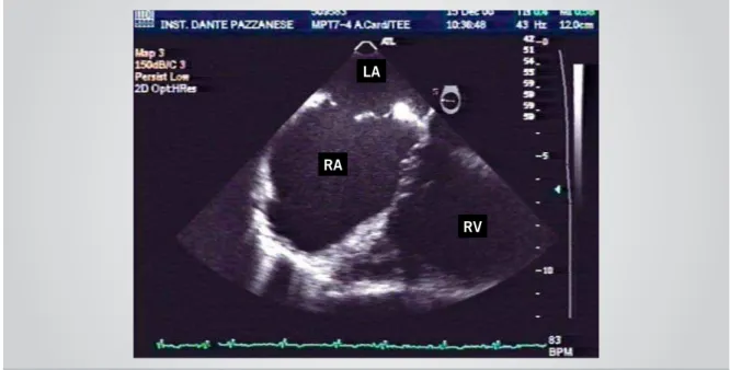
Complex anatomy ASD is considered when it has the following characteristics: stretched diameter > 26 mm; defi cient rims, measuring less than 4 mm in the anterior, posterior or inferior septal region; two orifi ces that are apart; multifenestrated interatrial septum and interatrial septum aneurysm (redundant and mobile interatrial septum with an excursion > 10 mm)7. Figure 1 shows
an ASD of complex anatomy.
Regardless of the type of probe utilized (biplane or multiplane), TEE must follow a standard methodology, in order to obtain all necessary information. Our group chooses to start with sectional images at the transverse plane (0o in the multiplane probe), slowly and progressively
moving the probe, from the plane that evidences the superior vena cava until the fl oor of the right atrium is reached, identifi ed by the presence of the coronary sinus and the Eustachian valve. Subsequently, we move on to the longitudinal plane (90o), with the section that contains
the left atrial appendix and the mitral valve, using a slow anti-clockwise rotation of the probe in order to obtain a scan of the atrial septum from the anterior to the posterior portion. This standardization allows the examiner to mentally reconstruct a three-dimensional model of the defect, as well as defi ne the characteristics of complex defects with precision5, 8.
Echocardiography during the
procedure
Atrial septal defects occlusion is continuously monitored by TEE or ICE. When this is accomplished
Fig. 1 - Complex ASD with multiple orifi ces shown by TEE. RA: right atrium; LA: left atrium; RV: right ventricle RA
LA
by TEE, even in adults, it is necessary to utilize general anesthesia. The main steps of the echocardiography during the implant are:
1. Measurement of the stretched diameter. In addition, it is necessary to verify whether the balloon is obstructing the defect completely and if there are no additional defects with the occlusion of the main defect. The choice of the Amplatzer device size depends on the stretched diameter. The balloon-catheter separates, by compression, the thin rims formed by the septumprimum, occupying the orifi ce delimitated by the rigid rims, constituted by the septum secundum, which will function as the device support. The balloon-catheter is insuffl ated until the septum shows resistance, demonstrating a clear waist in the balloon. It is important to remember that the balloon must be insuffl ated only enough to prevent fl ow through the defect, avoiding septal hyperextension and consequently, the overestimation of the device size. Measurements of the balloon diameters in the echocardiography and angiography are similar, as long as they are adequately performed. The correct determination of the diameter is essential for an adequate device choice. Although the Amplatzer device can be easily withdrawn and changed by another, the choice of a device with inadequate dimensions results in increased procedure costs.
2. To guide the interventionalist regarding the localization of the left atrium disc before the release of the remaining device components, so it will be near the interatrial septum, without protruding through the defect.
3. To demonstrate the device positioning after the release of all its components. The device can only be released when the presence of interatrial septum tissue between its two discs is demonstrated, and when there is no functional involvement of cardiac venous and valve structures.
4. Scanning for a possible residual shunt, grading it when present. In the period that follows the release, it is possible to identify a low velocity central fl ow through the device mesh, which normally disappears on the day after the implant5.
Under special circumstances, such as large ASDs in small children, or those with dimensions > 35 mm in adults, some authors have discontinued the stretched diameter technique for device selection. In these cases, the measurement of the upper-lower length of the interatrial septum is performed, in the four-chamber view (00 by TEE). The device is chosen so that the left disc
diameter does not exceed total septum length (Dr. Zahid Amin, personal communication). This new technique, in addition to allowing the percutaneous procedure, prevents the overestimation of the device size, which eventually could result in the erosion of the aorta wall to the right or left atrium, aortic-sinus fi stula or pericardial effusion with cardiac tamponade9. Following the previously
described indications, it is only possible to perform the percutaneous occlusion of these defects if the rims around
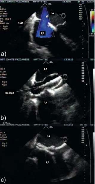
Fig. 2 - TEE during percutaneous occlusion of an ASD with a Helex septal occluder a) fossa ovalis type ASD, located in the central part of the septum, with the fl ow directed from the left atrium to the right; b) Balloon interposition through the defect to measure the stretched diameter; c) Helex occluder well-positioned in the septum, occluding the defect. RA: right atrium; LA: left atrium; ASD: atrial septal defects
the orifi ce measure at least 5 mm, except for that close to the aorta, which may be absent, although it does not contraindicate the procedure. Figure 2 shows a fossa ovalis ASD with morphological characteristics that are ideal for percutaneous treatment, the stretched diameter and the fi nal result after a Helex device is release.
Echocardiography in the follow-up of percutaneous closure of ASD
The follow-up of patients who underwent device closure of ASD is carried out through sequenced clinical evaluations and transthoracic echocardiogram on the day after the implantation, one, three and twelve months after, and every year, thereafter. The main parameters evaluated by echocardiography are device positioning, presence of residual shunt and measurement of right cardiac chamber
RA LA
RA LA
LA
RA ASD
dimensions3. By assessing the diastolic diameter of the
right ventricle at the longitudinal paraesternal axis before the implantation and during the late follow-up, we have observed that the right ventricle returns to its normal dimensions within the fi rst year post-procedure4.
In the beginning of our experience, we routinely repeated TEE after the third month post-implantation. In face of the excellent late results, not only in our institution but worldwide regarding the low late complication rates, the disappearance of small residual shunts and the reduction of the device profi le, we have followed patients with transthoracic echocardiogram only, being TEE utilized only in case of complications. Although complications such as aortic fi stula to the right or left atriums, pericardial effusion secondary to atrial perforation and thrombus or vegetation formation in the device have been rarely described in literature, none of these abnormalities were observed in our experience, which consists of 144 cases, including patients treated with Amplatzer and Helex devices. Compromise of the atrioventricular valve function and pulmonary or systemic vein return were not observed either.
Percutaneous occlusion of PFO
Approximately 25% of the general population has a PFO10. In almost all cases, it does not have pathological
implications, being just a variation of the normal condition, casually observed during the routine echocardiographic assessment. However, young patients (< 55 yrs) with stroke of undetermined cause after extensive investigation (including neurological, cardiac, vascular, hematological and rheumatological causes), PFO is found in up to 60% of the cases. Based on this epidemiological difference, it was postulated that PFO, functioning as a valve, could allow inversion of shunt at the atrial level (from right to left) in situations of high pressures in the right atrium, associating to embolic events for the systemic circulation (paradoxical embolism)11. Thus, looking for
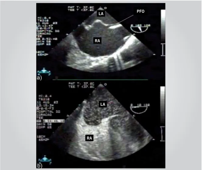
PFO is mandatory in individuals who have evidence of cerebral ischemia without other apparent causes. This assessment is performed with TEE. In addition to the anatomical visualization of the orifi ce in the fossa ovale, it is necessary to document the right to left shunt, using contrast. To obtain a contrast rich in microbubbles, physiological saline solution is agitated with air (9 ml of saline solution and 1 ml of air) and injected rapidly in a peripheral vein. At this moment, the patient is requested to perform the Valsalva maneuver to increase pressure on the right side of the heart. The presence of right to left shunt is confi rmed when the contrast, after fi lling up the right atrial cavity, is found inside the left atrium with an amount of at least 5 microbubbles, during the fi rst three cardiac cycles after the injection5.
In these patients, percutaneous occlusion of the PFO has been carried out safely and effectively, to prevent recurrences of thromboembolic phenomena12.
Despite the lack of prospective and randomized studies comparing pharmacological approach (anti-platelet and anti-coagulant drugs) with percutaneous treatment, previously published observational longitudinal studies have shown that occlusion with devices is at least, as effective as the clinical treatment, being more effective in some subgroups of patients, such as those presenting with more than one previous episode of stroke or recurrence during pharmacological treatment13. Some anatomical
aspects of the interatrial septum are particularly important for appropriate selection of the diameter and shape of the device that will be used, and must be adequately observed during the echocardiographic study. They are: a) the presence of interatrial septum aneurysm (defi ned by excursion > 10 mm of the septum); b) tunnel-shaped foramen ovale, and c) the presence of other orifi ces in the interatrial septum. Echocardiographic assessment of interatrial septum is also mandatory for professional divers, as there is a fi ve-fold increased risk of suffering decompression sickness for those who have PFO compared to those with intact interatrial septum14.
Recently, PFO has been associated to migraine with aura and its occlusion has demonstrated reduction on intensity and frequency of the migraine episodes15.
Percutaneous occlusion of PFO is, in general, quite rapid and simple, and can be carried out under echocardiographic monitoring, according to the interventionalist’s choice. Figure 3 shows echocardiographic pictures of a PFO and right-left fl ow demonstrated by the presence of microbubbles in the left atrium.
Drug prophylaxis with aspirin is maintained for 6 to 12 months after percutaneous closure. Treatment can be withdrawn only when a new TEE with microbubbles does not reveal any right to left shunting at atrial level. This evaluation is normally carried out six months after the procedure.
Percutaneous occlusion of ASD
and PFO guided by ICE
Currently available in the Brazilian market, the AcuNav has revolutionized interventional cardiology,
especially in the areas of electrophysiology and percutaneous treatment of septal defects. The AcuNav
is an ultrasound transducer performed to undergo the venous system, which results in high-resolution images and is capable of 12 cm penetration in the heart. The current commercialized catheter is a 90 cm long 10 French catheter, introduced in the venous system through an 11 French sheath. It has a transducer coupled to the distal extremity, which allows capture of section images of 90o. Its tip can be moved to the anterior-posterior and
Sequoia™, Aspen™ and Cypress™ (Acuson Corporation, Mountain View, CA) equipment, having been approved for intracardiac and intraluminal use by the FDA (Food and Drug Administration).
The main advantages of ICE for percutaneous treatment of ASD and PFO are the acquisition of better quality images, as the transducer is placed inside the right atrium, the best view of the posterior-inferior portion of the interatrial septum (the rim that is the hardest to visualize at the TEE), lower exposition to radiation by reducing the duration of the procedure, necessity of only one operator (the interventionalist can obtain the echocardiographic images him or herself) and the preclusion of general anesthesia in adolescents and adults, since TEE is not required16-20.
Recent studies have shown that all measurements obtained on ICE have excellent correlation with those obtained by TEE, with better demonstration of guidewires, catheters and device discs21, 22. Due to such advantages,
ICE monitoring of percutaneous occlusion of ASD and
PFO has become the standard of care for adolescents and adults in North America and Europe. In developing countries, however, due to the high cost of the transducer, it has been more uncommonly employed. Theoretically the transducer must be used only once. In day by day practice however, the equipment can be re-sterilized and re-utilized around 10 times, depending how carefully its handled (Carlos Zabal, personal communication). Figure 4 shows some images obtained during our initial experience with ICE at Dante Pazzanese Institute of Cardiology and Hospital do Coração.
Percutaneous occlusion of
ventricular septal defect (VSD)
Ventricular septal defect is the most frequent congenital heart disease, corresponding to around 20% of them23. In approximately 80% of the cases, it
is located in the membranous portion of the septum, with variable extensions to the inlet and outlet, being thus denominated perimembranous VSD23. With the Fig. 3 - TEE demonstrating a) patent foramen ovale (PFO), associated to an aneurysm of the interatrial septum; b) contrast study, delineating the aneurysm and showing a large amount of microbubbles crossing from the right atrium to left atrium. RA: right atrium; LA: left atrium
RA LA
RA LA
development of Amplatzer devices appropriate for this septal region, percutaneous closure of VSD was extended from the muscular region to the membranous area24, 25. Although virtually all perimembranous VSDs can
be occluded percutaneously, some important features must be carefully assessed by echocardiography. Differently from ASD patient selection, the pre-procedure evaluation is performed by transthoracic echocardiography. In our clinical practice, we have performed TEE only in adults with very unfavorable echocardiographic windows, in whom transthoracic evaluation of the defect is not accurate.
Patient selection
As mentioned previously, some anatomical and functional aspects must be evaluated before the indication of percutaneous treatment of VSD: A) Localization – if the communication is perimembranous, it is important to defi ne if it is located at the ventricular infl ow or outfl ow region. B) Distance from the aortic valve leafl ets: Although the absence of tissue separating the aortic valve leafl ets from the orifi ce is not an absolute contraindication for the procedure, it is known that, in this situation, it becomes more complex and requires more care. In general, it is preferable that the defect is at least 2 mm far from the aortic valve. C) Presence of accessory tissue around the defect: sometimes there is a small amount of tricuspid tissue tags around the orifi ce, which does not signifi cantly reduce fl ow through it. In these cases, the prosthesis must be attached to the muscular septum. On the other hand, there are defects that present large amounts of tissue around it, forming an aneurysm, which signifi cantly reduces the original fl ow orifi ce. These defects may be occluded with smaller devices, which are positioned inside the aneurysmatic sac and do not compromise tricuspid and aortic valves function. D) Presence of aortic valve prolapse. It is known that 2 to 7% of the VSDs can be associated to a prolapse of the right coronary leafl et of the aortic valve, leading to different degrees of aortic insuffi ciency. Although this fi nding is a contraindi-cation to the percutaneous procedure in study protocols in the United States, device implantation under these circumstances has been carried out safely and effectively with excellent medium-term results. In our experience of 29 cases, three patients had such complication, all with successful device implantation. Two of them persisted with no aortic insuffi ciency and one showed a minimal progression of the previously mild existing insuffi ciency. Monitoring serially these patients with echocardiography is mandatory. E) Presence of associated defects, such as subaortic membrane and right ventricular muscle band which might need surgical approach. Lesions such as pulmonary or aortic valve stenosis, persistent duct arteriosus and aortic coarctation can also be treated percutaneously, depending on their anatomical features. F) Presence of the left chambers overload that justifi es the intervention. G) Absence of fi xed pulmonary arterial hypertension.
Muscular VSDs located in the trabecular and apical regions of the septum can also be treated percutaneously. Although there are fewer anatomical variations, the presence of multiple orifi ces is common, particularly in the ventricular apex. When there is a single orifi ce, the Amplatzer device is the best choice, as it has small retention discs that occupy little space of the interventricular septum. In case of multiple orifi ces, CardioSEAL devices are more appropriate, as long as implanted in the most central orifi ce.
Recently, hybrid approach for the management of very
Fig. 4 - ICE during a percutaneous occlusion of an ASD with an Amplatzer septal occluder demonstrating: a) fossa ovalis type ASD; b) release of the left atrium device disc; c) the fi nal result after device release. RA: Right atrium; LA: left atrium; AO: aorta
RA
RA LA
RA
LA Prothesis
young patients (< 6 mo) with large muscular defects has been utilized. These patients are taken to the operating room for a thoracotomy to expose the heart and, through a right ventricular free wall puncture a guidewire is introduced in the left ventricle through the VSD. A short sheath is then advanced over the guidewire allowing the device positioning. The procedure is performed under TEE monitoring. This innovative and revolutionary approach, in which interventionalist, echocardiographer and cardiac surgeon work together, prevents the deleterious effects of extracorporeal circulation, resulting in occlusion rates > 90%26.
Echocardiography during
percutaneous closure of VSD
Multiplane probes are more suitable for this procedure, as intermediate planes between 00 and 1800 can provide
visualization of the VSD and device positioning. Similarly to ASD closure, echocardiography helps in the orifi ce measurement and device size choice, monitors the guidewires, catheters and sheaths position inside the heart and determines the location of the devices discs, before and after its release24. The primary functions of
the echocardiography also include careful evaluation of aortic and tricuspid valves, detection and grading of possible residual shunts. Although echocardiographic pictures are of great help for the procedure, fl uoroscopy and angiography are crucial during the intervention.
Echocardiography in the
follow-up after VSD percutaneous
occlusion
The follow-up of patients who underwent percutaneous occlusion of VSD is carried out through clinical assessments and serial transthoracic echocardiograms, when the following parameters must be evaluated: a) left cardiac cavity dimensions (normally after the fi rst year of treatment, these have returned to normal dimensions24);
positioning of the device in the interventricular septum; c) presence of residual shunt; d) evaluation and measurement of a possible fl ow gradient in the left ventricular outfl ow tract; and e) aortic and tricuspid valve function.
Our current experience consists of 29 cases treated percutaneously for perimembranous VSD and one for muscular VSD. Regardless of the morphological characteristics of the defect, all the procedures were carried out successfully. The presence of immediate residual shunt was noticed in 25% of the cases. Over a mean follow-up period of 10 ± 8.5 months, only two patients persisted with slight residual shunt, with one of them having less than one-month follow-up. Two other cases presented a gradient at the left ventricular outfl ow tract (17 and 25 mmHg on the day after the procedure) that disappeared during follow-up, possibly due to the spontaneous reduction of the device profi le and the
epithelization process which occurs within the fi rst six months after the implant24,27. Figure 5 shows a VSD before
and after the Amplatzer device implant.
Three-dimensional
echocardiography (3-D echo) in
the percutaneous occlusion of
septal defects
In the mid-90s, several studies showed the feasibility and accuracy of 3-D echo in the assessment of the heart28. At the same time, interventional cardiology was
established as an effective and safe technique for ASD occlusion. Consequently, several studies were published showing three-dimensional evaluation of the interatrial septum and its defects, and the connections between the implanted device and the adjacent cardiac structures29-33.
Three-dimensional reconstruction was initially performed from TEE pictures obtained every two to three degrees, from zero to 180o, synchronized to the patient’s cardiac
and respiratory cycle. Because of total immobilization of the patient was required, most of the studies were performed in the catheterization laboratory under general anesthesia32,33. Using the Hewlett-Packard Sonos (Philips,
Bothel, WA, EUA) and Echo-Scan TomTec Imaging Systems Inc., (Boulder, CO, EUA), 3 to 10 minutes were required to acquire the pictures, depending on the patient’s heart rate. Time for reconstruction was about 3 to 5 minutes, but it could take up to 40 minutes to prepare a set of images29,32.
3-D echo main advantages to evaluate ASD are the clear demonstration of ASD characteristics, with a particular visualization of the extension of the anterior-superior rim, defi ning the defect shape and allowing measurement of all orifi ce dimensions28,29,34, In this
image modality, it is possible evaluate the defect en face mimicking the surgeon’s view, with the advantage of being dynamic during the cardiac cycle. After device implantation, three-dimensional reconstruction can identify the device positioning, showing and locating possible arms or discs protrusion to the right atrium33.
Evaluation and measurement of residual defects can also be assessed by this method29.
Live 3-D echo has recently become available in the market. To date, only thoracic transducers are available, which does not provide ideal assessment of interatrial septum defects in older children and adults. Figure 6 shows an ASD assessed by TEE and the transthoracic 3-D view of the same defect.
Recently a report described the use of this technology to monitor an ASD occlusion in a six-year-old child weighing 22 kg, using subcostal views35. Catheters’ and
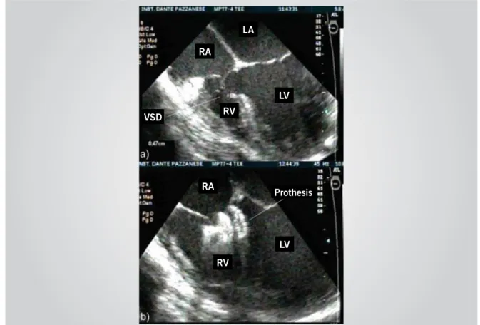
Fig. 5 - TEE during percutaneous occlusion of a perimembranous ventricular septal defect (VSD); a) the presence of a large VSD in the perimembranous region partially occluded by accessory tissue from septal leafl et of the tricuspid valve; b) Amplatzer device for perimembranous VSD, occluding the defect. RA: right atrium; LA: left atrium; RV: right ventricle; LV: left ventricle
Fig. 6 - a) Bidimensional TEE of an ostium secundum ASD; b) live 3-D reconstruction of the same defect that presents an aberrant shape. RA: right atrium; LA: left atrium; ASD: atrial septal defect
RA
LA
RA
RV
LV LV
RV
RA LA
Prothesis VSD
Decreased fl uoroscopy time, better outline of the defect shape, elimination of esophageal intubation and more reliability regarding the device positioning were highlighted as the main benefi ts of this type of monitoring35. With
the future development of a transesophageal transducer for live 3-D images, the method will be also useful in procedures for older children and adults.
C
ONCLUSIONS
As described in previous paragraphs, there are
different percutaneous techniques to treat septal defects. Technological developments have made the procedures feasible, effective and safe. Echocardiography has a primary role in identifying patients that benefi t from such procedures, in monitoring them and in the immediate and late post-occlusion follow-up. Similarly to the advances in the devices and percutaneous fi eld, echocardiography has evolved to provide more complete and precise information for the interventionalist, helping in the procedure and making it even safer and simpler.
R
EFERENCES
1. Sobrinho JHM, Fontes VF, Pontes Jr SC. Defeitos isolados do septo atrial. In: Sobrinho JHM, Fontes VF, Pontes Jr SC, editors. Cardiopatias Congênitas. São Paulo: Sarvier, 1990: 295-314.
2. Ferreira Martins JD, Anderson RH. The anatomy of interatrial communications--what does the interventionist need to know? Cardiol Young 2000; 10: 464-73.
3. Pedra SR, Pedra CA, Assef JE et al. Percutaneous closure of atrial septal defects. The role of transesophageal echocardiography. Arq Bras Cardiol 1999; 72: 59-69.
4. Fontes VF, Pedra SRFF, Braga SLNB, Pedra CAC. Fechamento Percutâneo da Comunicação Interatrial. Rev Soc Cardiol Estado de São Paulo 2002; 12: 293-305.
5. Pontes Jr SC. Importância da ecocardiografia transesofágica na avaliação morfológica dos defeitos septais atriais. Rev Bras de Ecocard 1999; 31: 25-37.
6. Pedra CA, Pedra SF, Esteves CA et al. Initial experience in Brazil with the Helex septal occluder for percutaneous occlusion of atrial septal defects. Arq Bras Cardiol 2003; 81: 435-52.
7. Pedra CA, Pedra SR, Esteves CA et al. Transcatheter closure of secundum atrial septal defects with complex anatomy. J Invasive Cardiol 2004; 16: 117-22.
8. Pontes Jr SC. Avaliação morfológica dos defeitos septais atriais. In: Assef JE, Belém M, Castro-Lima A, Torreão JAM, editors. Ecocardiografi a Transesofágica. Rio de Janeiro: Livraria e Editora Revinter Ltda, 2000: 164-76.
9. Chun DS, Turrentine MW, Moustapha A, Hoyer MH. Development of aorta-to-right atrial fi stula following closure of secundum atrial septal defect using the Amplatzer septal occluder. Catheter Cardiovasc Interv 2003; 58: 246-51.
10. Varma C, Benson LN, Warr MR et al. Clinical outcomes of patent foramen ovale closure for paradoxical emboli without echocardiographic guidance. Catheter Cardiovasc Interv 2004; 62: 519-525. 11. Yahia AM, Shaukat A, Kirmani JF, Qureshi AI. Age Is Not a Predictor
of Patent Foramen Ovale with Right-to-Left Shunt in Patients with Cerebral Ischemic Events. Echocardiography 2004; 21: 517-522. 12. Wahl A, Krumsdorf U, Meier B et al. Transcatheter treatment of atrial
septal aneurysm associated with patent foramen ovale for prevention of recurrent paradoxical embolism in high-risk patients. J Am Coll Cardiol 2005; 45: 377-80.
13. Windecker S, Wahl A, Nedeltchev K et al. Comparison of medical treatment with percutaneos closure of patent foramen ovale in patients with criptogenic stroke. J Am Coll Cardiol 2004; 44: 750-8. 14. Cartoni D, De Castro S, Valente G et al. Identifi cation of professional
scuba divers with patent foramen ovale at risk for decompression illness. Am J Cardiol 2004; 94: 270-3.
15. Morandi E, Anzola GP, Angeli S, Melzi G, Onorato E. Transcatheter closure of patent forâmen ovale: a new migraine treatment? J Interv Cardiol 2003; 16: 39-42.
16. Hijazi Z, Wang Z, Cao Q, Koenig P, Waight D, Lang R. Transcatheter closure of atrial septal defects and patent foramen ovale under intracardiac echocardiographic guidance: feasibility and comparison with transesophageal echocardiography. Catheter Cardiovasc Interv 2001; 52: 194-9.
17. Koenig PR, Abdulla RI, Cao QL, Hijazi ZM. Use of intracardiac echocardiography to guide catheter closure of atrial communications. Echocardiography 2003; 20:781-7.
18. Zanchetta M. On-line intracardiac echocardiography alone for Amplatzer Septal Occluder selection and device deployment in adult patients with atrial septal defect. Int J Cardiol 2004; 95: 61-8. 19. Z a n c h e t t a M , O n o r a t o E , R i g a t e l l i G e t a l . I n t r a c a r d i a c
echocardiography-guided transcatheter closure of secundum atrial septal defect: a new effi cient device selection method. J Am Coll Cardiol 2003; 42: 1677-82.
20. Zanchetta M, Rigatelli G, Pedon L et al. Transcatheter atrial septal defect closure assisted by intracardiac echocardiography: 3-year follow-up. J Interv Cardiol 2004; 17: 95-8.
21. Boccalandro F, Muench A, Salloum J et al. Interatrial defect sizing by intracardiac and transesophageal echocardiography compared with fluoroscopic measurements in patients undergoing percutaneous transcatheter closure. Catheter Cardiovasc Interv 2004; 62: 415-20. 22. Boccalandro F, Baptista E, Muench A, Carter C, Smalling RW. Comparison of intracardiac echocardiography versus transesophageal echocardiography guidance for percutaneous transcatheter closure of atrial septal defect. Am J Cardiol 2004; 93: 437-40.
23. Tynan M, Anderson RH. Ventricular Septal Defect. In: Anderson RH, Baker EJ, McCartney FJ, Rigby ML, Shinebourne EA, editors. Paediatric Cardiology. New York: Churchill, Livingstone, 2002: 983-1014. 24. Pedra CA, Pedra SR, Esteves CA et al. Percutaneous closure of
perimembranous ventricular septal defects with the Amplatzer device: technical and morphological considerations. Catheter Cardiovasc Interv 2004; 61: 403-10.
25. Pedra CA, Pedra SR, Esteves CA, Chamie F, Christiani LA, Fontes VF. Transcatheter closure of perimembranous ventricular septal defects. Expert Rev Cardiovasc Ther 2004; 2: 253-64.
26. Bacha EA, Cao QL, Starr JP, Waight D, Ebeid MR, Hijazi ZM. Perventricular device closure of muscular ventricular septal defects on the beating heart: technique and results. J Thorac Cardiovasc Surg. 2003; 126: 1718-23.
27. Cao QL, Du ZD, Joseph A et al. Immediate and six-month results of the profi le of the Amplatzer septal occluder as assessed by transesophageal echocardiography. Am J Cardiol 2001; 88: 754-9.
29. Acar P. Three-dimensional echocardiography in transcatheter closure of atrial septal defects. Cardiol Young 2000; 10: 484-92. 30. Acar P, Dulac Y, Roux D, Rouge P, Duterque D, Aggoun Y. Comparison
of transthoracic and transesophageal three-dimensional echocardiography for assessment of atrial septal defect diameter in children. Am J Cardiol 2003; 91: 500-502.
31. Acar P, Roux D, Dulac Y, Rouge P, Aggoun Y. Transthoracic three-dimensional echocardiography prior to closure of atrial septal defects in children. Cardiol Young 2003; 13: 58-63.
32. Maeno YV, Benson LN, McLaughlin PR, Boutin C. Dynamic morphology of the secundum atrial septal defect evaluated by three dimensional
transoesophageal echocardiography. Heart 2000; 83: 673-7. 33. M a e n o Y V, B e n s o n L N , B o u t i n C . I m p a c t o f d y n a m i c 3 D
transoesophageal echocardiography in the assessment of atrial septal defects and occlusion by the double-umbrella device (CardioSEAL). Cardiol Young 1998; 8: 368-78.
34. Marx GR, Sherwood MC, Fleishman C, Van Praagh R. Three-dimensional echocardiography of the atrial septum. Echocardiography 2001; 18: 433-43.