Symptomatic Severe Chronic Aortic Valve Disease. A
Comparative Study of Cardiac Magnetic Resonance
Imaging and Echocardiography
Marcelo Nigri, Carlos Eduardo Rochitte, Flávio Tarasoutchi, Guilherme S. Spina, José Rodrigues Parga,
Luiz Francisco Ávila, Roney Orismar Sampaio, José Antonio Franchini Ramires, Max Grinberg
Instituto do Coração do Hospital das Clínicas – FMUSP - São Paulo, SP - Brazil
Mailing Address: Marcelo Nigri • InCor - Av. Dr Enéas de Carvalho Aguiar, 44 - 05403-000 – São Paulo – SP - Brazil E-mail: [email protected] Received on 10/19/04 • Accepted on 08/09/05
O
BJECTIVETo show the real value of cardiac magnetic resonance imaging (CMRI) in the evaluation of patients with symptomatic chronic aortic valve disease.
M
ETHODSSeventy patients – 35 with aortic stenosis (AoS) and 35 with aortic regurgitation (AoR) with surgical indication, who underwent preoperative echocardiogram (ECHO) and CMRI to assess ventricular function, volumes, and left ventricular mass index using cine magnetic resonance imaging, were studied.
R
ESULTSNo statistically signifi cant difference was observed between the AoS and AoR groups when ECHO and CMRI variables were compared. When compared with the type of symptom, ECHO and CMRI variables showed the same pattern.
C
ONCLUSIONCMRI data were in agreement with ECHO data regarding the assessment of left ventricular volume and ejection fraction, and with the clinical presentation of patients with chronic aortic valve disease.
K
EY WORDSThe natural history of chronic aortic valve diseases – aortic stenosis (AoS), and aortic regurgitation (AoR) is associated with degrees of left ventricular remodeling that do not correspond1-3 to clinical manifestations.
Chronic aortic valve diseases, mainly of rheumatic etiology (which is prevalent in Brazil), with a signifi cant valve involvement may be stratified according to assessments based on cardiac imaging tests4-6.
Dyspnea on ordinary exertion as a manifestation of heart failure, as well as the presence of chest pain and syncope, generally result from left ventricular dysfunction, and less frequently from myocardial dysfunction.
Studies evaluating the clinical and morphological progression of aortic valve disease in the occasional presence of left ventricular dysfunction are lacking7-9. Thus, well-conducted studies still seek for predictive indexes using cardiac imaging methods that could bring forward the timing for surgery with increased safety. It is diffi cult to homogenize values of echocardiographic measurements due to the multiplicity of changes in ventricular remodeling. In this line of research, the left ventricular remodeling may be followed by an alteration in the clinical manifestation, which would warn us of the optimal timing for surgical treatment.
Thus, in patients with severe valvular heart diseases, the assessment of ventricular function using CMRI may be useful, because this is a diagnostic method that has become one of the main non-invasive supplementary tests in Cardiology in the past few years. Among its main advantages, we can point out the excellent anatomical resolution between the tissues, the acquisition of a three-dimensional rebuilding without using ionizing radiation, and non-nephrotoxic contrast medium (Gadolinium).
Thus, we attempted to associate clinical data with cardiac magnetic resonance imaging (CMRI) and transthroracic echocardiogram (ECHO), a supplementary method of the utmost importance used to track the assessment of left ventricular function in aortic valve diseases.
O
BJECTIVE
To assess the alterations in CMRI in the analysis of patients with symptomatic chronic aortic valve disease10 in comparison with echocardiographic parameters.
M
ETHODS
Seventy symptomatic patients (35 with AoS and 35 with AoR) from the Outpatient Clinic of the Medical Unit of Valvular Heart Diseases of the Instituto do Coração do Hospital das Clínicas, Faculdade de Medicina da Universidade de São Paulo (InCor/HC-FMUSP) were prospectively studied from May, 2001 to July, 2003. The mean age was 46.6 ± 12.4 years with predominance of males in 54 cases, and 75% of the patients with valvular
heart disease of rheumatic etiology, followed by bicuspid and degenerative valve.
The inclusion criteria for patients with severe chronic aortic valve disease and surgical indication were: clinical symptoms such as angina pectoris on exertion, syncope and dyspnea on moderate and mild exertion (paroxysmal nocturnal dyspnea, orthopnea) with a gradient between the left ventricle and the aorta above 50 mmHg by catheterization, and > 70 mmHg by ECHO, for AoS11. For AoR, the inclusion criteria were defi ned according to Spagnuolo et al12 as modified criteria, namely: cardiothoracic index > 0.50, presence of left ventricular hypertrophy as assessed by electrocardiogram, pulse pressure ≥ 80 mmHg, diastolic blood pressure ≤ 60 mmHg as assessed by ECHO, in that one single criterion was enough to admit the patient in the AoR group.
The exclusion criteria used in this study were: patients under eighteen and above 65 years of age; concurrent mitral valve disease; previous heart valve surgery; comorbidities (diabetes mellitus, high blood pressure, and dyslipidemia); and other heart diseases (aorta diseases, coronary artery diseases, myocardial diseases).
This project was analyzed and approved by the Ethics Committee of the Instituto do Coração do Hospital das Clínicas da Faculdade de Medicina da Universidade de São Paulo. All patients admitted in the study gave their written consent after obtaining information about the study and the method used.
Study schedule - Data on the occurrence of key symptoms such as angina pectoris, syncope and dyspnea on moderate and mild exertion (paroxysmal nocturnal dyspnea, orthopnea) were particularly recorded during the clinical history taking. Patients were then scheduled for diagnostic tests such as electrocardiograms, chest radiographs, ECHO, CMRI, and cardiac catheterization 11-13. ECHO was performed prior to CMRI.
Echocardiogram - Echocardiograms were interpreted according to recommendations of the American Association of Echocardiography.
All patients underwent ECHO and ventricular function, left ventricular end-diastolic (EDV) and systolic (ESV) volumes, and ventricular mass index (LVMI)14,15. Left ventricular ejection fraction (EF) was calculated using the Teichholz method16.
fraction)18. LVMI, EDV, ESV, and EF were calculated by detecting epicardial and endocardial borders in contiguous short-axis sections at the end-diastole and end-systole of cine magnetic resonance images using the Simpson’s rule19-20.
For all variables calculated, the level of statistical signifi cance of p = 0.05 was used.
R
ESULTS
Comparison between CMRI and ECHO
Aortic stenosis - The comparison of variables between the two methods – CMRI and ECHO in the AoS group is shown in table 1.
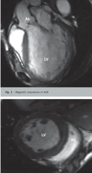
Fig. 1 – Magnetic resonance in AoR
Ao
LV
Fig. 2 – Magnetic resonance in AoS
LV
RV
S
TATISTICAL
ANALYSIS
Descriptive analysis - The calculation of means and standard deviation was performed for CMRI and ECHO variables.
Comparative analysis - The one-factor analysis of variance was used for comparisons of EF groups between ECHO and CMRI, and Bonferroni correction21 was used for multiple comparisons.
The logistic regression model was used to obtain a cut-off point for EF in relation to the clinical presentation.
Pearson’s correlation coeffi cient was used to study the correlation between EF and the clinical manifestation22.
Table 1 – Variables between CMRI and ECHO in AoS
Variables/AoS CMRI ECHO p
EF 0.59 ± 0.1 0.58 ± 0.1 NS
EDV (ml) 214 ± 112 193.4 ± 160 NS
ESV (ml) 127 ± 102 94.8 ± 92 NS
LVMI (g/m2) 166 ± 76 155 ± 60 NS
C M R I - C a r d i a c m a g n e t i c r e s o n a n c e i m a g i n g ; E C H O - Echocardiogram; AoS- Aortic stenosis; EF- Ejection fraction; EDV- End diastolic volume; ESV- End systolic volume; LVMI- Left ventricle mass index
No difference was observed in the calculation of variables between CMRI and ECHO in the AoS group.
Aortic regurgitation - The comparison of variables between CMRI and ECHO in the AoR group is shown in Table 2.
Table 2 – Variables between CMRI and ECHO in AoR
Variables/AoR CMRI ECHO p
EF 0.5 ± 0.1 0.5 ± 0.1 NS
EDV (ml) 393 ± 141 334.5 ± 157 NS
ESV (ml) 235.6 ± 131 183.5 ± 105 NS
LVMI (g/m2) 220 ± 70 195 ± 65 NS
CMRI- Cardiac magnetic resonance imaging; ECHO- Echocardio-gram; AoR- Aortic regurgitation; EF- Ejection fraction; EDV- End diastolic volume; ESV- End systolic volume; LVMI- Left ventricle mass index
Similarly to the AoS group, no signifi cant statistical difference of variables between the CMRI and ECHO was observed in the AoR group.
Analysis between CMRI and ECHO variables and clinical manifestations in AoS and AoR - The comparison between CMRI and ECHO variables and clinical manifestations in AoS is shown in Tables 3 and 4.
Only the EF variable in ECHO/CMRI showed a statistical signifi cance in the AoS group.
The comparison between CMRI and ECHO variables and clinical manifestations in AoR is shown in Tables 5 and 6.
D
ISCUSSION
Aortic valve diseases represented by AoS and AoR have different natural histories, though with similar clinical manifestations and diagnoses.
Table 3 – CMRI variables in AoS
CMRI Syncope/Angina (3) Dyspnea (32) p
EF 0.47 ± 0.1 0.39 ± 0.1 0.01
EDV (ml) 323 ± 141 344.2 ± 157 NS
ESV (ml) 235.6 ± 131 283.5 ± 105 NS
LVMI (g/m2) 220 ± 70 225 ± 65 NS
CMRI- Cardiac magnetic resonance imaging; EF- Ejection fraction; EDV- End diastolic volume; ESV- End systolic volume; LVMI- Left ventricle mass index
Table 4 – ECHO variables in AoS
ECHO Syncope/Angina (4) Dyspnea (31) p
EF 0.49 ± 0.1 0.40 ± 0.1 0.01
EDV (ml) 333 ± 141 355.2 ± 167 NS
ESV (ml) 245.6 ± 131 295.4 ± 115 NS
LVMI (g/m2) 225 ± 74 225 ± 68 NS
ECHO- Echocardiogram; EF- Ejection fraction; EDV- End diastolic volume; ESV- End systolic volume; LVMI- Left ventricle mass index
Table 5 – CMRI variables in AoR
CMRI Syncope/Angina (4) Dyspnea (31) p
EF 0.46 ± 0.1 0.37 ± 0.1 0.01
EDV (ml) 223 ± 141 354.5 ± 151 <0.01
ESV (ml) 122.6 ± 131 282.5 ± 103 0.001
LVMI (g/m2) 170 ± 70 220 ± 65 0.02
CMRI- Cardiac magnetic resonance imaging; AoR- Aortic regurgitation; EF- Ejection fraction; EDV- End diastolic volume; ESV- End systolic volume; LVMI- Left ventricle mass index
Table 6 – ECHO variables in AoR
ECHO Syncope/Angina (4) Dyspnea (31) p
EF 0.47 ± 0.1 0.36 ± 0.1 0.01
EDV (ml) 233 ± 141 374.5 ± 153 <0.01
ESV (ml) 127.6 ± 131 292.5 ± 103 0.001
LVMI (g/m2) 177 ± 70 228 ± 68 0.02
ECHO- Echocardiogram; AoR- Aortic regurgitation; EF- Ejection fraction; EDV- End diastolic volume; ESV- End systolic volume; LVMI- Left ventricle mass index
This is a pioneering study in which the advantages of CMRI in relation to ECHO were compared by studying the variables generally used to track a severe aortic valve disease. In addition, the literature lacks studies evaluating the interaction of these methods with the clinical presentation.
When the two diagnostic imaging methods – CMRI and ECHO – were used to calculate the variables, no differences between the clinical groups – AoS and AoR – were observed. Moriuchi et al23 studied 55 hypertensive patients by assessing end systolic and diastolic volumes, and left ventricular mass using CMRI and ECHO. No statistically significant differences were observed, which was similar to the data obtained in our study. Likewise, Shelton et al24 compared the left ventricular mass index of patients with hypertrophic cardiomyopathy between these two imaging methods and did not fi nd any advantages in the analysis of CRMI and ECHO (0.6 versus 0.8, respectively).
Also similar to our results, when analyzing the ejection fraction in AoR, Pflugfelder et al25 showed that no superiority was observed in the analysis of both methods. However, when the variables of the methods were analyzed with the clinical manifestations, the importance of the methods was remarkable in diagnosing the variables with the clinical presentation more precisely in the AoR group26. This can be correlated with a more intense ventricular dilation and, consequently, with a more intense ventricular maladaptation in the AoR group. Berko et al27 showed the contribution of imaging methods, emphasizing the ECHO in the diagnosis and prognosis of aortic valve disease, mainly related to patients with heart failure in the pre and postoperative of aortic valve surgery.
Likewise, Baxley et al28 showed, both qualitatively and quantitatively, the value of CMRI in patients with aortic valve disease, quantifying their heart failure symptoms and the status of the aortic valve prosthesis following surgery. Therefore, according to our results and the literature, CMRI is as effi cient as ECHO in providing qualitative and quantitative information in aortic valve diseases, and proved superior when planimetry is used to calculate the valve area24,25. Additionally, CMRI may be an alternative imaging method when ECHO is unable to clearly show the variable calculations due to limitations of the visual window.
In conclusion, no statistically signifi cant difference was found between the variables analyzed by ECHO and CMRI in both clinical groups.
R
EFERENCES
1. Zile MR. Chronic aortic and mitral regurgitation. Choosing the optimal time for surgical correction. Cardiol Clin 1991; 9: 239-53.
2. Bonow RO, Rosing DR, McIntosh CL, Jones M. The natural history of asymptomatic patients with aortic regurgitation and normal left
ventricular function. Circulation 1983; 68: 509-17.
4. Carabello BA, Williams H, Gash AK. Hemodynamic predictors of outcome in patients undergoing valve replacement. Circulation 1986; 74: 1309-16.
5. Bonow RO, Rosning DR, Kent KM, Eptein SE. Timing of operation for chronic aortic regurgitation. Am J Cardiol 1982; 50: 325-36.
6. Bonow RO. Chronic aortic regurgitation, role of medical therapy and optimal timing for surgery. Cardiology Clinics 1998; 16: 449-62.
7. Delahaye JP, Gevigney G. Can irreversible ventricular dysfunction be identifi ed in patients with heart valve disease Ann Cardiol Angeiol 1994; 43: 578-87.
8. Gaasch WH, Carroll JD, Levine HJ, Criscitiello MG. Chronic aortic regurgitation: prognostic value of left ventricular end-systolic dimension and end- diastolic radius/thickness ratio. J AM Coll Cardiol 1983; 1: 775-82.
9 Grinberg M, Tarasoutchi F, Bellotti G. O que signifi ca o “day before” na insufi ciência aórtica? Arq Bras Cardiol 1992; 58: 165-7.
10. Tarasoutchi F, Grinberg M, Parga J et al. Relação entre a função ventricular esquerda e desencadeamento de sintomas na insufi ciência aórtica crônica severa. Arq Bras Cardiol 1995; 64 (4): 301-9.
11. Karaian CH, Greenberg BH, Rahimtoola SH. The relation between functional class and cardiac performance in patients with chronic aortic insuffi ciency. Chest 1988; 88: 553-7.
12. Spagnuolo M, Kloth H, Taranta A, Doyle E, Pasternak B. Natural history of rheumatic aortic regurgitation. Criteria predictive of death, congestive heart failure, and angina in young patients. Circulation 1971; 44: 368-80.
13. Braunwald E. On the natural history of severe aortic stenosis (editorial). J Am Coll Cardiol 1990; 15: 1012.
14. Ta ke n a t a K , D a b e s t a n i A , G a r d i n J M . A s i m p l e d o p p l e r - echocardiographic method for estimating severity of aortic regurgitation. Am J Cardiol 1986; 57: 1340-3.
15. Pandial L, Oliver A, Vivaldi M et al. Doppler echocardiographic assessment of progression of aortic regurgitation. Am J Cardiol 1997; 80: 306-14.
16. Pacileo G, Calabro P, Limongelli G, Russo MG, Pisacane C, Sarubbi B. Left Ventricular Remodeling, Mechanics, and Tissue Characterization in Congenital Aortic Stenosis. J Am Soc Echoc 2003; 16: 214-20.
17. Shan K, Constantine G, Flamm SD. Role of MRI in Clinical Cardiology. Lancet 2004; 3632: 2162-71.
18. Smith HJ. Use of MRI in the diagnosis of cardiac disease. Tidsske Nor Laegeforen 2004; 124: 497-9.
19. Gerald MP, Lynne H, Mark D. Clinical use of cardiovascular magnetic resonance. Circulation 2003; 108: 647-53.
20. Thomson LE, Kim RJ, Judd RM. Magnetic resonance imaging for the assessment of myocardial viability. J Magn Reson Imaging 2004; 19: 771-88.
21. Rosner B. Fundamentals of Biostatistics. 2nd. Boston: PWS Publishers, 1986.
22. Leser W, Barbosa V, Baruzzi RG, Ribeiro MBD, Franco LJ. Elementos de Epidemiologia Geral. Rio de Janeiro: Atheneu, 2000.
23. Moriuchi M, Saito S. Evaluation of aortic regurgitation by cardiac cine magnetic resonance imaging: Analysis and comparision to Doppler echocardiography. Cardiology 1991; 78(4): 340-7.
24. Shelton DC, Shiow JL, Brown P et al. Practical value of cardiac magnetic resonance imaging for clinical quantifi cation of aortic valve stenosis.Circulation 2003; 108: 2236.
25. Pfl ugfelder PW, Higging CB.Comparison of cine MRI with Doppler echo for the evaluation of aortic regurgitation. Am JR 1989; 52(4): 729-35.
26. Magid NM, Opio G, Wallerson DC, Young MS, Borer JS. Heart failure due to chronic experimental aortic regurgitation. Am J Physiol 1994; 267: 552-6.
27. Berko BA. The role of this noninvasive test in the geriatric population. Geriatrics 2003; 58(7): 30-4.