Myocardial Remodeling in Chronic Pressure or
Volume Overload in the Rat Heart
Luiz Shiguero Matsubara, Silvia Narikawa, Ana Lucia dos Anjos Ferreira, Sergio Alberto Rupp de Paiva,
Leonardo Mamede Zornoff, Beatriz Bojikian Matsubara
Departamento de Clínica Médica, Faculdade de Medicina de Botucatu, Unesp - Botucatu, SP - Brazil
M a i l i n g A d d r e s s : Luiz Shiguero Matsubara • R u a C a r l o s G u a d a g n i n i , 1 3 0 8 - 1 8 6 1 0 - 1 2 0 – B o t u c a t u , S P - B r a z i l E-mail: [email protected] Received on 09/30/04 • Accepted on 05/18/05
O
BJECTIVETo compare cardiac structural changes in experimental pressure and volume overload models.
M
ETHODSThe study analysis included renovascular hypertensive rats (RVH, n=8), normotensive rats with volume overload caused by an aortocaval fi stula (ACF, n=10) and control rats (CONT, n=8). After four weeks, tail cuff blood pressure (SBP) was recorded. Rats were killed, the hearts were excised and the right and left ventricles (RV&LV) were weighed (RVW&LVW). Using histological sections, myocyte cross sectional areas (MA). LV wall thickness (LVWT) LV cavity diameter (LVD), normalized LVWT (LVWT/LVD) and collagen volume fraction (CVF) were measured. The comparisons were made using the ANOVA and Tukey test for a signifi cance level of 5%.
R
ESULTSTail cuff blood pressure (mmHg) was higher in the RVH group (RVH = 187 ± 22; CONT = 125 ± 10; ACF = 122 ± 6, p<0.05). LV hypertrophy was observed in the RVH and ACF groups. The ACF group presented a signifi cant increase in size of LVD, compared to CONT and RVH. The absolute and normalized ventricular wall thickness were similar among the groups. The RVH group presented a signifi cant increase in CVF compared to CONT group and ACF group.
C
ONCLUSIONCardiac remodeling patterns following volume or pressure overload are distinct, suggesting that their implications on ventricular dysfunction are not interchangeable.
K
EY WORDSMyocardium is comprised of myocytes, vessels and collagen interstitial matrix. The equilibrium of these three compartments maintain cardiac form and function. Composition changes of these compartments refl ect the process of myocardial remodeling that is closely related to cardiac dysfunction. The remodeling occurs in response to stimuli caused by mechanical or humoral agents on cardiac tissue1-3. In clinical practice, hypertrophy is more commonly seen in association with hemodynamic overload imposed by hypertension or volume overload. In the case of pressure overload, there is a synthesis of sarcomeres in parallel which increases the ventricle wall thickness resulting in concentric hypertrophy. In the case of volume overload, there is an increase of sarcomeres in series, associated with the slippage of myocyte bundles, resulting in eccentric hypertrophy. The presence of cardiac hypertrophy is an independent factor of higher morbidity and mortality caused by cardiovascular events4,5. However, this association has been studied extensively and is well established in chronic pressure overload, secondary to systemic hypertension6.
The description given for this condition is that the disproportional increase of the collagen interstitial matrix as well as the ventricular geometry change would reduce ventricular compliance leading to myocardial dysfunction7. Additionally, the slower growth of vessel density reduces the coronary reserve, potentially leading to myocyte oxygen and nutrient defi ciencies causing further ventricular dysfunction. Therefore, the increased myocardial interstitial collagen in LV hypertrophy which may or may not be associated with relative ischemia, is the main cause of myocardial dysfunction, particularly diastolic dysfunction1,8. However, very few studies has been conducted regarding to the consequences of hypertrophy secondary to volume overload. It has been demonstrated that rats with arteriovenous fi stula present unchanged interstitial collagen concentration9. In this experimental model, myocardial fi brosis is only described in conjunction with overt heart failure10.
Considering the distinct cardiac remodeling patterns resulting from different hemodynamic overloads, the objective of the study was to compare cardiac structural modifi cations in volume and pressure overload in rats.
M
ETHODS
The experimental protocol was submitted to the institution’s Animal Research Ethics Committee for evaluation and was approved by protocol number 187/2001.
Male Wister rats weighing 250 to 300 grams were used in the study. Randomly selected animals were anesthetized with sodium pentobarbital (50 mg/kg, i.p.) and submitted to a median laparotomy exposing the aorta and renal artery branches. The left renal artery was dissected to place a 7 x 1 mm silver clip with a 0.35 mm opening. The distal portion was closed ensuring that the
clip was fi xed in the desired position in order to induce unilateral renal ischemia. Next, the abdominal cavity was closed in layers with continuous sutures. Eight rats were used for the experimental group with renovascular hypertension (RVH).
The sex-and-age matched rats were also randomly selected and anesthetized as described above and submitted to a median laparotomy to create an infra-renal aortocaval fi stula (ACF group). The procedure described by Garcia & Diebold11 was used and consisted of the dissection of the abdominal aorta and inferior vena cava from the renal artery to the origin of the iliac arteries. Next, the vessels were obstructed using a vascular clip placed just below the renal artery and the anterior wall of the aorta was punctured with a 1.5 mm external diameter, 16 gauge needle. The needle was advanced approximately 1 cm into the aorta and, passing through the median vessel wall into the vena cava, creating a aortocaval fi stula. After withdrawal of the needle, the puncture orifi ce was sealed with a drop of cyanoacrylate glue. Next, the vascular clip was removed to reestablish blood fl ow, taking care to verify fi stula patency through visualization, by transparency, of the blood fl ow into the vena cava. The abdominal cavity was closed in layers using continuous sutures.
Finally eight Wistar rats of the same sex and age as described before were kept-without surgery procedures and were used as controls (CONT). The animals were housed in cages in groups of two or three and placed in a vivarium with a twelve hour light cycle, temperature and humidity controls. The rats received a standard diet and water ad libitum. The experimental groups and the controls were studied after four weeks of evolution. Tail cuff blood pressure (SBP) was taken using an automatic sphygmomanometer (Narco BioSystem, Austin, Texas, USA). Next, the rats were anesthetized with sodium pentobarbital (50 mg/kg, i.p.) and submitted to a median thoracotomy. The heart was removed and the ventricles were separated and weighed.
ventricles walls. This care was taken in order to have the best uniformity in the myocyte shape among the groups. The average cross sectional areas obtained for each group were used as an indicator of cell hypertrophy.
The slides stained with Picro Sirius red were used to quantify the interstitial collagen volume fraction using video densitometry. Images of the cardiac tissue were taken and analyezed using the system described above. The elements of the cardiac tissue were identified according to color level. Therefore, the collagen fi bers were visualized in red and the myocytes in yellow. The collagen volume fraction (CVF) was calculated automatically and corresponded to the sum of the collagen areas divided by the sum of the collagen tissue and myocyte areas. On average, thirty fi elds were analyzed using a lens with magnifi cation of 40X. Perivascular collagen was excluded from the analysis.
Remodeling of the ventricular chamber was evaluated using the slides stained with HE, measuring the left ventricular internal diameter (LVS) and the myocardial wall thickness (LVWT) and calculating the ratio wall thickness and ventricular diameter (LVWT/LVD).
Statistical analysis – The variables were presented as mean ± stardard deviation (SD) and comparison between the groups were made by analysis of variance (ANOVA), followed by multiple comparison Tukey test. The analyses were made using the statistical package SigmaStat for Windows, version 2.03 (SPSS, San Raphael, CA, USA), and a 5% signifi cance level was accepted.
R
ESULTS
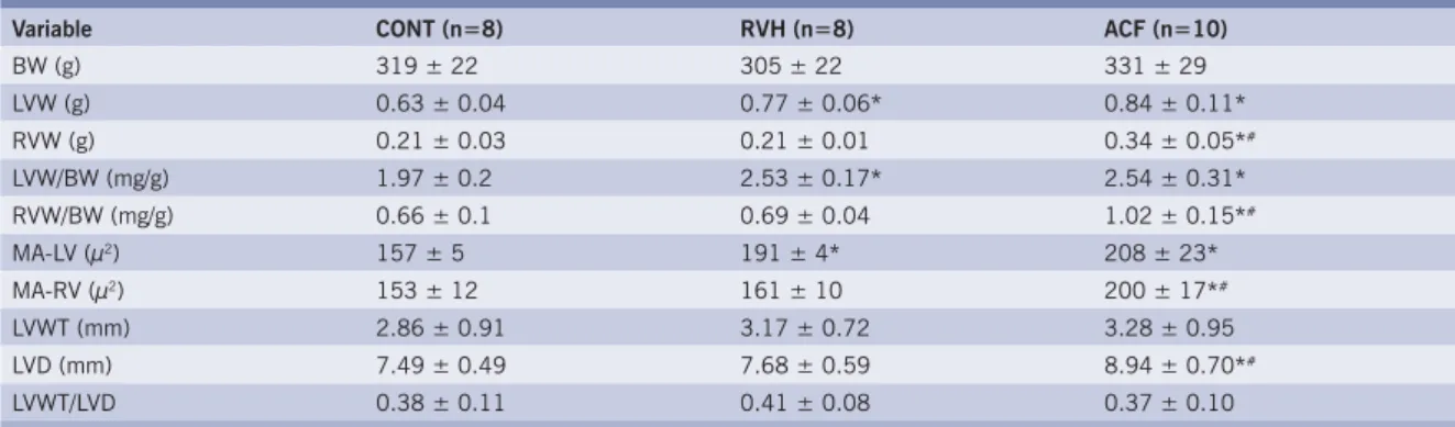
Tail cuff blood pressure was signifi cantly higher in the RVH group compared to CONT and ACF groups (fi g. 1). The morphometric data are presented in table 1. After four weeks left ventricular weight, LVW corrected to body weight and myocyte cross sectional area were similar in RVH and ACF groups, indicating similar degree
of LV hypertrophy. In the volume overload model (ACF) there was an increase in the right ventricle weight, RVW corrected by body weight and right ventricle myocyte cross sectional area compared to the CONT and RVH groups.
RVH group showed an increased collagen volume fraction when compared to CONT and ACF groups (fi g. 2). LV wall thickness was statistically similar among the experimental groups. LV cavity diameter was signifi cantly larger in the ACF group in relation to the other two groups. Left ventricular wall thickness and normalized wall thickness cavity diameter (LVWT/LVD) were comparable among the groups.
D
ISCUSSION
The objective of the present study was to compare cardiac remodeling secondary to either chronic volume or pressure overload.
In the pressure overload model, all indices showed left ventricular hypertrophy. The LV weight and myocyte sectional area grew signifi cantly in relation to the control group. The maintenance of normalized wall thickness suggests that the hypertrophy was still developing and had not yet modifi ed the ventricular chamber geometry to the concentric pattern. Therefore, it is reasonable to assume that in this period, the increased afterload in the hypertensive animals was not totally compensated by the developing hypertrophy. Nevertheless, the absence of right ventricle hypertrophy, in this experimental group, suggests that the myocyte cellular growth is more dependent on mechanical stimulus than on the trophic effects of neurohormonal activation. This results is agreement with other authors that evaluated similar models12. It is known that unilateral renal ischemia (Goldblatt - 2 kidney, 1 clip model) is the experimental model of renovascular hypertension where the activation of the rennin-angiotensin-aldosterone system is the leading factor in the genesis of hypertension13, hypertrophy and myocardial
Table 1 – Mean and standard deviations of the variables for the renovascular hypertension (RVH), aortocaval fi stula (ACF) and control (CONT) rat groups
Variable CONT (n=8) RVH (n=8) ACF (n=10)
BW (g) 319 ± 22 305 ± 22 331 ± 29
LVW (g) 0.63 ± 0.04 0.77 ± 0.06* 0.84 ± 0.11*
RVW (g) 0.21 ± 0.03 0.21 ± 0.01 0.34 ± 0.05*#
LVW/BW (mg/g) 1.97 ± 0.2 2.53 ± 0.17* 2.54 ± 0.31*
RVW/BW (mg/g) 0.66 ± 0.1 0.69 ± 0.04 1.02 ± 0.15*#
MA-LV (µ2) 157 ± 5 191 ± 4* 208 ± 23*
MA-RV (µ2) 153 ± 12 161 ± 10 200 ± 17*#
LVWT (mm) 2.86 ± 0.91 3.17 ± 0.72 3.28 ± 0.95
LVD (mm) 7.49 ± 0.49 7.68 ± 0.59 8.94 ± 0.70*#
LVWT/LVD 0.38 ± 0.11 0.41 ± 0.08 0.37 ± 0.10
interstitial fi brosis14, as well as vascular remodeling15. Sharifi et al16 observed increased angiotensin-converting enzyme activity both in the plasma and cardiac tissue of rats with renovascular hipertension, with two and twelve weeks of evolution, and concluded that the increased angiotensin-converting enzyme tissue activity has an important role in the alterations of the target organ in this experimental model. Therefore, both the mechanical effects of pressure overload and the humoral effects of the activation of the local and systemic renin-angiotensin system are relevant in the modulation of the ventricular remodeling.
Myocardial interstitial fi brosis was observed in the RVH group in accordance with previous results from our laboratory8 and other researchers17,18. This component of cardiac remodeling would be the result of both humoral fi broblast stimulation leading increased collagen synthesis17,19,20 and cellular necrosis21.
The rats submitted to the aortocaval fi stula presented a distinct hypertrophy pattern in comparison with hypertensive rats. The myocyte cellular area, ventricular weight and right ventricular weight normalized to body weight were higher than in CONT and RVH groups indicating combined right and left ventricular hypertrophy. Diastolic myocyte distension would be the main mechanical stimulus for right ventricle hypertrophy. However, the role of pulmonary hypertension caused by increased volume should not be discharged22. Therefore, it must be considered that the stimulus for right ventricular hypertrophy in aortocaval fi stula could be multifactorial, that is volume and pressure overload on pulmonary circulation22. Liu et al22 studying a similar model, showed that the myocyte increases both in width and length which helps maintain the muscular form. During the compensated phase, this growth contributes to the maintenance of the chamber geometry without ventricular dilation. Afterwards, when the capacity of the muscle to be hypertrophied is exhausted, the cavity radius increases
disproportionally and leads to heart failure23.
In the present study the LVWT/LVD ratio was preserved, which suggests that in the four week of the experiment, eccentric hypertrophy was still compensated. This stage of the remodeling allows the maintenance of wall tension and preservation of cardiac performance despite the hemodynamic overload.
The collagen interstitial matrix is also a factor to be studied in the remodeling process of volume overload secondary to aortocaval fi stula. It is known that the quantity of interstitial collagen is dependent on the interaction between the synthesis process and protein degradation. Pressure overload and the angiotensin II humoral stimulus are the main stimulating agents for collagen synthesis while muscular distension caused by volume overload contributes to its degradation24, in a process that probably involves collagenase activation due to mastocyte degranulation25.
Af ter per forming the aor tocaval fistula the metalloproteases are rapidly activated and remain signifi cantly elevated during the following fi ve days, returning to basal conditions in fourteen days25. Concurrent with the increase of metalloprotease activity, there is a signifi cant reduction in the collagen interstitial matrix24. In this study, it was observed that the ACF group maintained the amount of interstitial collagen, suggesting an equilibrium between degradation and interstitial matrix synthesis after four week. The most relevant implication of these observations in that the results of medical literature that show a direct relationship between myocardial hypertrophy and cardiovascular morbidity and mortality do not necessarily apply to the hypertrophy pattern caused by volume overload.
Our results show that pressure or volume overloads cause distinct patterns of cardiac remodeling suggesting that their implications on ventricular dysfunction are not interchangeable.
Fig. 1 – Graphical representation of the mean and standard deviations of tail cuff blood pressure for the renovascular hypertension (RVH), aortocaval fi stula (ACF) and control (CONT) rats groups. Analyzed using ANOVA, followed by the Tukey test
Fig. 2 – Graphical representation of the mean and standard deviations of the interstitial collagen fraction (CVF) for the renovascular hypertension (RVH), aortocaval fi stula (ACF) and control (CONT) rat groups. Analyzed using ANOVA, followed by the Tukey test
Grupos
CONT HRV FAV
0 1 2 3 4
p<0,05 p<0,05
Groups
CONT RVH ACF 0
50 100 150 200 250
p<0,05 p<0,05
p<0.05 p<0.05
Tail Cuff Blood P
ressure (mmHg)
CVF (%)
p<0.05 p<0.05
Groups Groups
A
CKNOWLEDGEMENTS
We would like to thank Fapesp for the fi nancial support (Proc. n.01/03459-7).
R
EFERENCES
1. Brilla CG, Matsubara L, Weber KT. Advanced hypertensive heart disease in spontaneously hypertensive rats - Lisinopril-mediated regression of myocardial fi brosis. Hypertension 1996; 28: 269-75.
2. Swynghedauw B. Molecular mechanisms of myocardial remodeling. Physiol Rev 1999; 79: 215-62.
3. Weber KT. Targeting pathological remodeling - Concepts of cardioprotection and reparation. Circulation 2000; 102: 1342-5.
4. Devereux RB. Therapeutic options in minimizing left ventricular hypertrophy. Am Heart J 2000; 139: S9-S14.
5. Ciardullo AV, Azzolini L, Bevini M et al. A diagnosis of left ventricular hypertrophy on ECG is associated with a high cardiovascular risk: fi ndings from a 40-to 69-year-old cohort in general practice. Fam Pract 2004; 21: 63-5.
6. Messerli FH. Hypertension and sudden cardiac death. Am J Hypertens 1999; 12: 181S-188S.
7. Janicki JS. Myocardial Collagen remodeling and left ventricular diastolic function. Braz J Med Biol Res 1992; 25: 975-82.
8. Matsubara LS, Matsubara BB, Okoshi MP, Franco M, Cicogna AC. Myocardial fi brosis rather than hypertrophy induces diastolic dysfunction in renovascular hypertensive rats. Can J Physiol Pharmacol 1997; 75: 1328-34.
9. Namba T, Tsutsui H, Tagawa H et al. Regulation of fi brillar collagen gene expression and protein accumulation in volume-overloaded cardiac hypertrophy. Circulation 1997; 95: 2448-54.
10. Brower GL, Janicki JS. Contribution of ventricular remodeling to pathogenesis of heart failure in rats. Am J Physiol-Heart Circul Physiol 2001; 280: H674-H683.
11. Garcia R, Diebold S. Simple, rapid and effective method of producing aortocaval shunts in the rats. Circulation Researc 1990; 24: 430-2.
12. Baba HA, Iwai T, Bauer M, Irlbeck M, Schmid KW, Zimmer HG. Differential effects of angiotensin II receptor blockade on pressure-induced left ventricular hypertrophy and fi brosis in rats. J Mol Cell Cardiol 1999; 31: 445-55.
13. Stroth U, Unger T. The renin-angiotensin system and its receptors. J Cardiovasc Pharmacol 1999; 33: S21-S28.
14. Matsubara BB, Matsubara LS, Franco M, Padovani JC, Janicki JS. The effect of non-antihypertensive doses of angiotensin converting enzyme
inhibitor on myocardial necrosis and hypertrophy in young rats with renovascular hypertension. Int J Exp Pathol 1999; 80: 97-104.
15. Rosendorff C. The renin-angiotensin system and vascular hypertrophy. J Am Coll Cardiol 1996; 28: 803-12.
16. Sharifi AM, Akbarloo N, Heshmatian B, Ziai A. Alteration of local ACE activity and vascular responsiveness during development of 2K1C renovascular hypertension. Pharmacol Res 2003; 47: 201-9.
17. Hocher B, George I, Rebstock J et al. Endothelin system-dependent cardiac remodeling in renovascular hypertension. Hypertension 1999; 33: 816-22.
18. Saam T, Ehmke H, Haas C, Ritz E, Amann K. Effect of endothelin blockade on early cardiovascular remodeling in the one-clip-two-kidney hypertension of the rat. Kidney Blood Press Res 2003; 26: 325-32.
19. Challah M, Nicoletti A, Arnal JF et al. Cardiac Angiotensin-Converting Enzyme Overproduction Indicates Interstitial Activation in Renovascular Hypertension. Cardiovasc Res 1995; 30: 231-9.
20. Nicoletti A, Heudes D, Mandet C, Hinglais N, Bariety J, Michel JB. Infl ammatory cells and myocardial fi brosis: Spatial and temporal distribution in renovascular hypertensive rats. Cardiovasc Res 1996; 32: 1096-107.
21. Okoshi MP, Matsubara LS, Franco M, Cicogna AC, Matsubara BB. Myocyte necrosis is the basis for fi brosis in renovascular hypertensive rats. Braz J Med Biol Res 1997; 30: 1135-44.
22. Liu Z, Hilbelink DR, Crockett WB, Gerdes AM. Regional Changes in Hemodynamics and Cardiac Myocyte Size in Rats With Aortocaval Fistulas .1. Developing and Established Hypertrophy. Circ Res 1991; 69: 52-8.
23. Brower GL, Henegar JR, Janicki JS. Temporal evaluation of left ventricular remodeling and function in rats with chronic volume overload. Am J Physiol-Heart Circul Physiol 1996; 40: H2071-H2078.
24. Chancey AL, Brower GL, Peterson JT, Janicki JS. Effects of matrix metalloproteinase inhibition on ventricular remodeling due to volume overload. Circulation 2002; 105: 1983-8.