269
IMAGES IN NEUROLOGY
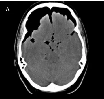
Tension pneumocephalus and rhinorrhea
related to chronic sinusitis
Pneumoencéfalo hipertensivo e rinorreia relacionados à sinusite crônica
Victor de Almeida Kosac
1, André PC Matta
2, Frederico M Prado
1, Osvaldo JM Nascimento
2, Gabriela DJ Matta
3,
Tereza CS dos Santos
41Medical residents of Neurology, Department of Neurology, Federal Fluminense University, Niterói RJ, Brazil;
2MD, PhD, Professor of Neurology, Department of Neurology, Federal Fluminense University, Niterói RJ, Brazil;
3Ophthalmologist, Post-graduating program in Neurology and Neuroscience, Federal Fluminense University, Niterói RJ, Brazil;
4Radiologist at Antonio Pedro Hospital, Federal Fluminense University, Niterói RJ, Brazil.
Correspondence: Victor de Almeida Kosac; Rua Doutor Paulo César 25 / apto. 1.608; 24240-000 Niterói RJ - Brasil; E-mail: [email protected] Conflict of interest: There is no conflict of interest to declare.
Received 18 March 2012; Received in final form 26 November 2012; Accepted 03 December 2012. 1. Lefranc M, Peltier J, Demuynkc F, et al. Tension pneumocephalus and
rhinorrhea revealing spontaneous cerebrospinal fluid fistula of the anterior cranial base. Neurochirurgie 2009;55:340-344.
2. Webber-Jones JE. Tension pneumocephalus. J Neurosci Nurs 2005;37: 272-276.