Intramural duodenal hematoma secondary to pancreatitis:
case report and review of the literature
João Henrique Botto de Oliveira
I, Raiza Samenica Esper
I, Rodrigo Campos Ocariz
I, Flora Specian Sartori
I, Lucas Marcelo Dias
Freire
II, Elinton Adami Chaim
III, Francisco Callejas-Neto
IV, Everton Cazzo
VDepartment of Surgery, Centro Médico de Campinas (CMC), Campinas (SP), Brazil
ABSTRACT
CONTEXT: Spontaneous intramural duodenal hematoma is uncommon and is usually associated with coagulopathy, anticoagulant therapy and endoscopic procedures. The aim here was to describe a case of intramural duodenal hematoma caused by chronic exacerbation of pancreatitis.
CASE REPORT: A 46-year-old male with chronic alcoholic pancreatitis was admitted to hospital due to abdominal pain, melena and low hemoglobin. An intramural duodenal hematoma with active bleeding was detected and selective angioembolization was warranted. The patient evolved with a perforated duo-denum and underwent laparotomy with exclusion of the pylorus and Roux-en-Y gastrojejunostomy. He was discharged nine days later.
CONCLUSION: Intramural duodenal hematoma is a rare complication of pancreatitis. Selective emboliza-tion is the preferred treatment for hemorrhagic complicaemboliza-tions of pancreatitis. However, the risk of visceral ischemia and perforation should be considered.
IMD. Resident Physician, Department of Surgery,
Centro Médico de Campinas (CMC), Campinas (SP), Brazil
IIMD. Attending Physician, Endovascular Surgery
Unit, Centro Médico de Campinas (CMC), Campinas (SP), Brazil
IIIMD, PhD. Full Professor, Department of Surgery,
Faculdade de Ciências Médicas da Universidade Estadual de Campinas (FCM/UNICAMP), Campinas (SP), Brazil.
IVMD, MSc. Assistant Professor, Department of
Surgery, Faculdade de Ciências Médicas da Universidade Estadual de Campinas (FCM/ UNICAMP), Campinas (SP), Brazil.
VMD, PhD. Adjunct Professor, Department of
Surgery, Faculdade de Ciências Médicas da Universidade Estadual de Campinas (FCM/ UNICAMP), Campinas (SP), Brazil.
KEY WORDS:
Pancreatitis. Duodenum. Hematoma.
Embolization, therapeutic. Duodenal diseases.
INTRODUCTION
he irst description of an intramural duodenal hematoma was made by McLauchlan in 1838. his condition is usually associated with blunt abdominal trauma. Spontaneous intramural duodenal hematoma is uncommon and has been linked to coagulopathy, anticoagulant ther-apy and endoscopic procedures.1-3 Other causes include several pancreatic diseases, colla-genosis, peptic ulcers and pancreaticoduodenal aneurysm.4-7 To date, the exact mechanism leading to intramural hematoma in cases of pancreatitis has not yet been fully elucidated and the prognosis has not yet been completely deined, mainly due to its scarcity.1,3,5-9
his study sought to describe a case of an intramural duodenal hematoma caused by chronic exacerbation of pancreatitis.
CASE REPORT
he patient (JCAP) was a 43-year-old white male who had been a chronic abuser of alcohol for 25 years (two liters of distilled liquor/day), with an antecedent of acute pancreatitis ive years before the present case report. He had been complaining of strong typical pain for three days, along with melena.
At admission to hospital, the following test results were noted: leukogram = 15,000 u/l, hemoglobin = 16 g/dl and amylase = 99 IU/l. A computed tomography (CT) scan showed signs of chronic pancreatitis and a bulky submucosal duodenal hematoma from the bulb to the third portion of the duodenum with intramural active bleeding in the region of the gastroduodenal artery (Figure 1A). Upper digestive tract endoscopy revealed a large submucosal hematoma in the duodenum, without any active bleeding into the lumen (Figure 2).
duodenal rupture from the bulb to the second portion of the duo-denum, along with signs of acute pancreatitis. he duodenal rup-ture was closed, along with exclusion of the pylorus, and Roux-en-Y gastroenteroanastomosis was carried out.
he patient then progressed with a high-output duodenal is-tula. Treatment consisting of parenteral nutrition, octreotide and antibiotic therapy was started, and this led to regression over a nine-day period. A control CT scan demonstrated regression of the hematoma, while the signs of chronic pancreatitis continued to be present, but without evidence of exacerbation (Figure 1C). A con-trasted upper gastrointestinal radiographic series showed exclusion of the pylorus and patent gastroenteroanastomosis. he patient was then followed up for six months, with uneventful evolution.
DISCUSSION
Duodenal hematomas have been described as complications of both acute and chronic pancreatitis. Acute pancreatitis is a com-mon disease, with an incidence of 20 to 40 cases/100,000 person-years of life and a mortality rate close to 5%, and the vast majority of the cases are of biliary etiology.10 On the other hand, chronic pancreatitis is a progressive inlammatory disorder characterized by irreversible destruction of the pancreatic parenchyma and may
Figure 2. Upper gastrointestinal endoscopy.
Figure 3. Arterial embolization: A) active bleeding in the region of the gastroduodenal artery; B) post-embolization control.
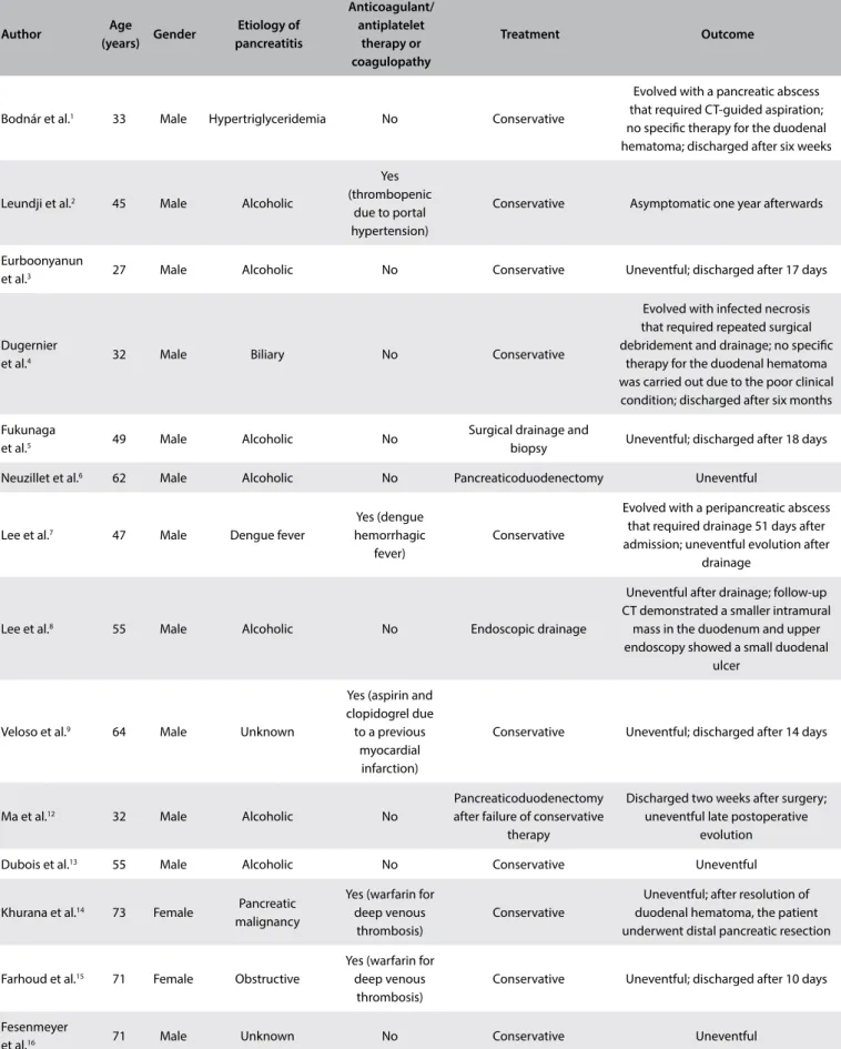
Table 1. Database search results for duodenal hematomas caused by pancreatitis
Electronic databases Search strategies Results
MEDLINE (PubMed)
(Duodenum) AND (Hematoma) AND
(Pancreatitis)
13 case reports
LILACS (BVS)
(((Duodenum) OR (Duodeno)) AND (Hematoma) AND ((Pancreatitis) OR (Pancreatite) OR (Pancreatítis)))
1 case report Figure 1. Computed tomography scans: A) at admission; B) post-embolization; C) post-surgical control.
A
B
C
One day ater this procedure, there was worsening of the pain. A CT scan showed signs of visceral perforation (Figure 1B). Emergency laparotomy was warranted and revealed the following: an ischemic duodenum with a bulky wall hematoma and a 6-cm
Author Age
(years) Gender
Etiology of pancreatitis
Anticoagulant/ antiplatelet
therapy or coagulopathy
Treatment Outcome
Bodnár et al.1 33 Male Hypertriglyceridemia No Conservative
Evolved with a pancreatic abscess that required CT-guided aspiration; no speciic therapy for the duodenal hematoma; discharged after six weeks
Leundji et al.2 45 Male Alcoholic
Yes (thrombopenic
due to portal hypertension)
Conservative Asymptomatic one year afterwards
Eurboonyanun
et al.3 27 Male Alcoholic No Conservative Uneventful; discharged after 17 days
Dugernier
et al.4 32 Male Biliary No Conservative
Evolved with infected necrosis that required repeated surgical debridement and drainage; no speciic
therapy for the duodenal hematoma was carried out due to the poor clinical condition; discharged after six months
Fukunaga
et al.5 49 Male Alcoholic No
Surgical drainage and
biopsy Uneventful; discharged after 18 days
Neuzillet et al.6 62 Male Alcoholic No Pancreaticoduodenectomy Uneventful
Lee et al.7 47 Male Dengue fever
Yes (dengue hemorrhagic
fever)
Conservative
Evolved with a peripancreatic abscess that required drainage 51 days after admission; uneventful evolution after
drainage
Lee et al.8 55 Male Alcoholic No Endoscopic drainage
Uneventful after drainage; follow-up CT demonstrated a smaller intramural
mass in the duodenum and upper endoscopy showed a small duodenal
ulcer
Veloso et al.9 64 Male Unknown
Yes (aspirin and clopidogrel due to a previous
myocardial infarction)
Conservative Uneventful; discharged after 14 days
Ma et al.12 32 Male Alcoholic No
Pancreaticoduodenectomy after failure of conservative
therapy
Discharged two weeks after surgery; uneventful late postoperative
evolution
Dubois et al.13 55 Male Alcoholic No Conservative Uneventful
Khurana et al.14 73 Female Pancreatic malignancy
Yes (warfarin for deep venous
thrombosis)
Conservative
Uneventful; after resolution of duodenal hematoma, the patient underwent distal pancreatic resection
Farhoud et al.15 71 Female Obstructive
Yes (warfarin for deep venous
thrombosis)
Conservative Uneventful; discharged after 10 days
Fesenmeyer
et al.16 71 Male Unknown No Conservative Uneventful
CT = computed tomography.
be associated with disabling chronic pain and permanent loss of exocrine and endocrine function. he majority of such cases are alcohol-related. heir prevalence is 3-4 cases/100,000 people.11
One of the rarest and most fatal complications of pancreatitis is spontaneous bleeding of intestinal vessels. A review of the literature was conducted through an online search for the MeSH terms “duo-denum”, “hematoma” and “pancreatitis” in MEDLINE (via PubMed) and LILACS (via BVS) in papers published over the last 20 years (Table 1). We included original studies that reported single cases or case series of this disease. All the papers were checked according to their titles and abstracts (screening). Full papers were obtained from journals available through the website of the Commission for Improvement of Higher Education Personnel (Comissão de Aperfeiçoamento de Pessoal de Nível Superior, CAPES), of the Brazilian Ministry of Education. Articles that were unavailable were requested from their authors. Articles presenting potentially relevant studies were read and analyzed to assess the inclusion cri-teria. We excluded articles that consisted of in vitro or animal
stud-ies, articles in which the participants’ characteristics did not match those mentioned above, poster session abstracts, review articles and other types of publications. Articles that described traumatic or iatrogenic duodenal hematomas were also excluded. Other papers were used for contextualization and discussion.
Ater extensive online research, we identiied 14 studies, which were all single case reports. Table 21-9,12-16 summarizes the main articles found and their reported outcomes. Figure 4 presents a low diagram of the articles selected.
Vascular erosion occurs due to extravasation of proteolytic enzymes. Potentially fatal bleeding, characterized by a decrease of two hemoglobin points, are rare complications occurring in about 1% to 5% of patients with acute or chronic pancreatitis. he vessels most afected are the gastroduodenal, pancreatoduodenal, splenic, gastroepiploic and let gastric arteries, because of their proximity to the pancreas, along with small branches of the inferior mesenteric arteries. he symptoms include pain, melena, hematemesis and ret-roperitoneal bleeding. Upper endoscopy, CT scans and angiography are involved in making the diagnosis.17
he majority of the previously reported cases showed that con-servative therapy was possible, but this method is associated with lengthier hospital stay and a need for transfusion of blood components. Interventional therapies may include surgical repair using endoscopic and endovascular techniques. he beneits of endoscopy include its minimally invasive nature, compared with other treatment options, and the absence of radiation exposure. However, it has limitations, given that it cannot be used for areas of bleeding that are inaccessible to the intestinal lumen.8 Arterial embolization seems to be efective for management of bleeding. Embolization is considered successful when both radiologically and clinically there is evidence of bleeding control characterized by hemoglobin stabilization and absence of signs and symptoms of shock.18,19 Laparotomy for bleeding therapy should only be considered for hemodynamically unstable patients. Advances in endovascular radiology have led to this method becoming the pre-ferred treatment option.18
he present case demonstrates the need for a high degree of suspicion that the diagnosis could comprise bleeding caused by pancreatitis, as well as the need to remain open to the possibility of a severe and rare complication ater embolization (visceral per-foration). his complication requires rapid intervention, because the risk of mortality that it presents is up to 10 times greater than that commonly observed in cases of acute pancreatitis.20,21 he currently available evidence consists solely of single case reports, which thus precludes inal conclusions regarding the optimal ther-apy. Hence, further research is necessary.
CONCLUSION
Intramural duodenal hematoma is a rare complication of pancre-atitis. Selective embolization is the preferred treatment, but the risk of visceral ischemia and perforation should be considered.
REFERENCES
1. Bodnár Z, Várvölgyi C, Tóth J, Sápy P, Kakuk G. Intramural duodenal
hematoma complicating acute necrotizing pancreatitis. Gastrointest
Endosc. 2000;52(6):791-3.
2. Leundji H, Cuingnet P, Simon M, Boruchowicz A. [Duodenal hematoma
associated with thrombopenia in chronic alcoholic pancreatitis].
Gastroenterol Clin Biol. 2002;26(2):185-6. Figure 4. Flow diagram of the narrative review of the literature.
Studies included in the review
(n = 14) Full-text articles
assessed for eligibility (Total n = 14)
Records excluded (n = 1)
Reasons:
- Redundant data: 1 Records screened
(n = 15)
Records excluded (n = 11)
Reasons: - Not pertinent: 11 Records identiied through database
search (PubMed and LILACS) (n = 26)
Included
Elig
ibilit
y
S
cr
eening
Iden
tiica
3. Eurboonyanun C, Somsap K, Ruangwannasak S, Sripanaskul A.
Spontaneous Intramural Duodenal Hematoma: Pancreatitis,
Obstructive Jaundice, and Upper Intestinal Obstruction. Case Rep
Surg. 2016;2016:5321081.
4. Dugernier TL, Breuskin FM. Duodenal air dissection secondary to
intramural hematoma in necrotizing pancreatitis. Endoscopy.
2002;34(12):1024.
5. Fukunaga N, Ishikawa M, Yamamura Y, Ichimori T, Sakata A. Spontaneous
intramural duodenal hematoma complicating acute pancreatitis.
Surgery. 2011;149(1):143-4.
6. Neuzillet C, Facchiano E, Palazzo L, et al. Intramural duodenal hematoma
as a complication of paraduodenal pancreatitis. Clin Res Hepatol
Gastroenterol. 2011;35(2):140-2.
7. Lee CY, Tsai HC, Lee SS, et al. Dengue hemorrhagic fever presenting
with hemorrhagic pancreatitis and an intramural hematoma of the
duodenal wall: a case report and review of the literature. Southeast
Asian J Trop Med Public Health. 2013;44(3):400-8.
8. Lee JY, Chung JS, Kim TH. Successful endoscopic decompression
for intramural duodenal hematoma with gastric outlet obstruction
complicating acute pancreatitis. Clin Endosc. 2012;45(3):202-4.
9. Veloso N, Amaro P, Ferreira M, Romãozinho JM, Soia C. Acute pancreatitis
associated with a nontraumatic, intramural duodenal hematoma.
Endoscopy. 2013;45 Suppl 2 UCTN:E51-2.
10. Forsmark ChE, Vege SS, Wilcox CM. Acute Pancreatitis. N Engl J Med.
2017;376(6):598-9.
11. Gestic MA, Callejas-Neto F, Chaim EA, et al. Tratamento cirúrgico da
pancreatite crônica com a técnica de Frey: panorama atual [Surgical
treatment of chronic pancreatitis with frey procedure: current situation].
ABCD Arq Bras Cir Dig. 2011;24(4):305-11.
12. Ma JK, Ng KK, Poon RT, Fan ST. Pancreatic-induced intramural duodenal
haematoma. Asian J Surg. 2008;31(2):83-6.
13. Dubois J, Guy F, Porcheron J. A pancreatic-induced intramural duodenal
hematoma: a case report and literature review. Hepatogastroenterology.
2003;50(53):1689-92.
14. Khurana T, Shah A, Ali I, Islam R, Siddiqui AA. Intramural Duodenal
Hematoma with Acute Pancreatitis in a Patient with an Overt Pancreatic
Malignancy. ACG Case Rep J. 2014;1(4):209-11.
15. Farhoud S, Stephani SM, Bromberg SH. Pancreatite aguda devida a
hematoma intramural do duodeno por uso de anticoagulante [Acute
pancreatitis due to intramural hematoma of the duodenum by the use
of anticoagulants]. Arq Gastroenterol. 2001;38(1):53-6.
16. Fesenmyer ME, Nelson DB. Intramural duodenal hematoma due to
pancreatitis. J Clin Gastroenterol. 1998;26(4):350-2.
17. Vujic I. Vascular complications of pancreatitis. Radiol Clin North Am.
1989;27(1):81-91.
18. Fitzpatrick J, Bhat R, Young JA. Angiographic embolization is an
efective treatment of severe hemorrhage in pancreatitis. Pancreas.
2014;43(3):436-9.
19. Phillip V, Rasch S, Gaa J, Schmid RM, Algül H. Spontaneous bleeding
in pancreatitis treated by transcatheter arterial coil embolization: a
retrospective study. PLoS One. 2013;8(8):e72903.
20. Kim SI, Jin YJ, Cho SG, et al. Duodenal perforation and esophageal
ischemia following transarterial chemoembolization for hepatocellular
carcinoma: A case report. Medicine (Baltimore). 2016;95(27):e3987.
21. Cheah WK, So J, Chong SM, Goh P. Duodenal ulcer perforation following
cyanoacrylate injection. Endoscopy. 2000;32(5):S23.
Sources of funding: None
Conlict of interest: None
Date of irst submission:May 2, 2017
Last received: May 2, 2017
Accepted: May 29, 2017
Address for correspondence:
Everton Cazzo
Departamento de Cirurgia da Facudade de Ciências Médicas da
Universidade Estadual de Campinas (FCM/UNICAMP)
Rua Alexander Fleming, s/no
Cidade Universitária Zeferino Vaz — Campinas (SP) — Brasil
CEP 13085-000
Tel. (+55 19) 3521-9450