ARTICLE
Management of desmoid-type fibromatosis
involving peripheral nerves
Tratamento da fibromatose tipo desmoide envolvendo nervos periféricos
Mario G. Siqueira1, Paulo L. Tavares1, Roberto S. Martins1, Carlos O. Heise1, Luciano H.L. Foroni1, Marcelo
Bordalo2, Roberto Falzoni3
Desmoid-type ibromatosis (DTF) is a ibrous prolifera-tion from fascia and musculoaponeurotic tissues localized mainly in the abdomen. Owing to its slow growing and no risk of metastatization, these rare neoplastic lesions are re-garded as benign, but the tendency of local invasion and the high rate of recurrence leads to signiicant morbidity by de-struction of adjacent vital structures. here is no consensus about the best modality of treatment for these lesions, that ranges from wait-and-see and clinical treatment to radical surgery and radiotherapy. Due to the rarity of these tumors, evidence regarding the best treatment protocol for each case is drawn from case reports and small series of patients in the literature. he lesions involving or compressing peripheral nerves are even rarer and its ideal management has not been deined. In this report, we present four cases of desmoid-type
ibromatosis involving the peripheral nerve. he diferent ways to manage these lesions are critically analyzed, and an attempt to deine therapeutic suggestions is made.
CASE REPORT
Case 1
Male, 22-years-old, with a slow growing mass in the medi-al region of the right arm, immediately proximmedi-al to the medimedi-al epicondyle of the humerus, for seven years. In 1998, he was sub-mitted to surgical treatment in another institution. he
resec-tion was reported as complete and the histological diagnosis was neuroibroma. In 2002, the tumor recurred and the same surgeon resected a 12 centimeters mass with irm adhesions
1Peripheral Nerve Surgery Unit, Department of Neurosurgery, University of São Paulo Medical School, São Paulo, SP, Brazil;
2Department of Radiology, University of São Paulo Medical School, São Paulo SP, Brazil;
3Department of Pathology, University of São Paulo Medical School, São Paulo SP, Brazil.
Correspondence: Mario G. Siqueira; Rua Maestro Cardim 592 / Conj. 1101; 01323-001 São Paulo SP - Brasil; E-mail: [email protected] Conflict of interest: There is no conflict of interest to declare.
Received 17 December 2011; Accepted 30 December 2011
ABSTRACT
Desmoid-type fibromatosis is an uncommon and aggressive neoplasia, associated with a high rate of recurrence. It is characterized by an infiltrative but benign fibroblastic proliferation occurring within the deep soft tissues. There is no consensus about the treatment of those tu-mors. We present a surgical series of four cases, involving the brachial plexus (two cases), the median nerve and the medial brachial cutane-ous nerve. Except for the last case, they were submitted to multiple surgical procedures and showed repeated recurrences. The diagnosis, the different ways of treatment and the prognosis of these tumoral lesions are discussed. Our results support the indication of radical surgery followed by radiotherapy as probably one of the best ways to treat those controversial lesions.
Key words: desmoid-type fibromatosis, desmoid tumor, fibromatosis, peripheral nerve.
RESUMO
A fibromatose do tipo desmoide é uma lesão tumoral agressiva e rara, associada a alto índice de recorrência. É caracterizada pela fibroblás-tica infiltrativa, porém benigna, que ocorre no interior de tecidos moles profundos. Não existe consenso com relação ao tratamento desses tumores. Apresentamos uma série cirúrgica de quatro casos comprometendo o plexo braquial (dois casos), o nervo mediano e o nervo cutâ-neo medial do braço. Com exceção do último caso, todos foram submetidos a múltiplos procedimentos cirúrgicos e apresentaram recorrên-cias repetidas. São discutidos o diagnóstico, as diferentes formas de tratamento e o prognóstico dessas lesões tumorais. Nossos resultados apoiam o conceito de que cirurgia radical seguida por radioterapia é uma das melhores formas de se tratar essas controvertidas lesões.
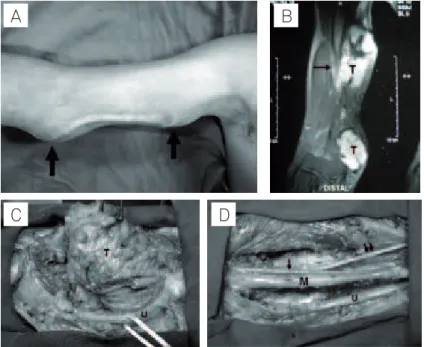
to the periosteum of the humerus and to other deep tissues, including the median and ulnar nerves. Once again, the histo-logical diagnosis was of a neuroibroma. In 2004, a new recur-rence, provoking local pain with irradiation to the distal ulnar nerve distribution, brought the patient to our institution. At examination, we observed two irm masses, one immediately proximal to the elbow and the other about 5 centimeters prox-imal and apart to the irst one, both in the medial surface of the right arm (Fig 1). here were no neurological deicits. Two hardened masses with 10 and 11 centimeters in their largest diameters, with great adhesion to the adjacent muscles and apparent invasion in some areas, were founded at surgery. he ulnar nerve was dislocated inferior and medially, and the me-dian nerve and the brachial artery were inside the proximal tu-mor. he resection was apparently complete and no additional neurological deicits were observed. he pain disappeared in the immediate postoperative period. he histological diagnosis now was of DTF. he patient refused any adjuvant treatment and one year after the last surgery a new lesion developed at the proximal limit of the surgical scar, without neurological im-pairment. Two solid masses, with 4 centimeters each in their largest diameters, extending to the axillary region, involving and compressing the median and ulnar nerves, were resect-ed in the proximal arm. he limit of the lesions with the adja-cent ibrosis and muscles was totally imprecise in some areas. After histological conirmation of recurrent DTF, the patient was submitted to complementary treatment with radiother-apy. After two years, the tumor recurred outside the radiation ield, now in the infraclavicular area, and was removed once
again. Since this last surgery, performed two years ago, the pa-tient is doing well.
Case 2
Female, 33-years-old, had a previous resection of a left subcutaneous axillary nodule in another institution. We have no description of the procedure or of the mass histopa-thology. Fourteen months later, she was referred to us with a new axillary nodule, at the same site, complaining of pain, hypoesthesia and paresthesias in the medial aspect of the arm (medial brachial cutaneous nerve territory) and show-ing a tshow-inglshow-ing sensation evoked by percussion of the nodule. MRI demonstrated a nodular mass (12x8x7 mm) in the left axilla, well circumscribed, related to nervous structures, but without iniltrative characteristics. Intraoperatively, the nodule was involved by ibrous tissue, probably related to the previous procedure, and was compressing the medial brachial cutaneous nerve. he nerve was anatomically in-tact under the lesion. Total surgical removal was performed and, surprisingly, the histopathological diagnosis was DTF. he pain resolved and the patient is recovering her sensi-tive deicit. he patient is well, without recurrences, sixteen months after surgery.
Case 3
Female, 34-years-old, was submitted to resection of a right supraclavicular mass two years before in another institution, with a diagnosis of DTF. One year later, she had a recurrence in the same place, without associated neurological deicits or
A
C
D
B
pain. he tumor was partially resected and the patient was re-ferred to our hospital. On the irst examination, the patient was neurologically intact and a voluminous mass was ob-served in her right supraclavicular region. Paresthesias were elicited by manipulation of the mass. he MRI showed a large lobulated mass (8.5x8.0x5.0 cm) compressing the anterior sca-lenus muscle and iniltrating the multiidus, levator scapulae and posterior scalenus muscles (Fig 2). he upper and middle trunks of the brachial plexus were involved, and the tumor dis-located the lower trunk, as well as the proximal segments of the internal jugular and subclavian veins. Gadolinium intrave-nous injection provoked a heterogeneous enhancement of the mass. A radical resection of the lesion was performed, with-out compromising the neurological status of the patient, and DTF diagnosis was conirmed. he surgery was soon followed by radiotherapy. he patient is still doing well after ive years of follow-up, with no evidences of recurrence.
Case 4
Female, 19-years-old, had a right subscapular mass re-moved in 2005 and a right neck mass resected in 2006 in anoth-er institution. In both lesions the diagnosis of DTF was made. Five months later, the patient observed a growing mass in the area of the neck surgery. Except for mild local pain, the patient was asymptomatic. MRI demonstrated recurrence of both le-sions, the subscapular measuring 6.5x4.0x2.0 cm and the cervi-cal lesion with 9.0x7.0x3.0 cm. he posterior mass was located between the trapezius muscle and the right scapula. he right cervical lesion was medial to the sternocleidomastoid muscle and posterior to the internal jugular vein and carotid artery. he
cervical mass was iniltrating the anterior and middle scalenus muscle and involved the C7 brachial plexus root and the phren-ic nerve. Both lesions were radphren-ically resected, and, after the histological diagnosis of DTF, the patient was referred to the radiotherapy department. he patient is neurologically in-tact almost four years after the last surgery and there is no evi-dence of recurrence.
DISCUSSION
Over the years, many terms have been used to describe this tumor, including: desmoid tumor, well-diferentiated nonmetastasizing ibrosarcoma, ibrosarcoma, Grade I ibro-sarcoma, aggressive ibromatosis and desmoid-type ibroma-tosis (DTF), the last term being the designation of choice of the World Health Organization1. DTFs typically evolve over
a period of three years and stabilize thereafter. Recurrences or progression most commonly occurs between 14 and 17 months after surgical resection2. It is estimated that these
tumors account for 0.03% of all neoplasms and comprise around 3% of all soft tissue neoplasms3,4, with an annual
oc-currence of 2.4 to 4.5 new cases per million persons3, but the
real incidence of DTF is diicult to ascertain, mainly because of the multiple denominations attributed to these lesions. In the largest published series of peripheral nerve tumors (Louisiana State University Health Sciences Center – 543 tu-mors), these lesions comprised only 2.0% of the cases3,5.
Macroscopically, DTF is a rubbery gray-whitish lesion with a scar appearance, without a capsule, that can reach large
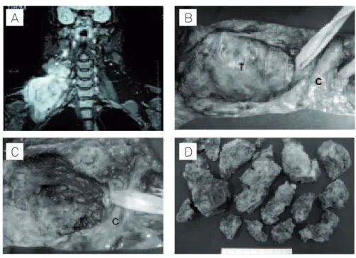
Fig 2. Case 3. (A) RM, Coronal T2-weighted MR image, with fat-supression, demonstrating a large mass with high signal intensity in the supraclavicular region, involving/dislocating the brachial plexus. (B). Intraoperative photograph demonstrating a large tumoral mass (T) in the supraclavicular region. Penrose drains identifying brachial plexus elements. C: clavicle. (C) Intraoperative photograph after removal of the tumor. C: clavicle. (D) Macroscopic aspect of the tumor removed.
A
D
B
C
dimensions (up to 20 cm). Its histology is characterized by a local iniltrative process composed by mature ibroblasts or myoibroblasts in an abundant collagen matrix, without any evidence of cellular anaplasia or abnormal mitoses (Fig 3). Although regarded as benign lesions, DTFs are locally iniltra-tive. hey usually invade the adjacent muscles and occasion-ally penetrate the trabecular spaces of bones in the area. hey can also invade, involve or compress nerves and vessels, as in the cases we are reporting, and sometimes can grow inside scars. he destruction of adjacent vital structures and organs can cause extensive morbidity. he more aggressive lesions are characterized by a larger number of myoibroblasts, while the less aggressive show a larger number of mastocites. here is no report of regional linfonode involvement6.
First described in the abdominal wall of pregnant women, DTFs can occur virtually in any area of the body, but two thirds of these lesions are originated from the anterior aponeurosis of the rectus abdominis muscle2, and are frequently associated
with the autosomal-dominant familial adenomatous polypo-sis of the colon (Gardner syndrome). he majority of the extra-abdominal DTFs, frequently called aggressive ibromatosis, is originated from muscles/aponeurosis in the shoulder and pel-vic girdles, and are solitary and sporadic7. Multicentric tumors,
like our case #4, are even more rare2. here are few reports of
DTFs originated inside nerves, probably from stroma cells8.
he DTFs resemble a ibrosarcoma for its iniltrative capacity, but they do not metastasize. hese slow growing neoplasias, although with extremely rare occurrence of malignant degen-eration, present a high incidence of recurrence, like in our cas-es #1, 3 and 4, owing to its high propensity for local invasion9.
he diiculty in performing an oncologic resection in most cases facilitates this high level of recurrence. Although the re-section seems to be radical in many cases, the histological ex-amination, as in our cases #1, 3 and 4, frequently demonstrates invasion of the adjacent tissues. he possible risk factors for re-currence include patient age, gender, tumor size, location, sta-tus of disease (primary or recurrent), surgical margins, limb/
girdle or intra-abdominal location, omission of radiotherapy, radiation dose less than 50 Gy and insuicient radiation ield size (case #1).
he etiology of the DTFs is unknown, despite trauma and endocrinological factors had been frequently implicated in its pathogenesis10.he age and sex of our patients is in
ac-cordance with the literature, as the process tends to involve a younger population ( from puberty to 40 years) and to afect women more often. here is no ethnic prevalence.
he clinical presentation varies depending on the local-ization of the lesion. Usually, the patients search for medical care because of a slow growing mass, sometimes painful, that may compromise adjacent nerve and vascular structures, as in our cases.
Image evaluation of DTFs can be done by computed to-mography (CT) or magnetic resonance imaging (MRI), the MRI being considered superior in evaluating the pattern of tumor growth, as well as the involvement of nearby struc-tures. On CT scans, the lesion appears hyperattenuated with signiicant enhancement after contrast injection11. In MRI,
the lesion appears hypointense to isointense relative to mus-cle on T1-weighted imaging, and with a signal intensity simi-lar to fat on T2-weighted images12.
here is no consensus regarding the best treatment DTFs. Since it is a heterogeneous disease, treatment should be in-dividualized to achieve local tumor control with concurrent-ly acceptable morbidity and preservation of quality of life. At present, the management of patients with these tumors usually includes surgery, radiation therapy and cytotoxic and noncytotoxic chemotherapy.
All our patients were submitted to surgical resection, which is currently the mainstay treatment. When technically feasible, the ideal treatment would be an en bloc radical
sur-gical excision of the lesion, with negative microscopic margins, even if this approach carries the risk of some functional and/or cosmetic compromise. Owing to the diiculty for the surgeon to clearly delineate the margins of the lesion due to the iniltrative
Fig 3. Photomicrograph of tissue from desmoid-type fibromatosis (Case 1), demonstrating: (A) Neoplastic proliferation of fibroblastic cells permeating skeletal muscle tissue and adipose tissue (100x HE); (B) Fibroblastic neoplastic cells in different stages of maturation, without atypias, necrosis or mitosis (400x HE).
properties of the tumor, a margin of 2 to 3 cm beyond de edg-es of palpable tumor to achieve local control is advocated3, but
in some special locations, like in the brachial plexus region (cases # 3 and 4), this oncological approach is not possible with-out sacriicing important nerves and vessels7,13. he extension
of the irst resection seems to be the most important associ-ated factor relassoci-ated to the level of recurrence14. Except for case
#2, all the others were operated before admission in our institu-tion and we had no reliable informainstitu-tion about the extension of the irst or second resection. In the subtotal surgical resections, with positive microscopic margins, the incidence of recurrence can reach levels of 90%15, but, even when resection with a wide
surgical margin is achieved, the local recurrence rate remains high. Positive surgical margins after surgical resection are con-troversial as risk factors for tumor recurrence16. he importance
of evaluating the risk/beneit ratio of obtaining a more extensive tumor resection at the expense of putting neurovascular struc-tures at risk should be emphasized.
Despite its low mitotic index, DTFs have some radiosensitiv-ity, and complementary treatment with radiotherapy (RT) has been used as the irst treatment in patients who are not good surgical candidates17 and in some recurrence cases16. Although
the results are quite variable, this type of treatment seems to lead to some local control18,19. he usual radiation doses
(ap-proximately 50 Gy) and the application of modern, conformal radiation treatment planning techniques for the minimization of normal tissue dose, keeps the risk of complications at a low level20. Having in mind that the patients are usually young and
otherwise healthy, and considering the normal tissue toxicity and potential late radiation efects, some researchers claim that RT should be pursued only in patients with gross residual dis-ease21. But, the higher incidence of recurrence in the cases that
did not receive RT made some authors to change the approach and proceed with RT after the initial surgery22. Except for case
#2, a small and circumscribed lesion, all our patients received postoperative radiotherapy. Taking into account the long post-operative period without recurrence in cases #3 and #4, and the recurrence outside the radiation ield in case #1, this modality of treatment was apparently beneicial to our patients.
here are several recent reports in the literature indicating for the possibility of adjuvant medical therapy with cytotoxic
chemotherapeutic agents used for sarcomas (methotrexate, cyclophosphamide, ifosamide, etoposide, cisplatin, vinblastine and doxorubicin) and with hormonal (noncytotoxic) chemo-therapy (tamoxifen, testolactone, medroxyprogesterone)23-25.
Hormonal therapy has been tried based on the female pre-dominance of the disease and the inding that some of these tumors exhibit estrogen receptors. he evidence regarding the eicacy of these agents is drawn from small number of patients. When using this type of drug, one must remember that the probability of complications is reasonable and that the possibility of remission or stabilization of the disease in the literature does not surpass 50%25. his type of therapy
should be reserved for the unresectable and recurrent cases. A drug that deserves special mention is the imatinib mesy-late, a recently developed oral anticancer agent designed to selectively inhibit tyrosine kinases implicated in oncogenesis. Although the precise target and predictive factors for response to treatment in patients with DTFs are unknown, it seems that imatinib is active in the treatment of recurrent and progressive lesions, providing objective response and long-term stable dis-ease in a large proportion of patients26.
here are also reports in the literature of the use of non-ste-roid anti-inlammatory drugs, oral contraceptives, vitamin K, warfarin and vitamin C, isolated or combined, as adjuvant ther-apy for these lesions27. Despite many of these therapies do not
present deinitive prove of its eicacy, they are frequently used in partially resected and recurrent lesions.
Recently, new modalities of treatment of DTFs like intraop-erative electron radiotherapy28, radiofrequency ablation29 and
cryoablation30, have been reported. Although these treatments
yielded good local control rates, their eicacy must be validated in a larger number of patients.
In summary, desmoid-type ibromatosis are neoplasms with an unpredictable biologic behavior, being diicult to af-irm that they can be controlled. A treatment strategy should be idealized for each patient, however the analysis of our se-ries and of the recent literature lead us to the conclusion that a radical surgical resection followed by radiation therapy seems to be the best protocol available at the moment. Long-term follow-up with imaging studies is always necessary to detect possible tumor recurrence.
1. Goldblum J, Fletcher JA. Desmoid-type fibromatoses. In: Fletcher CDM, Unni KK, Mertens F (Eds). World Health Organization classification of tumours: pathology & genetics of tumours of soft tissue and bone. Lyon: IARC Press; 2002:83-84.
2. Stoeckle E, Coindre JM, Longy M, et al. A critical analysis of treatment strategies in desmoids tumors: a review of a series of 106 cases. Eur J Surg Oncol 2009;35:129-134.
3. Kim DH, Murovic JA, Tiel RL, Moes G, Kline DG. A series of 146 peripheral non-neural sheath nerve tumors: 30-year experience at Louisiana State University Health Sciences Center. J Neurosurg 2005;102:256-266.
4. Myhre-Jensen O. A consecutive 7-year series of 1331 benign soft tissue tumors. Clinicopathologic data. Comparison with sarcomas. Acta Orthop Scand 1981;52:287-293.
5. Kim DH, Murovic JA, Tiel RL, Moes G, Kline DG. A series of 397 peripheral neural sheath tumors: 30-year experience at Louisiana State University Health Sciences Center. J Neurosurg 2005;102:246-255.
6. Plaat BE, Balm AJ, Loftus BM, Gregor RT, Hilgers FJ, Keus RB. Fibromatosis of the head and neck. Clin Otolaryngol Allied Sci 1995;20:103-108.
7. Enzinger FM, Weiss SW. Soft tissue tumors. 2 ed. St. Louis: CV Mosby; 1988:152-154.
8. Ferraresi S, Garozzo D, Bianchini E. Aggressive fibromatosis (desmoid tumor) of the radial nerve: favorable resolution. Case report. J Neurosurg 2001;95:332-333.
9. Miralbell R, Suit HD, Mankin HJ, Zuckerberg Lr, Stratcher MA, Rosenberg AE. Fibromatoses: from postsurgical surveillance to combined surgery and radiation therapy. Int J Radiat Oncol Biol Phys 1990;18:535-540.
10. Tonelli F, Valanzano R, Brandi ML. Pharmacologic treatment of desmoid tumors in familial adenomatous polyposis: results of an in vitro study. Surgery 1994;115:473-479.
11. Abdelkader M, Riad M, Williams A. Aggressive fibromatosis of the head and neck (desmoid tumors). J Laryngol Otol 2001;115:772-776. 12. Vandevenne JE, De Schepper AM, De Beuckeleer L, et al. New concepts
in understanding evolution of desmoid tumors: MR imaging of 30 lesions. Eur Radiol 1997;7:1013-1019.
13. Goubier JN, Teboul F, Oberlin C. Tumeurs desmoides et plexus brachial. Chir Main 2003;22:203-206.
14. Gaposchkin CG, Bilsky MH, Ginsberg R, Brennan MF. Function-sparing surgery for desmoid tumors and other low-grade fibrosarcomas involving the brachial plexus. Neurosurgery 1998;42:1297-1301. 15. Acker JC, Bossen EH, Halperin EC. The management of desmoid
tumors. Int J Radiat Oncol Biol Phys 1993;26:851-858.
16. Huang PW, Tzen CY. Prognostic factors in desmoids-type fibromatosis: a clinicopathological and immunohistochemical analysis of 46 cases. Pathology 2010;42:147-150.
17. Nuyttens JJ, Rust PF, Thomas CR Jr, Turrisi AT. Surgery versus radiation therapy for patients with aggressive fibromatosis or desmoids tumors: a comparative review of 22 articles. Cancer 2000;88:1517-1523. 18. Goy BW, Lee SP, Eilber F, et al. The role of adjuvant radiotherapy in the
treatment of resectable desmoid tumors. Int J Radiat Oncol Biol Phys 1997;39:659-665.
19. Plukker J, Oort I, Vermey A, et al. Aggressive fibromatosis:
therapeutic problems and the role of adjuvant radiotherapy. Br J Surg 1995;82:510-514.
20. Micke O, Seegenschmiedt MH. Radiation therapy for aggressive fibromatosis (desmoids tumors): results of a national patterns of care study. Int J Radiat Oncol Biol Phys 2005;61:882- 891.
21. Wong SL. Diagnosis and management of desmoids tumors and fibrosarcoma. J Surg Oncol 2008;97:554-558.
22. Seinfeld J, Kleinschmidt-DeMasters BK, Tayal S, Lillehei KO. Desmoid-type fibromatosis involving the brachial plexus. Neurosurg Focus 2007;22:E22.
23. Wilcken N, Tattersall MH. Endocrine therapy for desmoid tumors. Cancer 1991;68:1384-1388.
24. Constantinidou A, Jones RL, Scurr M, Al-Muderis O, Judson I. Advanced aggressive fibromatosis: Effective palliation with chemotherapy. Acta Oncol 2011;50:455-461.
25. Wilcken N, Tattersall MH. Endocrine therapy for desmoid tumors. Cancer 1991;68:1384-1388.
26. Chug R, Wathen JK, Patel SR, et al. Efficacy of imitinib in aggressive fibromatosis: Results of a phase II multicenter Sarcoma Alliance for Research through Collaboration (SARC) trial. Clin Cancer Res 2010;16:4884-4891.
27. Wadell WR, Kirsch WM. Testolactone, sulindac, warfarin, and vitamin K, for unresectable desmoid tumors. Am J Surg 1991;161:416-421 28. Timke C, Oertel S, Hensley FW, et al. Intraopertaive electron
radiotherapy for the management of aggressive fibromatosis. Int J Radiat Oncol Biol Phys 2010;76:1154- 1160.
29. Ilaslan H, Schils J, Joyce M, Marks K, Sundaram M. Radiofrequency ablation: another treatment option for local control of desmoids tumors. Skeletal Radiol 2010;39:169-173.