DOI: 10.1590/0004-282X20150105
ARTICLE VIEW AND REVIEW
Neonatal brachial plexus palsy: a permanent
challenge
Paralisia do plexo braquial neonatal: um desafio permanente
Carlos Otto Heise1,2, Roberto Martins3, Mário Siqueira3
Neonatal brachial plexus palsy (NBBP) is an ancient
dis-ease. here are references to this condition back to the the
Old Testament, and Galen’s histories. he irst scientiic de
-scription was made by the Scottish obstetrician William Smellie, in 17681. he classical neurologic description of the
upper brachial plexus lesion was done by Duchenne in 1872 and Erb in 18742. Augusta Klumpke, the irst woman in France
to be interne des hôpitaux, described the lower plexus lesion
in 1885, including the ocular autonomic involvement3. he
most famous patient with this condition was Kaiser Wilhelm II, who ruled Germany during the First World War. Kennedy performed the irst surgery in 19032, but it was not until the
convincing results reported by Gilbert in the 80’s that surgical treatment became an option for these patients4.
he incidence of NBBP varies from 0.5 to 3.0 cases per 1000 live births5,6,7. Despite the advances in modern obstet
-rics, its incidence has not declined during the last decades. In
Sweden, there has been actually an increase in its incidence
for unknown reasons, but higher birth weight in the popu
-lation has been probably contributed to this fact7. here is
no data of Brazilian incidence, but it is probably in the lower spectrum due to the high proportion of cesarean sections in our country. Nevertheless, we have seen over 400 cases in our hospital in the last 14 years. he recent eforts of Brazilian government to increase the proportion of vaginal birth may actually increase our incidence of NBPP.
RISK FACTORS AND PREVENTION
here are several well-known risk factors for NBPP, how
-ever, its occurrence remains an essentially unpredictable event8. Most cases have no recognizable cause and just a mi
-nority of deliveries with identiiable risk factors will result in
brachial plexus lesions.
he irst point relates to the existence of congenital le -sions9. Although there are several convincing reports, these
seem to account for a very small portion of cases. hey have a diferent natural history, since limb atrophy is usually present since birth. Needle electromyography was once used to identify
1Universidade de São Paulo, Faculade de Medicina, Hospital das Clínicas, Departamento de Neurologia, Sao Paulo SP, Brazil; 2Instituto Fleury, Departamento de Neuroisiologia, Sao Paulo SP, Brazil;
3Universidade de São Paulo, Faculade de Medicina, Hospital das Clínicas, Departamento de Neurocirurgia, Sao Paulo SP, Brazil;
Correspondence: Carlos Otto Heise; HCFMUSP, Departamento de Neurologia; Av. Dr. Enéas de Carvalho Aguiar, 255/5º andar; 05403-001 São Paulo SP, Brasil; E-mail: carlos.heise@grupoleury.com.br
Conflict of interest: There is no conlict of interest to declare. Received 03 May 2015; Accepted 25 May 2015.
ABSTRACT
Neonatal brachial plexus palsy (NBPP) has an incidence of 1.5 cases per 1000 live births and it has not declined despite recent advances in obstetrics. Most patients will recover spontaneously, but some will remain severely handicapped. Rehabilitation is important in most cases and brachial plexus surgery can improve the functional outcome of selected patients. This review highlights the current management of infants with NBPP, including conservative and operative approaches.
Keywords: obstetric paralysis, brachial plexus, birth injuries, peripheral nerve surgery, brachial plexus surgery.
RESUMO
A paralisia neonatal do plexo braquial (PNPB) tem uma incidência de 1,5 casos por 1000 nascidos vivos e não tem diminuído a despeito dos recentes avanços em obstetrícia. A maioria dos pacientes recupera-se espontaneamente, mas alguns permanecerão com sequelas graves. A reabilitação é importante na maioria dos casos e a cirurgia do plexo braquial pode melhorar o resultado funcional em pacientes selecionados. Esta revisão destaca o manejo atual de lactentes com PNPB, incluindo as terapêuticas conservadora e cirúrgica.
this situation based on the false assumption that an acute le
-sion would take about two weeks to generate abnormal mus
-cle spontaneous activity10, but there is experimental evidence
that this time frame is considerably shorter in newborns11.
he majority of NBPP is related to brachial plexus stretch
-ing dur-ing the delivery. he relative contributions of obstetrics
maneuvers and uterine propulsion have been fervently debat
-ed due to its legal implications12,13. here are documented cas
-es of NBPP without fetal head traction and the term “obstetric paralysis” has been condemned by several authors14. he main
risk factor for NBPP is shoulder dystocia which is reported in at least half of the cases15. he fetal shoulder gets stuck under the
pubic symphysis, opening the angle between the clavicle and cervical spine, and creating an upward tension gradient. his explains the higher incidence of upper brachial plexus lesions.
Birth weight is the most important fetal factor for NBPP, and it is clearly related to shoulder dystocia. A birth weight higher than 4.5 kg carries a ten-fold risk increase for brachial
plexus lesions6. Maternal diabetes mellitus is also related to
this, but also seems to have some independent risk contribu
-tion. Other maternal risk factors include obesity, short stat
-ure, and previous shoulder dystocia13.
Forceps extractions are related to a higher risk for NBPP6;
however, it is not clear if this is due to fetal traction or just an associated factor present in a diicult delivery situation. Pelvic deliveries are related to severe and often bilateral le
-sions, which are probably caused by cervical hyperexten -sion16. Cesarean sections have a protective efect, but cannot
avoid NBPP completely6.
he indication of cesarean section for macrosomic babies would be a rational approach for prevention; however, fetal ultrasound is not very accurate for detection of large fetus7,17.
A cost-efective analysis indicated that it would take 3695 ce
-sarean sections to prevent a single permanent NBPP in pa
-tients with estimated birth weight higher than 4.5 kg18. he
American College of Obstetricians and Gynecologists recom
-mends cesarean section for estimated birth weight higher than 5 kg13, which correspond to less than 4% of our cases.
CLINICAL PICTURE
he clinical presentation can be classiied according to
the anatomic structures compromised. NBPP is a closed
supraclavicular lesion that afects sequentially the upper
(C5-C6), middle (C7) and lower (C8-T1) brachial plexus
trunks. he right side is afected in two thirds of the cases due to the most common fetal presentation. Bilateral cases
are seen in up to 5%, but are usually asymmetric19.
Isolated lesion of the upper trunk (C5-C6), also known as Erb’s palsy or Narakas grade I injury, occurs in about half of cases. he typical limb posture is called “waiter’s tip”, in which
the arm is adducted and internally rotated, the elbow is
ex-tended, and the wrist is lexed20 (Figure 1). he Moro relex
is absent in the afected side, but the grasp relex is normal. Motor deicit includes shoulder abduction, external rotation
and elbow lexion. Biceps tendon relex is lost, but pain sen
-sibility is usually preserved.
Upper and middle trunk (C5-C7) lesions, or Narakas grade II injury, accounts for one third of the cases. In addition to the motor deicits seen in Erb´s palsy, elbow and wrist extension
are also compromised19. Finger lexion is present, but usually
weaker than the healthy side. All tendon relexes are absent in the afected limb. Pain sensibility may be lost in the thumb or middle inger, and this is related to a poor prognosis21.
Total plexus lesions (C5-T1) are seen in the remaining 17% of the cases. Some patients can still show minor in
-ger movements and are classiied as Narakas grade III inju
-ry. Narakas grade IV picture is of a complete lail arm, with abnormal sensibility, and sympathetic ocular involvement known as Claude-Bernard-Horner syndrome3,20 (Figure 2).
Isolated lower plexus lesions, known as Klumpke’s palsy, are
extremely rare22. Most reported cases were probably total
plexus lesions which recovered upper plexus function after a while. hese patients develop a late posture of elbow lexion, wrist extension and supination known as “beggar’s hand”23.
Figure 1. Patient with an upper brachial plexus lesion on the right side showing the classical “waiter’s tip” posture. The arm is adducted and internally rotated, the elbow is extended, and the wrist is lexed.
ANCILLARY EXAMS
he diagnosis of NBPP is clinically obvious and no ancillary
exam is necessary3. Plain X rays can be helpful for detection of
concurrent lesions, such as clavicular fracture or phrenic pa -ralysis2. Electrodiagnosis and image studies can be useful for
prognostic and surgical planning providing data to character
-ize the root viability. To that extend the lesion can be divided in preganglionic or postganglionic injury according to the local
-ization related to dorsal root ganglion (DRG). In the pregangli
-onic injury the lesion is proximal to the DRG and is associated with root avulsion or intraforaminal root injury. Accordingly,
the root cannot be used as donor to reconstruct the brachial
plexus. he postganglionic injury is distal to the DRG and the repair can be performed by interposing nerve grafts from the viable root (or roots) to the distal plexus.
Nerve conduction studies and electromyography were
commonly performed at three months of age as part of pre
-operative investigation. Technical issues and overly optimis
-tic results led several surgeons to abandon this procedure6,24,25.
However, recent reports have shown that electrodiagnosis can be useful for prognostic estimation if performed earlier. Motor
nerve conduction studies can estimate the percentage of mo
-tor axonal degeneration, which correlates with the functional
outcome26. Preservation of sensory potentials in a patient with
severe paralysis is indicative of pre ganglionar lesion, which carries a grim prognosis27. Biceps needle electromyography at
one month of age has been used as part of Leiden´s University algorithm for surgical indication in these patients28.
Detection of nerve root avulsions is the main indication for image studies29. he classical inding is the pseudomenin
-gocele, but the correlation of this marker with root avulsion is not perfect30. Modern image studies can detect intraspinal
nerve root continuity31. he ideal method for evaluation is
still a matter of controversy. Some prefer computed tomog
-raphy myelogram due to a higher resolution. On the other hand, magnetic resonance image (MRI) is less invasive, al
-lows multiplanar reconstructions, and can evaluate extraspi
-nal lesions. Some authors report similar resolution between
these methods. MRI is currently the method of choice in pe
-diatric patients32.
PROGNOSIS AND SURGICAL INDICATION
Data about NBPP prognosis is surprisingly confuse. he proportion of patients with complete recovery varies among diferent studies from 7% to 97%33,34. Ancient publications had
a grim perspective, which were followed by an overly optimis
-tic view35. Unfortunately, there is no perfect study to address
this issue. he ideal design would be a population based on a prospective study, with patients enrolled soon after birth, followed for at least three years with no surgical interven
-tion and with less than 10% of losses, and with a complete
and reproducible inal evaluation36. Recent studies indicate
a more balanced perspective: about 50% of the patients will be completely recovered2,5, while about 15% will be severely
handicapped. hese would be the ideal candidates for sur
-gical intervention. he remaining 35% of the patients will
have a satisfactory outcome, but with some shoulder func
-tional limitation37. External rotation is usually the main
prob-lem, and these patients show excessive shoulder abduction (trumpet sign) while attempting to put hand to mouth.
Early surgery provides a larger time window for nerve re
-generation and theoretically would have a better outcome. On the other hand, since the rate of spontaneous recovery is high, many children would be submitted to an unnecessary procedure. here is no agreement of which infants should be
operated and when it should be done. he most popular cri
-terion was introduced by Gilbert, based on the prognostic studies conducted earlier by Tassin: infants without biceps function at three months of age should be operated4. his
view has been endorsed by many other nerve surgeons, al
-though some studies have criticized this approach due to a low speciicity38. Note that “biceps function” was originally
related to biceps palpable contraction, but others use elbow
lexion24,26. Electric muscle activity detected by needle elec
-tromyography is not considered biceps function.
For patients with total plexus lesions, there is little con
-troversy about the indication of early surgery37. Some
actu-ally prefer to operate earlier than three months, while most wait up to this age due to anesthetic safety2. For patients with
C5-C6 or C5-C7 lesions, some surgeons prefer to wait a little
longer29, up to six months of age, which is probably a more
cost-efective approach39. Clinical evaluation should be not
only based on elbow lexion, but also include shoulder ab
-duction, elbow extension and wrist and inger extension24.
Surgery after twelve months of age is usually not very efec
-tive, although some late selective distal nerve transfers can still ofer good results40.
SURGERY
he supraclavicular approach usually provides adequate ield for exploration and reconstruction of the brachial plexus
structures41. Combined infraclavicular approach through a del
-topeitoral incision is rarely necessary in cases of lower trunk lesions, but section of the clavicle is usually not performed. Intraoperative nerve stimulation is crucial for the identiica
-tion of viable neural structures, but recording of nerve ac-tion potentials across the sites of lesions has not been proved to be advantageous in this particular situation12,40,42.
he typical lesion found is the neuroma in continuity (Figure 3), in which there is an internal rupture of axons and dis
-organization of the supporting connective tissue, correspond
-ing to lesions type 3 or 4 in the Sunderland classiication43. his
but very few axons are able to efectively cross the lesion site12.
here are three possible surgical approaches in this situation: ex
-ternal neurolysis, nerve grafting, and nerve transfers2. External
neurolysis consists in removing scaring around the nerve. It has not been proved to be an efective isolated procedure42, but it is
a necessary step for other reconstructive strategies.
Nerve grafts are used to connect nerve stumps after the removal of the neuroma in continuity42. he sural nerve is
usually harvested for this purpose, but other nerves can be
used as well44. he grafts provide a path for nerve regener
-ation, but clinical results will take many months to appear, since the axonal sprouts will have to grow from the lesion site to the target muscle. After crossing the cooptation site, the axon grows at a rate of 1 to 5 mm/day45.
Nerve transfers were originally developed for nerve repair when a viable proximal nerve stump was not available, such as in cases of root avulsions37. he donor nerve may be part
of the brachial plexus itself (intraplexual transfer) or a nearby nerve outside the plexus (extraplexual transfer)2. Examples of
intraplexual transfers include the use of the medial pectoral nerve, transfer from a triceps motor branch to the axillary nerve, or from a fascicle of the ulnar nerve to the biceps mo -tor branch (Oberlin procedure)46. Examples of extraplexual
transfers include from the accessory nerve to the suprascap
-ular nerve or from the intercostal nerves to the musculocu
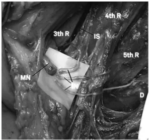
-taneous nerve (Figure 4)12,37. he phrenic nerve is also a possi
-ble donor in adults, but is not used in infants. Nerve transfers provide a more distal source of motor axons with a single
cooptation site37. his means that recovery is usually faster
and that late procedures can still be efective.
REHABILITATION
Limb immobilization has been associated with shoulder
deformities and is not recommended47, except if bone frac
-tures are also present. Some advocate that immobilization may be useful for pain treatment during the irst week48, but
it is diicult to evaluate pain in these patients. It seems that NBPP is not painful, at least in older patients49. his picture
is very diferent from that seen in adults with brachial plexus lesions, who usually show severe neuropathic pain, especially after root avulsions.
Physical therapy and occupational hand therapy are
im-portant, but it is essential to involve the parents in the re
-habilitation program. Passive range-of-motion exercises are critical to avoid muscle contractures and should be done several times on a daily basis50. It is a good idea to include it
in other routine activity such as changing dippers. As soon as the child shows intentional voluntary control, it is impor
-tant to stimulate the afected limb to avoid developmental
apraxia19,48. Encouraging bimanual activities is an interesting
strategy for that51. Wrist splinting can help to enhance hand
function in cases of wrist drop48, as long as it does not prevent
limb use during daytime. Aberrant reinnervation can result
in biceps-triceps cocontraction19, which can be treated with
botulinum toxin52. his can also be used to prevent muscle
contracture of the shoulder internal rotators40.
LONG TERM COMPLICATIONS
Internal rotation contractures and posterior humeral
sub-luxation are by far the most common long term complication
in NBPP2,37. It is related to muscular imbalance due to poor
ac-tive external rotation37,40. It leads to a progressive shoulder de
-formity according to the Waters classiication, ranging from mild glenoid retroversion (Waters grade II) to a complete pos
-terior luxation with false glenoid and proximal humeral de
-formity (Waters grade VII)53. Early referral to an orthopedic
Figure 4. Surgical view of intercostals nerves transfer to musculocutaneous nerve in the right thoracic region. All brachial plexus roots were avulsioned during the supraclavicular approach. 3th R: third rib; 4th R: fourth rib; 5th R: ifth rib; D: distal; IN: intercostal nerves; IS: intercostal space; MN: musculocutaneous nerve.
surgeon is important to avoid glenoid dysplasia and possi -bly shoulder pain37. Other orthopedic deformities can also be
seen, such as scapular winging, elbow lexion contracture, ra
-dial head luxation, ixed pronation or supination posture, and
claw hand deformity2. Growth imbalance between the upper
limbs is common in severe cases of NBPP2.
Little attention has been devoted to sensory disturbanc
-es, since the prognosis of sensory deicits is usually good49.
However, some children can develop a self-mutilating bit
-ing behavior54. his is more common after brachial plexus
surgery and is probably related to some kind of uncomfort
-able paresthesia55. his is only temporary and it is crucial to
prevent the child from eating of their own ingers and assure the parents that it will pass after a few months.
CONCLUSION
NBPP is a common situation and there is no perspective of adequate prevention in the near future. Most afected newborns will recover spontaneously, but some might be severely handi
-capped without appropriated care. Early referral to specialized centers with multidisciplinary approach should be provided to all patients that do not recover after a couple of weeks.
References
1. Shenaq SM, Bullocks JM, Dhillon G, Lee RT, Laurent JP. Management of infant brachial plexus injuries. Clin Plast Surg. 2005;32(1):79-98. doi:10.1016/j.cps.2004.09.001
2. Terzis JK, Kokkalis ZT. Pediatric brachial plexus reconstruction. Plast Reconstr Surg. 2009;124:370e-85e. doi:10.1097/PRS.0b013e3181bcf01f
3. Kay SP. Obstetrical brachial palsy. Br J Plast Surg. 1998;51(1):43-50. doi:10.1054/bjps.1997.0166
4. Gilbert A, Razaboni R, Amar-Khodja S. Indications and results of brachial plexus surgery in obstetrical palsy. Orthop Clin North Am. 1988;19(1):91-105.
5. Evans-Jones G, Kay SP, Weindling AM, Cranny G, Ward A, Bradshaw A et al. Congenital brachial palsy: incidence, causes, and outcome in the United Kingdom and Republic of Ireland. Arch Dis Child Fetal Neonatal Ed. 2003;88:f185-9. doi:10.1136/fn.88.3.F185
6. Gilbert WM, Nesbitt TS, Danielsen B. Associated factors in 1611 cases of brachial plexus injury. Obstet Gynecol. 1999;93(4):536-40. doi:10.1016/S0029-7844(98)00484-0
7. Mollberg M, Hagberg H, Bager B, Lilja H, Ladfors L. High birthweight and shoulder dystocia: the strongest risk factors for obstetrical brachial plexus palsy in a Swedish population-based study. Acta Obstet Gynecol Scand. 2005;84(7):654-9.
doi:10.1111/j.0001-6349.2005.00632.x
8. Wolf H, Hoeksma AF, Oei SL, Bleker OP. Obstetric brachial plexus injury: risk factors related to recovery. Eur J Obstet Gynecol Reprod Biol. 2000;88(2):133-8. doi:10.1016/S0301-2115(99)00132-3
9. Alfonso DT. Causes of neonatal brachial plexus palsy. Bull NYU Hosp Jt Dis. 2011;69(1):11-6.
10. Koenigsberger MR. Brachial palsy at birth: intrauterine or due to delivery trauma? Ann Neurol. 1980;8(2):228.
11. Gonik B, McCormick EM, Verweij BH, Rossman KM, Nigro MA. The timing of congenital brachial plexus injury: a study of electromyography indings in the newborn piglet. Am J Obstet Gynecol. 1998;178(4):688-95. doi:10.1016/S0002-9378(98)70478-8
12. Malessy MJ, Pondaag W. Obstetric brachial plexus injuries. Neurosurg Clin N Am. 2009;20(1):1-14. doi:10.1016/j.nec.2008.07.024
13. Doumouchtsis SK, Arulkumaran S. Are all brachial plexus injuries caused by shoulder dystocia? Obstet Gynecol Surv. 2009;64(9):615-23. doi:10.1097/OGX.0b013e3181b27a3a
14. Jennett RJ, Tarby TJ, Kreinick CJ. Brachial plexus palsy: an old problem revisited. Am J Obstet Gynecol. 1992;166(6 Pt 1):1673-6. doi:10.1016/0002-9378(92)91555-O
15. Christoffersson M, Rydhstroem H. Shoulder dystocia and brachial plexus injury: a population-based study. Gynecol Obstet Invest. 2002;53(1):42-7. doi:10.1159/000049410
16. Ubachs JM, Slooff AC, Peeters LL. Obstetric antecedents of surgically treated obstetric brachial plexus injuries. Br J Obstet Gynaecol. 1995;102(10):813-7. doi:10.1111/j.1471-0528.1995.tb10848.x
17. Gonen R, Spiegel D, Abend M. Is macrosomia predictable, and are shoulder dystocia and birth trauma preventable? Obstet Gynecol. 1996;88(4 Pt 1):526-9. doi:10.1016/0029-7844(96)00230-X
18. Rouse DJ, Owen J, Goldenberg RL, Cliver SP. The effectiveness and costs of elective cesarean delivery for fetal macrosomia diagnosed by ultrasound. JAMA. 1996;276(18):1480-6. doi:10.1001/jama.1996.03540180036030
19. Dijk JG, Pondaag W, Malessy MJ. Obstetric lesions of brachial plexus. Muscle Nerve. 2001;24(11):1451-61. doi:10.1002/mus.1168
20. Alfonso I, Alfonso DT, Papazian O. Focal upper extremity neuropathy in neonates. Semin Pediatr Neurol. 2000;7(1):4-14. doi:10.1016/S1071-9091(00)80005-4
21. Heise CO, Martins RS, Foroni LH, Siqueira MG. Prognostic value of thumb pain sensation in birth brachial plexopathy. Arq Neuropsiquiatr. 2012;70(8):590-2. doi:10.1590/S0004-282X2012000800006
22. Al-Qattan MM, Clarke HM, Curtis CG. Klumpke’s birth palsy. Does it really exist? J Hand Surg Eur Vol. 1995;20(1):19-23. doi:10.1016/S0266-7681(05)80008-7
23. Al-Qattan MM, Al-Khawashki H. The “beggar’s” hand and the “unshakable” hand in children with total obstetric brachial plexus palsy. Plast Reconstr Surg. 2002;109(6):1947-52. doi:10.1097/00006534-200205000-00026
24. Clarke HM, Curtis CG. An approach to obstetrical brachial plexus injuries. Hand Clin. 1995;11(4):563-80.
25. Heise CO, Siqueira MG, Martins RS, Gherpelli JL.
Clinical-electromyography correlation in infants with obstetric brachial plexopathy. J Hand Surg Am. 2007;32:999-1003. doi:10.1016/j.jhsa.2007.05.002
26. Heise CO, Siqueira MG, Martins RS, Gherpelli JL. Motor nerve-conduction studies in obstetric brachial plexopathy for a selection of patients with a poor outcome. J Bone Joint Surg Am. 2009;91(7):1729-37.
27. Vanderhave KL, Bovid K, Alpert H, Chang KW, Quint DJ, Leonard JA Jr et al. Utility of electrodiagnostic testing and computed tomography myelography in preoperative evaluation of neonatal brachial plexus palsy. J Neurosurg Pediatr. 2012;9(3):283-9. doi:10.3171/2011.12.PEDS11416
29. O’Brien DF, Park TS, Noetzel MJ, Weatherly T. Management of birth brachial plexus palsy. Childs Nerv Syst. 2006;22(2):103-12. doi:10.1007/s00381-005-1261-y
30. Chow BC, Blaser S, Clarke HM. Predictive value of computed tomographic myelography in obstetrical brachial plexus palsy. Plast Reconstr Surg. 2000;106(5):971-7. doi:10.1097/00006534-200010000-00001
31. Smith AB, Gupta N, Strober J, Chin C. Magnetic resonance neurography in children with birth-related brachial plexus injury. Pediatr Radiol. 2008;38(2):159-63. doi:10.1007/s00247-007-0665-0
32. Medina LS, Yaylali Y, Zurakowski D, Ruiz J, Altman NR, Grossman JA. Diagnostic performance of MRI and MR myelography in infants with a brachial plexus birth injury. Pediatr Radiol. 2006;36(12):1295-9. doi:10.1007/s00247-006-0321-0
33. Adler B, Patterson RL. Erb’s palsy. Long-term results of eighty-eight cases. J Bone Joint Surg A. 1967;49(6):1052-64.
34. Nocon JJ, McKenzie DK, Thomas LJ, Hansell RS. Shoulder dystocia: an analysis of risks and obstetric maneuvers. Am J Obstet Gynecol. 1993;168(6 Pt 1):1732-7. doi:10.1016/0002-9378(93)90684-B
35. Gordon M, Rich H, Deutschberger J, Green M. The immediate and long term outcome of obstetric trauma. Am J Obstet Gynecol. 1973;117(1):51-6.
36. Pondaag W, Malessy MJ, van Dijk JG, Thomeer RT. Natural history of obstetric brachial plexus palsy: a systematic review. Dev Med Child Neurol. 2004;46(2):138-44. doi:10.1111/j.1469-8749.2004.tb00463.x
37. Hale HB, Bae DS, Waters PM. Current concepts in the management of brachial plexus birth palsy. J Hand Surg Am. 2010;35(2):322-31. doi:10.1016/j.jhsa.2009.11.026
38. Smith NC, Rowan P, Benson LJ, Ezaki M, Carter PR. Neonatal brachial plexus palsy: Outcome of absent biceps function at three months of age. J Bone Joint Surg Am. 2004;86(10):2163-70.
39. Brauer CA, Waters PM. An economic analysis of timing of microsurgical reconstruction in brachial plexus birth palsy. J Bone Joint Surg Am. 2007;89(5):970-8. doi:10.2106/JBJS.E.00657
40. Ruchelsman DE, Pettrone S, Price AE, Grossman JA. Brachial plexus birth palsy: an overview of early treatment considerations. Bull NYU Hosp Jt Dis. 2009;67(1):83-9.
41. Bahm J, Ocampo-Pavez C, Noaman H. Microsurgical technique in obstetric brachial plexus repair: a personal experience in 200 cases over 10 years. J Brachial Plex Peripher Nerve Inj. 2007;2(1):1. doi:10.1186/1749-7221-2-1
42. Lin JC, Schwntker-Colizza A, Curtis CG, Clarke HM. Final results of grafting versus neurolysis in obstetrical brachial plexus palsy. Plast Reconstr Surg. 2009;123(3):939-48. doi:10.1097/PRS.0b013e318199f4eb
43. Laurent JP, Lee RT. Birth-related upper brachial plexus injuries in infants: operative and nonoperative approaches. J Child Neurol. 1994;9(2):111-7. doi:10.1177/088307389400900202
44. Spinner RJ, Kline DG. Surgery for peripheral nerve and brachial plexus injuries or other nerve lesions. Muscle Nerve. 2000;23(5):680-95. doi:10.1002/(SICI)1097-4598(200005)23:5<680:: AID-MUS4>3.0.CO;2-H
45. Robinson LR. Traumatic injury to peripheral nerves. Muscle Nerve. 2000;23(6):863-73. doi:10.1002/(SICI)1097-4598(200006)23:6<863::
AID-MUS4>3.0.CO;2-0
46. Siqueira MG, Socolovsky M, Heise CO, Martins RS, Di Masi G. Eficacy and safety of Oberlin’s procedure in the treatment of
brachial plexus birth palsy. Neurosurgery. 2012;71(6):1156-60. doi:10.1227/NEU.0b013e318271ee4a
47. Peixinho M, Serdeira A. Luxação ântero-inferior da articulação do ombro na paralisia obstétrica secundária à imobilização contínua. Rev Hosp Clin Fac Med S Paulo. 1971;26(2):49-54.
48. Eng GD, Binder H, Getson P, O’Donnell R. Obstetrical brachial plexus palsy outcome with conservative management. Muscle Nerve1996;19(7):884-91.
doi:10.1002/(SICI)1097-4598(199607)19:7<884::AID-MUS11>3.0.CO;2-J
49. Anand P, Birch R. Restoration of sensory function and lack of long-term chronic pain syndromes after brachial plexus injury in human
neonates. Brain. 2002;125(1):113-22. doi:10.1093/brain/awf017
50. Yang LJ. Neonatal brachial plexus palsy: management and
prognostic factors. Semin Perinatol. 2014;38(4):222-34. doi:10.1053/j.semperi.2014.04.009
51. Dumont CE, Forin V, Asfazadourian H, Romana C. Function of the
upper limb after surgery for obstetric brachial plexus palsy. J Bone Joint Surg. 2001;83(6):894-900. doi:10.1302/0301-620X.83B6.11389
52. Rollnick JD, Hierner R, Schubert M, Shen ZL, Johannes S, Tröger
M et al. Botulinum toxin treatment of cocontractions after birth-related brachial plexus lesions. Neurology. 2000;55(1):112-4. doi:10.1212/WNL.55.1.112
53. Waters PM, Smith GR, Jaramillo D. Glenohumeral deformity secondary to brachial plexus birth palsy. J Bone Joint Surg Am. 1998;80(5):668-77.
54. Al-Qattan MM. Self-mutilation in children with obstetric brachial plexus palsy. J Bone Joint Surg Eur Vol. 1999;24(5):547-9.
doi:10.1054/jhsb.1999.0222
55. McCann ME, Waters P, Goumnerova LC, Berde C. Self-mutilation in young children following brachial plexus birth injury. Pain.