w w w . r b o . o r g . b r
Original
Article
Anatomical
study
on
the
innervation
of
the
elbow
capsule
夽
Cristina
Schmitt
Cavalheiro
∗,
Mauro
Razuk
Filho,
João
Rozas,
João
Wey,
Antonio
Marcos
de
Andrade,
Edie
Benedito
Caetano
FaculdadedeCiênciasMédicasedaSaúdedeSorocaba(FCMS),PontifíciaUniversidadeCatólicadeSãoPaulo(PUC-SP),Sorocaba,SP, Brazil
a
r
t
i
c
l
e
i
n
f
o
Articlehistory:
Received22September2014 Accepted10November2014 Availableonline19October2015
Keywords:
Elbowjoint Jointcapsule Cadaver Anatomy
a
b
s
t
r
a
c
t
Objectives: Toputforwardananatomicaldescriptionoftheinnervationoftheelbowcapsule, illustratedthroughmorphologicalanalysisondissections.
Methods:Thirtyelbowsfromfreshfixedadultcadaversaged32–74years,ofbothsexes,were dissected.
Results:Amongthedissectedarms,weobservedthatthemediannervedidnothaveany branchesintwoarms,whileithadonebranchinfivearms,twobranchesintwoarms, threebranchesintenarms,fourbranchesinninearmsandfivebranchesintwoarms.The radialnervedidnothaveanybranchesintwoarms,whileithadonebranchintwoarms, twobranchesinninearms,threebranchesintenarms,fourbranchesinfivearmsandfive branchesintwoarms.Theulnarnervedidnothaveanybranchesinthreearms,whileit hadonebranchinsixarms,twobranchesinfourarms,threebranchesinfivearms,four branchesinsevenarms,fivebranchesinfourarmsandsixbranchesinonearm.
Conclusions: Weobservedbranchesoftheradial,ulnarandmedialnervesintheelbowjoint, andacloserelationshipbetweentheircapsularandmotorbranches.
©2015SociedadeBrasileiradeOrtopediaeTraumatologia.PublishedbyElsevierEditora Ltda.Allrightsreserved.
Estudo
anatômico
da
inervac¸ão
da
cápsula
do
cotovelo
Palavras-chave:
Articulac¸ãodocotovelo Cápsulaarticular Cadáver Anatomia
r
e
s
u
m
o
Objetivos:Promover a descric¸ão anatômica da inervac¸ão da cápsula do cotovelo com ilustrac¸ãopormeiodamorfologiadasdissecac¸ões.
Métodos:Foramdissecados30cotovelosdecadáveresadultosfrescosefixados,comidade entre32e74anos,deambosossexos.
夽
WorkperformedattheFaculdadedeCiênciasMédicasedaSaúdedeSorocaba(FCMS),PontifíciaUniversidadeCatólicadeSãoPaulo (PUC-SP),Sorocaba,SP,Brazil.
∗ Correspondingauthor.
E-mails:[email protected],[email protected](C.S.Cavalheiro).
http://dx.doi.org/10.1016/j.rboe.2015.10.001
674
rev bras ortop.2015;50(6):673–679Resultados: Observamos,dentreosbrac¸osdissecados,doiscomnenhumramodonervo mediano,cincocomumramo,doiscomdoisramos,10comtrêsramos,novecomquatro ramosedoiscomcincoramos.Quandosetratadonervoradial,doisbrac¸osnão apresen-taramramos,doismostraramdoisramos,novecontinhamdoisramos,10contaramcomtrês ramos,cincotinhamquatroramosedoistinhamcincoramos.Emrelac¸ãoaonervoulnar, tivemostrêsbrac¸ossemramosarticulares,seiscomumramo,quatrocomdoisramos,cinco comtrêsramos,setecomquatroramos,quatrocomcincoramoseumcomseisramos.
Conclusões: Constatamosramosdonervoradial,ulnaremedialnaarticulac¸ãodocotovelo, assimcomoarelac¸ãopróximaentreosseusramoscapsularesemotores.
©2015SociedadeBrasileiradeOrtopediaeTraumatologia.PublicadoporElsevier EditoraLtda.Todososdireitosreservados.
Introduction
Thefirstmentionsofthenervebranchesoftheelbowcapsule datefrom1844,indescriptionsofabranchofthecutaneous nerveperforatingthebrachialmuscleandreachingthe cap-sule; branches of the median nerve penetrating the elbow joint;andbranchesoftheulnarnervebranchingoutbetween medialepicondyleandtheolecranon.Abranchoftheradial nerveextending tothe longhead ofthe tricepsand head-ing toward the olecranon and posterior capsule was also described.
In 1857, small branches of the musculocutaneous and mediannerveextendingtotheanteriorpartofthecapsuleand variablebranchesoftheanteriorinterosseousnerve appear-ingbetweentheradiusandulnaandinnervatingthecapsule aroundtheradialheadweredescribed.Withregardtothe pos-teriorpartofthecapsule,abranchderivedfromthe radial nervethatoriginatedfromthemusclebranchofthelateral andmedialheadofthetricepsbrachiimusclewasdescribed. Astudyconductedin1877reportedthepresenceofsmall filamentsfromthemediannervegoingtotheanteromedial regionofthecapsuleandbranchesofulnarorigingoingtothe posteromedialcapsule.
Insubsequentyears,studiesbegantodescribethissubject withgreaterprecisionthroughdissections.Fromdissections onsevenadultelbowsandfivefetalelbows,thecontributions ofthefourmainnervesinnervatingtheelbowcapsule(ulnar, median,musculocutaneousandradial)weredemonstrated.A studyin1949onlymentionedramificationsgoingtothe ole-cranonprocess,anddidnotdescribecapsularbranchesofthe radialnerve.
Thepresent-day mainanatomytextbooks,suchasGray, Hollinshead,LatarjetandLiard,donotcitetheradialnerve.1–8
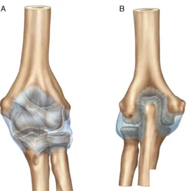
Theelbowcapsuleisextensiveandcoatsthedistal extrem-ityofthehumerusandproximalextremityoftheulnaand radius.Anteriorlyandproximally,itisinsertedabovethefossa ofthecoronoidprocessand capitellum.Distally,it adheres mediallytothecoronoidprocessoftheulnaandlaterallyto theannularligamentoftheradius(Fig.1A).
Posteriorlyandproximally,thecapsuleadheresabovethe olecranonfossa,goesaroundthemarginandcontinuesacross theentiremedialandlateralcolumn,whereitcoversallofthe sigmoidfossa(Fig.1B).
The anterior joint capsule is usually thinner and more transparent. It remains under tension when the elbow is
extendedandrelaxeswhentheelbowisflexed.Thegreatest capacityofthejointcapsuleis30–35mlat80◦offlexion,when
itisfullydistended.
Inrelationtothemusculocutaneousnerve,itisknownthat theareathatitinnervatesistheanteriorcapsule.Thisnerve issuesasmallbranchfromitsmaintrunk,whichpenetrates themiddlethirdofthebrachialmuscleandgoesindeeperto reachtheanteriorpartofthehumerusandsupplythe perios-teum.Itthenreachestheelbowcapsule,whereitdividesinto avariablenumberofbranches(Fig.2A).Thisnerveisthemost constant supplierofthecapsule,bothmacroscopically and microscopically.Insomecases,thiscapsulebranchmayform anastomoses withbranches ofthemedian nerveand then continuetothe capsule(Fig.2B).Theregionofthe muscu-locutaneousnervemaybejuxtaposedbothtomedianandto lateralareas.9–12
Before passingbetween theheads ofthepronator teres muscle, themediannervebranches outintosmall sections thatgotothecapsularregionoftheanteriormedialepicondyle
Fig.1–Anteriorlimitsoftheelbowcapsule(A).Posterior
Fig.2–Ramificationofthemusculocutaneousnerveon
reachingtheelbowcapsule(A).Thecapsulebranchmay
formanastomoseswithbranchesofthemediannerveand
thencontinuetothecapsule(B).
(Fig. 3A). A joint branch may also occur, which develops more proximally to the elbow, posteriorly to the bifurca-tionofthebrachialartery,and joinsthemusculocutaneous nervetoinnervatetheanteriorcapsule(Fig.3B).Theanterior interosseousnervegivesrisetoasmallfilamentthatsupplies
Fig.3–Ramificationofthemediannerveinsmallsections
thatgofromtheanteriormedialepicondyletowardthe
capsuleregion(A).Unionofthemediannervewiththe
musculocutaneousmuscleforinnervatingtheanterior
capsule(B).
Fig.4– Branchesoftheulnarnervebeginninginthegroove
betweenthemedialepicondyleandtheolecranon(A).Joint
branchesoriginatingabovethecubitaltunnel(B).
theposteroinferiorpartofthecapsule,adjacenttotheulna. Thus,themediannerveusuallyinnervatestheanteromedial partofthejointcapsuleandthisareamaybeoverlainbythe musculocutaneousnerve.13–15
Theulnarnerveusuallyappearsasthreebranches,which beginatthesulcusbetweenthemedialepicondyleand the olecranon(Fig.4A).Jointbranchesthatariseseveral centime-ters abovethecubital tunnelhavebeen described (Fig. 4B). Thesesupplytheposteromedialpartofthecapsulesandthe neighborhoodofthemedialepicondyleandolecranon,both inthecubitaltunnel.Thisareamaybeoverlainbytheradial nerve.16–18
Adescendingbranchisissuedfromthemaintrunkofthe radialnerve,andthisfollowsthelateralheadofthetriceps muscle. Whenit reachestheolecranon,itbifurcates tothe capsuleintheregionoftheolecranonfossa(Fig.5A).Thereis alsoasmallfilamentthatarisesfromthebranchgoingtothe anconeusmuscleandinnervatestheposterolateralregionof thecapsule(Fig.5B).Insomecases,theposteriorandproximal capsules,whichinvolvetheextremityoftheolecranon,are innervatedbythinbranchesfromtheulnarcollateralnerve, whichisabranchofthemaintrunkoftheradialnerve(Fig.5C).
Thisregionmaybeoverlainbyulnarinnervation.Regarding theanteriorcapsule,afterthispassesthroughthe intramus-cularseptumofthesupinatormuscle,itgenerallydividesinto smallbranchesthatmayformanastomoseswithstructures ofthemusculocutaneousnerve(Fig.5D).18–25
676
rev bras ortop.2015;50(6):673–679Para M, Ancôneo
Para M, Ancôneo
A
B
D
C
Fig.5–Bifurcationoftheradialbranchtothecapsuleinthe
regionoftheolecranonfossa(A).Innervationofthe
posterolateralregionofthecapsule(B).Branchofthemain
trunkoftheradialnerve(C).Anastomosesoframifications
oftheradialnervewithstructuresofthemusculocutaneous
nerveencompassingtheanteriorcapsule(D).
andlaterallybytheradialnerve,andthereisacentralareaof mutualinnervation(Fig.6B).
Atthisjuncture,dissectionsonfreshandfixedcadavers becomenecessaryinordertoproveandenrichthesestudies withgreaterdetail.
Just like all joint capsules, the elbow capsule isclosely linked tothe bones and is surrounded by muscles, which clearly showsthe difficulty inprecisely establishingwhich structuresreachit,especiallythenervesthatbranchout to structuresadjacenttothecapsule.Thus,carefulanddetailed dissectionisneeded,especiallywithregardtotheupperlimbs, inwhichtheanatomyisrichindetailsandvariations.
Therearefewstudiesrelatingtoinnervationoftheelbow capsule, and the results have presented divergences and havesometimesbeen incomplete.Therefore, elucidationof thisdifficulttopicwas soughthere,through ananatomical descriptionbasedondissectionof30elbowsfromcadavers, basedontheabovereviewoftheliterature.
N.M. Cutâneo
N.M. Mediano
N. Radial
N. Radial
Áear Mútua N.M. Cutâneo+ N.Mediano
Áear Mútua N.Ulnar+ N. Radial
Áear Mútua N.M. Radial+ M.Cutâneo
N.Ulnar
B
A
Fig.6–Innervationoftheanteriorcapsule(A).Innervation
oftheposteriorcapsule(B).
The objective ofthis study was to providean anatomi-caldescriptionofthe innervationoftheelbowcapsuleand to illustrate datathat are poorly elucidated in the current medicalliterature,from themorphologyofthedissections. Moreover,thisstudysoughttodemonstratethemorphology ofthemainnervesandtheirramificationstotheelbow cap-sule, identifythelocationsofnerveinsertionsoftheelbow capsuleandcomparethefindingsfromdissectionswiththe informationinthemedicalliterature.
Materials
and
methods
Thirty elbows from fresh and fixedadult cadaversofboth sexes weredissected.There were12 rightarmsand18 left armsandtheagesrangedfrom32to74years.
Forthedissections,routinelaboratorymaterialswereused: non-sterile latex gloves,scalpels,anatomical tweezers, rat-tooth tweezers,Kelly tweezers, Iris Golgranscissors, Mayo scissors,needleholdersandcottonthread.
Astandard medialcutaneous incisionwas made,which resultedinexposureofthesubcutaneoustissueandenabled access to the intermuscular septum. Theulnar nerve was foundtobepositionedbehindtheproximalextremityofthe septum.Thiswaspreservedandthenwasdissectedalongthe anteriorsurfaceofthemedialheadofthetricepsmuscle.
In theelbow, the ulnarnerve passesbehind the medial epicondyleofthehumerusandismedialtotheulnar collat-eralligamentandolecranon.Itfollowsthehumerusdistally, reachesadeeppositionandrestsontheshortflexorofthe fingers.Thedissectionwasterminatedatthislevel.
Themediannervewasexposed throughusingthe origi-nalincision.Theanteriorskinflapwaspulledbacklaterallyin ordertoexposetheanteriorstructuresoftheelbowregion.The cutaneousandsubcutaneoustissueswereremovedwithout damagingthedeepertissues.Themediannervewas investi-gatedanteriorly,whereitfunctionsincloseproximitytothe brachialartery.
Fig.7–Mediannerve(A)anditsfourjointbranches(B–E).
muscle islateral to this.In the elbow region, the nerve is locatedina deep plane,besidethe brachial artery. Atthis point, the bicipital aponeurosisislocatedanteriorly tothe nerve.
Thebicepstendonwaspulledbacklaterallytoachieve ade-quateexposureofthemediannerve.Thus,thebranchesof thenervewereobservedatthislevelfordissectingtheradial nerve.
Alateralapproachtotheelbowwasthenusedseparately. Attheproximalextremityofthecadavers,theradialnerve waseasilyfound,togetherwiththebrachialarteryinthespiral grooveofthehumerus,betweenthelateralandmedialheads ofthetriceps.Thenervewasseentopassthroughthebrachial muscleand,whenthiswaslimitedtothetendon,itcrossed thecapsuleoftheelbowjoint.Uponreachingthesupinator muscle,itseparatedintotwoparts:theposteriorinterosseous branchandthesuperficialbranchoftheradialnerve.The mus-culocutaneousnerve wasnot preciselydissected inall the cadaversandthuswasexcludedfromthisstudy.
Thisstudywasapprovedbytheresearchethicscommittee.
Results
Amongthe dissectedarms,weobservedtwoarmswithno branches of the median nerve, five with one branch, two withtwobranches,tenwiththreebranches,ninewithfour branchesandtwowithfivebranches.Fig.7showsasample fromdissectionofthemediannerve.
Withregardtotheradialnerve,twoarmsdidnotpresent anybranches,twohadonebranch,ninehadtwobranches,10 hadthreebranches,fivehadfourbranchesandtwohadfive branches.Fig.8showsadissectedarmwiththeradialnerve anditsbranchesexposed.
Inrelationtotheulnarnerve,therewerethreearmswithno jointbranches,sixwithonebranch,fourwithtwobranches, fivewiththreebranches,sevenwithfourbranches,fourwith fivebranchesandonewithsixbranches.Fig.9presentsa dis-sectedarmshowingtheulnarnerveanditsrespectivejoint branches.
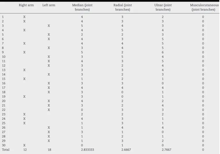
Table1correlatesthenumberofjointbranchesfoundin thenervesstudied,ineacharmdissected.
FromFig.10,thefrequenciesofthedifferentnumbersof joint branches in each nerve can be seen.It is clear that themostfrequentfindingswerethreejointbranchesforthe
Fig.8–Radialnerve(A)anditsfourjointbranches(B–E).
Fig.9–Ulnarnerve(A)anditsthreejointbranches(B–D).
12
10
8
6
4
2
0
Median
Radial Ulnar
Number of arms
0 branches
4 branches
1 branch
5 branches 2 branches
6 branches 3 branches
Fig.10–Numberofcadaverswiththerespectivenumberof
branchesfoundineachnerve.
mediannerve,threefortheradialnerveandfourfortheulnar nerve.
Discussion
Therearefewanatomicalorclinicalstudiesintheliterature, in relation to the patterns or routes ofinnervation of the elbowjointstructures.Morrey6cited“afewsmallbranches”
678
rev bras ortop.2015;50(6):673–679Table1–Resultsfoundfromthedissections.
Rightarm Leftarm Median(joint branches)
Radial(joint branches)
Ulnar(joint branches)
Musculocutaneous (jointbranches)
1 X 4 3 2 0
2 X 4 3 3 0
3 X 5 4 3 0
4 X 4 5 4 0
5 X 2 2 3 0
6 X 4 3 5 0
7 X 4 4 5 0
8 X 3 4 5 0
9 X 5 2 6 0
10 X 3 5 4 0
11 X 4 3 5 0
12 X 3 2 4 0
13 X 3 3 4 0
14 X 3 2 3 0
15 X 1 2 1 0
16 X 3 3 0 0
17 X 4 4 4 0
18 X 3 0 1 0
19 X 3 0 1 0
20 X 4 2 2 0
21 X 3 2 4 0
22 X 0 3 3 0
23 X 2 2 2 0
24 X 4 3 1 0
25 X 1 3 1 0
26 X 1 4 4 0
27 X 3 1 0 0
28 X 1 2 1 0
29 X 1 3 2 0
30 X 0 1 0 0
Total 12 18 2.833333 2.6667 2.7667 0
mediannerve that originate before the motorbranches to thepronatorteres,radialcarpalflexorandlongpalmar mus-cles.
Gonzalez et al.10 suggested that the ulnar nerve emits
branchestotheelbowinapositionposteriortothemedial epicondyle of the humerus, while Watchmaker et al.26
only identified two branches in their study on 15 ulnar nerves.Thomasetal.11describedtheposteriorinterosseous
nerve, but not its anatomical relationship with the elbow joint.
Conclusion
Weobservedbranchesoftheradial,medianandulnarnerves intheelbowjointcapsule,whichdemonstratestheir impor-tanceintheinnervationofthisregion.
Therefore,thisstudy,togetherwithotherstudiesthat con-sidertherolesofthemedian,ulnarandradialnervesinthe innervationoftheelbowcapsule,constituteausefulbasisfor useofdenervationtechniquesontheelbowjointfor reliev-ing the pain ofarthritis and other chronic diseases inthe elbow.
Conflicts
of
interest
Theauthorsdeclarenoconflictsofinterest.
r
e
f
e
r
e
n
c
e
s
1.ZancolliEA.Structuralanddynamicbasisofhandsurgery.Br JSurg.2005;56(7):481–556.
2.LangmanJ,WoerdemanMW.Atlasofmedicalanatomy. Philadelphia:Saunders;1978.
3.LinellEA.Thedistributionofnervesintheupperlimb,with referencetovariablesandtheirclinicalsignificance.JAnat. 1921;55:79–112.
4.HollinsheadWH.Thebackandlimbs.In:Anatomyfor surgeons.NewYork:Harper&Row;1969.p.379.
5.TestutL,LatarjetA.Tratadodeanatomiahumana.9thed. Barcelona:Salvat;1949.
6.MorreyBF.Anatomyofelbowjoint.In:MorreyBF,editor.The elbowanditsdisorders.3rded.Philadelphia:Saunders;2000. p.13–42.
7.MorreyBF,AnKN.Articularandligamentouscontributionsto thestabilityoftheelbowjoint.AmJSportsMed.
1983;11(5):315–9.
8.GrayH.Anatomia.39ed.RiodeJaneiro:GuanabaraKoogan; 2004.
9.VieiraEA,CaetanoEB.Basesanatômicasfuncionaisda articulac¸ãodocotovelo;contribuic¸ãoaoestudodas estruturasestabilizadorasdoscompartimentosmediale lateral.RevBrasOrtop.1999;34(8):481–8.
10.GonzalezMH,LotfiP,BendreA,MandelbroytY,LieskaN.The ulnarnerveattheelbowanditslocalbranching:ananatomic study.JHandSurgBr.2001;26(2):142–4.
thesupinatormuscle.JHandSurgAm.2000;25(5): 936–41.
12.TanakaY,AokiM,IzumiT,WadaT,FujimiyaM,YamashitaT. Effectofelbowandforearmpositiononcontactpressure betweentheextensororiginandthelateralsideofthe capitellum.JHandSurgAm.2011;36(1):81–8.
13.SasakiK,TamakawaM,OndaK,IbaK,SonodaT,YamashitaT, etal.Thedetectionofthecapsulartearattheundersurface oftheextensorcarpiradialisbrevistendoninchronictennis elbow:thevalueofmagneticresonanceimagingand computedtomographyarthrography.JShoulderElbowSurg. 2011;20(3):420–5.
14.MullettH,SpragueM,BrownG,HausmanM.Arthroscopic treatmentoflateralepicondylitis:clinicalandcadaveric studies.ClinOrthopRelatRes.2005;439:123–8.
15.AnderssonG,DanielsonP,AlfredsonH,ForsgrenS.
Nerve-relatedcharacteristicsofventralparatendinoustissue inchronicAchillestendinosis.KneeSurgSportsTraumatol Arthrosc.2007;15(10):1272–9.
16.DanielsonP,AnderssonG,AlfredsonH,ForsgrenS.Marked sympatheticcomponentintheperivascularinnervationof thedorsalparatendinoustissueofthepatellartendonin arthroscopicallytreatedtendinosispatients.KneeSurgSports TraumatolArthrosc.2008;16(6):621–6.
17.AlbrechtPJ,HinesS,EisenbergE,PudD,FinlayDR,Connolly MK,etal.Pathologicalterationsofcutaneousinnervationand vasculatureinaffectedlimbsfrompatientswithcomplex regionalpainsyndrome.Pain.2006;120(3):
244–66.
18.SlaterH,Arendt-NielsenL,WrightA,Graven-NielsenT. Sensoryandmotoreffectsofexperimentalmusclepainin patientswithlateralepicondylalgiaandcontrolswithdelayed onsetmusclesoreness.Pain.2005;114(1–2):118–30.
19.LimAY,PereiraBP,KumarVP,DeConinckC,TakiC,BaudetJ, etal.Intramuscularinnervationofupper-limbskeletal muscles.MuscleNerve.2004;29(4):523–30.
20.MolinierF,LaffosseJM,BoualiO,TricoireJL,MoscoviciJ.The anconeus,anactivelateralligamentoftheelbow:new anatomicalarguments.SurgRadiolAnat.2011;33(7):617–21.
21.NishidaK,IwasakiN,MinamiA.Anconeusmuscleflapfor thetreatmentofsofttissuedefectsovertheolecranonafter totalelbowarthroplasty.JHandSurgEurVol.2009;34(4):538–9.
22.PraagmanM,ChadwickEK,vanderHelmFC,VeegerHE.The effectofelbowangleandexternalmomentonloadsharingof elbowmuscles.JElectromyogrKinesiol.2010;20(5):912–22.
23.TakigawaN,RyuJ,KishVL,KinoshitaM,AbeM.Functional anatomyofthelateralcollateralligamentcomplexofthe elbow:morphologyandstrain.JHandSurgBr.
2005;30(2):143–7.
24.ZhangLQ,NuberGW.Momentdistributionamonghuman elbowextensormusclesduringisometricandsubmaximal extension.JBiomech.2000;33(2):145–54.
25.PereiraBP.Revisitingtheanatomyandbiomechanicsofthe anconeusmuscleanditsroleinelbowstability.AnnAnat. 2013;195(4):365–70.
26.WatchmakerGP,LeeG,MackinnonSE.Intraneural