O R I G I N A L A R T I C L E UDC: 616.833-089 DOI:10.2298/VSP110301007S
Collateral branches of the brachial plexus as donors in nerve transfers
Bo
þ
ne grane brahijalnog pleksusa – donori u transferima nerava
Miroslav Samardžiü*, Lukas Rasuliü*, Novak Lakiüeviü†, Vladimir Bašþareviü*, Irena Cvrkota*, Mirko Miüoviü*, Andrija Saviü*
*Neurosurgical Clinic, Clinical Center of Serbia, Faculty of Medicine, University of Belgrade, Belgrade, Serbia, †Neurosurgical Clinic, Clinical Center of Montenegro,
Podgorica, Montenegro
Abstract
Background/Aim. Nerve transfers in cases of directly ir-reparable, or high level extensive brachial plexus traction inju-ries are performed using a variety of donor nerves with vari-ous success but an ideal method has not been established. The purpose of this study was to analyze the results of nerve transfers in patients with traction injuries to the brachial plexus using the thoracodorsal and medial pectoral nerves as donors. Methods. This study included 40 patients with 25 procedures using the thoracodorsal nerve and 33 procedures using the medial pectoral nerve as donors for reinnervation of the musculocutaneous or axillary nerve. The results were analyzed according to the donor nerve, the age of the patient and the timing of surgery. Results. The total rate of recovery for elbow flexion was 94.1%, for shoulder abduction 89.3%, and for shoulder external rotation 64.3%. The corresponding rates of recovery using the thoracodorsal nerve were100%, 93.7% and 68.7%, respectively. The rates of recovery with medial pectoral nerve transfers were 90.5%, 83.3% and 58.3%, respectively. Despite the obvious differences in the rates of recovery, statistical significance was found only be-tween the rates and quality of recovery for the musculocuta-neous and axillary nerve using the thoracodorsal nerve as do-nor. Conclusion. According to our findings, nerve transfers using collateral branches of the brachial plexus in cases with upper palsy offer several advantages and yield high rate and good quality of recovery.
Key words:
brachial plexus; wounds and injuries; nerve transfer; thoracic nerves; neurosurgical procedures; treatment outcome.
Apstrakt
Uvod/Cilj. Za nervne transfere u sluÿajevima nemoguý no-sti direktne rekonstrukcije visokih ekstenzivnih povreda brahijalnog pleksusa koje ne podležu direktnoj reparaciji ko-riste se razliÿiti donorni nervi. Ovo istraživanje imalo je za cilj da analizira rezultate nervnih transfera kod bolesnika sa trakcionom povredom brahijalnog pleksusa u kojima se kao donori koriste torakodorzalni i medijalni pektoralni nervi. Metode. Istraživanjem je bilo obuhvaýeno 40 bolesnika kod kojih je kao donor za reinervaciju muskulokutaneusa ili aksilarisa u 25 postupaka korišýen torakodorzalni nerv i u 33 postupka medijalni pektoralni nerv. Rezultati su analizirani prema donornom nervu, uzrastu bolesnika i vremenu kada je operacija izvršena. Rezultati. Ukupna stopa oporavka iz-nosila je 94,1% za fleksiju podlaktice, 89,3% za abdukciju ramena i 64,3% za spoljnu rotaciju ramena. Odgovarajuýe stope oporavka pri korišýenju torakodorzalnog nerva bile su 100%, 93,7% i 68,7%, a pri korišýenju medijalnog pektoral-nog nerva 90,5%, 83,3% i 58,3%, respektivno. Uprkos oÿ i-glednim razlikama u stopama oporavka, statistiÿki znaÿajne korelacije ustanovljene su samo za stope i kvalitet oporavka muskulokutaneusa i aksilarisa u sluÿajevima kada je kao do-nor korišýen torakodorzalni nerv. Zakljuÿak. Prema našim nalazima, nervni transferi u sluÿajevima gornje paralize u kojima se kao donori koriste boÿne grane brahijalnog plek-susa imaju nekoliko prednosti i daju visoku stopu i dobar kvalitet oporavka.
Kljuÿne reÿi:
plexus brachialis; rane i povrede; transfer živca; nn. thoracici; neurohirurške procedure;
leÿenje, ishod.
Introduction
In the past, nerve transfers were the treatment of choice in cases with spinal nerve root avulsion, or those with di-rectly irreparable proximal lesions, ie very proximal or
repair 2. The main advantage of this procedure over nerve grafting is a conversion of proximal high-level injury to a low-level one 1.
Nerve transfers have been attempted using a variety of donor nerves, but an ideal method has not been estabilshed. In general, there are two types of donors: extraplexal, in-cluding intercostal, spinal accessory, phrenic, motor branches of the cervical plexus, or collateral C7 spinal nerve, and intraplexal, including proximal spinal nerve stumps or collateral motor branches of the brachial plexus. In fact, the latter presents a distal form of the classic intraplexal nerve transfer.
Nerve transfers using these nerves were performed for the first time in 1920 by Vulipus and Stoffel (according to ref. 3 ), who transferred some of the branches to the pectoral muscles onto the musculocutaneous or axillary nerves. In 1929 Foerster performed transfer of nerves to the latissimus dorsi and subscapular muscles onto the axillary nerve, as re-ported by Narakas 4, 5. Thereafter, in 1948 Lurja (according to ref. 3 ) reported transfer of the pectoral and thoracodorsal nerves, particularly onto the musculocutaneous nerve in pa-tients with upper trunk injuries.
We used these two nerves in nerve transfer for the first time in 1980. Thoracodorsal to musculocutaneous and me-dial pectoral to the axillary nerve transfers were performed in a 24-year-old man four months after a motorcycle accident. Good recovery of both functions was obtained one year after
the surgery. The aim of this study was to analyze the results of nerve transfers in patients with traction injuries to the bra-chial plexus using the thoracodorsal and medial pectoral nerves as donors.
Methods
During the past 30 years, since January 1980, we per-formed nerve transfer using collateral branches of the bra-chial plexus as donors in 44 patients with upper palsy due to traction injury. The number of followed up patients was 40, or more precisely 33 with nerve transfers using the medial pectoral nerve, and 29 using the thoracodorsal nerve as do-nor. Both nerves were used simultaneoulsy in 22 of the fol-lowed up patients. The age of the folfol-lowed up patients ranged from 9 do 55 years, with 27 (67.5%) being less than 30 years of age.
The preoperative diagnosis was made on the basis of standard clinical, electrodiagnostic and radiologic tests. Us-ing these methods we diagnosed a variety of injury patterns to the C5, C6, and sometimes C7 spinal nerve roots or spinal nerves. Extended upper brachial plexus palsy was present in 11 patients.
Indications for nerve transfer included preoperatively documented avulsion of the C5 and C6 spinal nerve roots, or intraoperatively demonstrated directly irreparable very proximal lesion of the corresponding spinal nerves, and high lesions with a long nerve gap. Surgical procedures were per-formed 3 do 16 months (mean 5 months) after injuries, and 33 patients (82.5%) were treated within 6 months after in-jury. Moreover, 17 patients (42.5%) were treated within 3 months after the injury.
The extent of surgical exploration was adopted to the reliability of the preoperative diagnosis. In the majority of cases the brachial plexus was explored only infraclavicularly. In diagnostically unclear cases, we performed an additional supraclavicular exploration limited to the C5 and C6 spinal nerves and upper trunk.
In these 44 patients we performed 38 reinnervations of the musculocutaneous nerve, 13 using the thoracodorsal nerve and 25 using the medial pectoral nerve as donors, and 33 reinnervations of the axillary nerve, 20 using the thoraco-dorsal nerve and 13 using the medial pectoral nerve as do-nors (Table 1). Both nerves were used simultaneously in 24
patients, and in the remaining cases nerve transfers using these collateral branches were combined with the spinal ac-cessory or intercostal nerve transfers. The choice of donor and recipient nerve was based predominantly on the possi-bility for a direct nerve anastomosis. In seven nerve transfers we combined the thoracodorsal nerve with the subscapular or long thoracic nerves, and in another four transfers with the intercostal nerves. In eight nerve transfers the medial pecto-ral nerve was combined with the spinal accessory or inter-costal nerves. The main reason for these combinations was completion of the suture line in cases with recipient nerves of considerably larger diameter.
The medial pectoral nerve, either by itself or in combi-nation with a branch of pectoral ansa, or its two terminal branches joined in a common trunk by fibrin glue, and the thoracodorsal nerve were anastomosed to the recipient nerves either directly or using short nerve grafts. Grafts 2 do
Table 1 Summary of nerve transfers using collateral branches of the brachial plexus as donors
Recipient nerve Donor nerve
Musculocutaneous Axillary Total
Thoracodorsal 9 13 22
Thoracodorsal and intercostal 2 2 4
Thoracodorsal and subsapular or long thoracic 2 5a 7
Medial pectoral 21 9 30
Medial pectoral and spinal accessory or intercostal 4b 4c 8
Total 38 33 71
5 cm in length were used in five cases of medial pectoral nerve transfer. In one case with an extensive peripheral le-sion to the musculocutaneous nerve, we used an 8 cm long nerve graft in thoracodorsal nerve transfer. The epifascicular epineurium of the recipient nerves was removed in order to reduce fibrosis on the suture line. The anastomoses were completed with two epiperineural sutures on the upper side of the nerve, or with a circumferential suture using four to five stithces around the nerve. In the majority of cases, the suturing was combined with fibrin gluing.
The results of the surgery were related to the donor and recipient nerves according to the modification of grading system which we used in our previous reports 6, as follows: 1) ”bad” denotes no movement or weightless movement; 2) “fair” denotes movement against gravity with the ability to hold position, active abduction up to 45 degrees, elbow flexion up to 90 degrees, the range of external rotation from full internal rotation up to 45 degrees; 3) “good” denotes movement against resistance with the ability to repeat movements in succession, active abduction of more than 45 degrees, full range elbow flexion, external rotation up to 90 degrees. 4) “excellent” denotes near normal function with external rotation over 90 degrees.
Fair, good, and excellent results were considered to rep-resent recovery. According to our grading system, recovery roughly corresponds to M2 or more grade of recovery ac-cording to the Louisiana State University Medical Center grading system, and to M3 or more grade of recovery ac-cording to the British Medical Research Council system. The quality of recovery was estimated and the basis of propor-tions of excellent and good versus fair results. The follow-up period was at least two years.
Statistical analyses were performed using commercially available software (SPSS version 15.0, Inc., Chicago IL).
The following tests were performed: analysis of descriptive values (number and percentage of cases, mean, minimal and maximal values), Pearson’s Ȥ2-test, Fisher exact test, and Mann Whitney U-test. The dependent variables were elbow flexion, arm abduction and external rotation. The signifi-cance of the association of independent variables including the type of donor nerve, the age of the patient and the timing of surgery were tested. A p-value of 0.05 was considered significant.
Results
Functional recovery of elbow flexion was obtained in 32 (94.1%) out of 34 nerve transfers in total, with good quality in 25 (78.1%) of 32 functionally useful transfers. Using the thoracodorsal nerve as donor we obtained recovery in all 13 cases, with good quality of recovery in 12 (92.3%) of them (Table 2). Using the medial pectoral nerve as donor the rate of recovery was somewhat lower, ie 19 (90.5%) of 21 cases, and the quality of recovery was significantly lower, 13 (68.4%) excellent and good results among recoveries (Table 3, Fig. 1).
Shoulder abduction recovery was obtained in 25 (89.3%) out of 28 nerve transfers in total. The quality of re-covery was also lower compared to that for elbow flexion. Excellent and good results were obtained in 16 (64%) of 25 recovered cases. Using the thoracodorsal nerve as donor we achieved functional recovery in 15 (93.7%) of 16 cases with good quality of recovery in only 9 (60%) of 15 recoveries (Table 2, Fig. 2). The rate of medial pectoral nerve recovery was somewhat lower, 10 (83.3%) of 12 transfers, but the quality of recovery was better, 7 (70%) excellent and good results among recoveries (Table 3).
Table 2 The results of 29 nerve transfers using the thoracodarsal nerve as donor
Outcomes (number of cases)
Musculocutaneous Axillary
Donor nerve
Bad Fair Good Excellent Bad Fai r
Good Excellent
Thoracodorsal 1 6 2 5 4 1
Thoracodorsal
and intercostal 1 1 1 1
Thoracodorsal and subscapular or long thoracic
2 1 2* 1
Total 1 9 3 1 6 7 2
*one case combined with the long thoracic nerve
Table 3 The results of 33 nerve transfers using the medial pectoral nerve as donor
Outcomes (number of cases)
Musculocutaneous Axillary
Donor nerve
Bad Fair Good Excellent Bad Fair Good Excellent
Medial pectoral 1 6 7 3 2 3 2 1
Medial pectoral and spinal accessory or intercostal
1 1 2 3 1
A
B
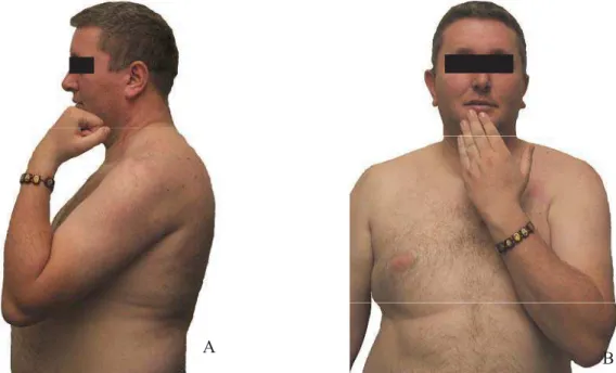
Fig. 1 – Excellent recovery of elbow flexion (A ) with preserved brachio-thoracic pinch (B) for a 19-year-old male patient. Reinnervation of the left musculocutaneous nerve using the medial pectoral nerve as donor was done 3 months after injury
A
B
Some shoulder external rotation recovery was obtained in 18 (64.3%) of 28 nerve transfers to the axillary nerve. Ex-cellent and good results were obtained in only 6 (33.3%) of 18 recoveries and were related to the good quality of recov-ery of the elbow flexion and shoulder abuction. The rates of recovery were similar for both nerves, 11 (68.7%) of 16 transfers using the thoracodorsal nerve, and 7 (58.3%) of 12 transfers using the medial pectoral nerve. The quality of re-covery was also similar, 4 (36.3%) excellent and good results for the thoracodorsal nerve and 2 (28.6%) for the medial pectoral nerve among recoveries (Table 4, Fig. 3).
The first signs of recovery appeared after 4 to 12 months (mean 7 months) for elbow flexion, and 4 to 13 months (mean 8 months) for shoulder abduction and exter-nal rotation using the thoracodorsal nerve as donor, or after 5 do 15 months (mean 9 months) and 4 do 12 monthas (mean 6 months) using the medial pectoral nerve as donor.
Recovery, ie maximal strength, was completed after 11 do 21 months (mean 13 months) for elbow flexion and after 9 to 24 months (mean 17 months) for shoulder abduction and external rotation when the thoracodorsal nerve was used as donor. In procedures using the medial pectoral nerve, com-plete recovery was achieved within 12 to 24 months (mean 17 months), and within 8 to 24 months (mean 15 months), respectively.
In general, statistical analyses showed no significant correlations among the age of the patient, the timing of sur-gery and the final outcome, probably because of the pre-dominance of the patients of less than 30 years of age and
surgical procedures performed within 6 months after the in-juries.
There was no significant difference in the results ob-tained using the thoracodorsal or medial pectoral nerves as donors, either for the musculocutaneous nerve or for the ax-illary nerve (Pearson’s Ȥ2 test, p = 0.213 and p = 0.858, re-spectively). The same was true for the various techniques of nerve transfer because there was no significant difference between solitary thoracodorsal or pectoral medial nerve transfer and transfer of these nerves in combination with other donors. However, on the basis of the recovery
percent-ages, there could be a slight trend toward better results for thoracodorsal nerve transfer. Similarly, there was no signifi-cant difference in the quality of recovery within these two nerves and various techniques used (Fisher test, p = 0.195, and p = 0.683 respectively). On the basis of the recovery percentages, there was a trend toward better quality of recov-ery for the musculocutanoeus nerve using the thoracodorsal nerve as donor, and for the axillary nerve using the medial pectoral nerve as donor.
In procedures using the thoracodorsal nerve as donor, the rate of recovery for the musculocutaneous nerve was sig-nificantly better than for the axillary nerve (Mann Whitney test, p= 0.038), and the quality of recovery was also signifi-cantly better (Fisher test, p = 0.04). There was no statistically significant difference between rates and quality of recovery for the musculocutaneous and axillary nerve in procedures using the medial pectoral nerve as donor (Mann Whitney test, p = 0.0671).
Table 4 The results of thoracodorsal and medial pectoral nerve transfers regarding shoulder external rotation
Result Donor nerve
Bad Fair Good Excellent
Thoracodorsal 2 4 2 2
Thoracodorsal and intercostal 1 1
Thoracodorsal and subsapular or long thoracic 2 2
Medial pectoral 3 3 2
Medial pectoral and intercostal or spinal accessory 2 2
Total 10 12 4 2
Regarding the rate and quality of recovery for the shoulder external rotation following reinnervation of the ax-illary nerve, there was no significant difference regarding the donor nerve used.
Comparing the average times of appearance of the ini-tial signs of recovery and maximal recovery, we observed no significant differences between the types of donor and re-cipient nerves.
Comparative reviews of thoracodorsal and medial pec-toral nerve transfer results are given in Table 5 and Table 6, respectively.
Discussion
Nerve transfers using collateral branches of the brachial plexus present a distal form of the intraplexal nerve transfer that generally yield better results because of the higher num-ber of motor finum-bers and more physiologic reconstruction. However, these offer some advantages to the classical
intra-plexal transfer, such as insignificant axonal mixing, the ab-sence of mass or cross innervation, anastomosis close to the target muscle and more precise evaluation of donor nerve functional validity compared to that of the proximal nerve stumps.
Collateral branches of the brachial plexus, particularly the thoracodorsal and medial pectoral nerves, are voluntary motor nerves with a significant number of motor fibers, close functional relationship with upper arm nerves, better cortical reintegration owing to central plasticity based on preexisting central and medullary synaptic connections 7, 19, anatomic
proximity to the recipient nerves that enables tension free di-rect anastomosis or rarely anastomosis using short nerve grafts close to motor end plate of the target muscle 1, 2, 10, 16. These nerves do not fulfill some other criteria, including the criterion that a motor donor nerve should be expendable or redundant, without significant diameter mismatching with the recipient nerve and preferrably innervating synergistic
Table 5 The results of thoracordorsal nerve transfer
Rate of useful recovery Series (reference number)
n/total (%) Axillary nerve as recipient
Borrero, 2007 7(a) 8/8 (100)
Dai, 1990 8 2/2 (100)
Haninec et al, 2007 9 6/7 (86)
Samardzic et al, 2005 10 14/15( 93.3)
Sulaiman et al, 2009 11 4/1 (36)
Musculocutaneous nerve as recipient
Dai, 1990 8 1/1 (100)
Haninec et al, 2007 9 3/3 (100)
Novak et al, 2002 12(b) 5/6 (83/3)
Richardson, 1997 13 4/4 (100)
Samardzic et al, 2005 10 12/12 (100)
Tung et al, 2003 14 (c) 1/1 (100)
(a) – nerve transfer of the thoracodorsal or lower subascapular nerve – not specified (b) – a modified technique using two branches of the thoracodorsal nerve
(c) – nerve transfer to the brachial muscle branch combined with the Oberlin procedure to the biceps muscle branch
Table 6 The results of medial pectoral nerve transfer
Rate of useful recovery Series (reference number)
n/total n (%) Axillary nerve as recipient
Ranalli et al,2008 15(a) 1/1 (100) Samardzic et al, 2002 16 9/11 (81.8) Musculocutaneous nerve as recipient
Blauw and Sloff, 2003 3 (b) 22/25 (88.8) Brandt and Mackinnon, 1993 17 4/5 (80)
Haninec et al, 2007 9 10/11 (91)
Samardzic et al, 2002 16 12/14 (85.7) Sulaiman et al, 2009 11 36/41 (87) Sulaiman et al, 2009 11(c) 14/14 (100) Tung et al, 2003 14(d) 6/6 (100)
Wellons et al,2009 18 16/20 (80)
(a) –
nerve transfer supplemented by additional spinal accessory to subascapular nerve transfer (b) – patients with obstetric brachial plexus palsy
muscles with the target muscle 1, 2. These problems are espe-cially important for the use of the medial pectoral nerve, but they may be overcome, at least partially, as we shall see later.
The first priorities in brachial plexus repair are restora-tion of full range and strong elbow flexion, shoulder stabil-ity, active arm abduction and some external rotation 5. Re-covery of all functions is equally important since these en-able elbow movements through a more functional range 20. The recovery of elbow flexion may be achieved through re-innervation of the musculocutaneous nerve using different technical methods. Since the biceps muscle acts as a primary forearm supinator and secondarily provides elbow flexion, and the brachialis muscle is the primary muscle providing elbow flexion, Tung et al. 14 proposed separate neurotization of both muscles in order to maximize the potential for recov-ery of strong function. On the other hand restoration of shoulder function is somewhat controversial. Several authors recommended reinnervation of the suprascapular nerve since the supra- and infrascapular muscles are important for initia-tion of the arm abducinitia-tion and some external rotainitia-tion. In a metaanalysis Merrel et al. 21 concluded that significantly better results were achieved an by the spinal accessory to su-prascapular nerve transfer than with nerve transfer to the ax-illary nerve, with 92% vs. 69% recoveries. Using this proce-dure, Terzis et al. 22, 23 reported good or excellent recovery of shoulder abduction (grade 3 or more) in 79% of the patients and in 55% of the patients for external rotation. Bertelli and Ghizoni 24 achieved average an range of arm abduction up to 122 degrees, and external rotation up to 118 degrees in pa-tients with upper palsy. Venkatramani et al. 25 obtained somewhat lower average ranges, 66 degrees for arm ab-duction and 44 degrees for external rotation. Suzuki et al. 26 obtained similar average shoulder abduction of 77.1 de-grees but shoulder external rotation was only 16.7 dede-grees. However, Malessy et al. 27 achieved grade M3 or M4 rein-nervation of the supraspinatus and infraspinatus muscles in 17% and only 8%, respectively. He concluded that reinner-vation of the shoulder in patients with upper brachial plexus palsy following suprascapular nerve neurotization is dissapointly low. In a significant number of our cases we obtained good arm abduction and some external rotation reinnervating only the axillary nerve. This could be ex-plained by reinnervation of the teres minor and posterior fi-bers of the deltoid muscle that act as shoulder external ro-tators. Furthermore, the reinnervated biceps contributes to shoulder stability through its long head and produces some active external rotation 28. Probably the best solution is dual nerve transfer to both the suprascapular and axillary nerves 29
. However, the first muscle to be reinnervated attracts a majority of axons and in this case the supraspinatus reduces the potential for reinnervation of the external rotator, the infraspinatus muscle 30.
The thoracodorsal nerve
The thoracodorsal nerve is a motor nerve that originates from the posterior cord and receives nerve fibers from the seventh, eight, and sometimes sixth cervical nerves. More
than 52% of motor fibers originate from the C7 nerve root 31. This nerve has cerebral centers integrated into the function of the upper extremity and innervates the latissimus dorsi mus-cle. The mean surgically useable length of the nerve is 12.3 cm with a range of 8.5 to 19.0 cm 32. The diameter of the nerve ranges from 2.1 to 3 mm 10, 33. The number of myeli-nated fibers ranges from 1,530 to 2,479 10. According to these characteristics, the thoracodorsal nerve may be consid-ered as an excellent donor in motor nerve transfers.
The number of motor axons in the thoracodorsal nerve is sufficient for reinnervation of the biceps and brachialis muscles without a need for neurolysis and exclusion 12, 14 or redirection of the lateral antebrachial cutaneous sensory nerve fibers. Similarly, we think that there is no need for augmentation by additional nerve transfer to the brachialis muscle as proposed by Tung et al. 14. However, nerve anas-tomosis should be done distally to the branches to the cora-cobrachialis muscle since this is not important for elbow flexion and shoud not be reinnervated 17. It should be empha-sized that in the majority of cases with extended upper bra-chial plexus palsy involving the C7 spinal nerve, or injuries to the middle trunk and posterior cord, the thoracodorsal nerve is not functional.
Regarding functional deficit after thoracodorsal nerve section, we believe that additional palsy of arm adduction and internal rotation due to the loss of the latissimus dorsi in severely disabled shoulder and arm movements presents an acceptable sacrifice 6, 10, 12. Similarly, Borrero 7, Novak et al. 12
and Tung et al. 11 did not register ill effects from the de-nervation of the latissimus dorsi muscle. Narakas 5 recom-mended the thoracodorsal nerve transfer exclusively for the axillary nerve, similarly to the first report by Foerster ac-cording to Narakas 4, 5, and Kline 15, but the results were not so encouraging 11. We obtained functional recovery in all 13 cases for the musculocutaneous nerve, and in 15 (93.7%) of 16 cases for the axillary nerve. Our results are supported by those published by Richardson 13, who obtained functional recovery of the biceps muscle in all four cases with nerve re-pair delayed for two years, as well as by Novak et al. 12, who reported successful reinnervation of the biceps muscle in all six cases using a modified technique, ie separate transfer of the thoracodorsal divisions to the biceps and brachialis branches of the musculocutaneous nerve. They obtained M4 or M5 grades in five of their cases. Dai et al. 8 obtained re-covery in one case of nerve transfer to the musculocutaneous nerve and in two cases for the axillary nerve. Finally, Tung et al. 14 used this nerve in one case, for reinnervation of the brachialis branch of the musculocutaneous nerve in combi-nation with Oberiln procedure and obtained good reinnerva-tion (Table 5). By contrast, Narakas 5 used the thoracodorsal nerve for reinnervation of the axillary nerve without any sig-nificant functional improvement: he obtained proper reinner-vation of the deltoid muscle, but not with muscle contrac-tions.
transfer to the axillary nerve are less impressive, especially regarding the quality of recovery, probably due to the functional complexity of the shoulder abduction, the role of the supraspinatus muscle that is not reinnervated in these cases, and essentially antagonistic function of the latissi-mus dorsi latissi-muscle, although this could be successfully re-trained 12.
Finally, the use of the thoracodorsal nerve will preclude the use of the latissimus dorsi muscle for secondary proce-dures.
The medial pectoral nerve
The medial pectoral nerve is a motor nerve that derives from the anterior division of the inferior trunk and receives nerve fibers from the 8th cervical and the 1st thoracic nerves. This nerve has also cerebral centers that are integrated into the function of the upper extremity and innervates with several branches the sternal part of the pectoralis major muscle 34.
The anatomic relationship of the lateral and medial pectoral nerves is variable and somewhat unclear. Classi-cally, they have been described as two distinct nerves linked with ansa that develops at the expense of the lateral nerve 3, 16, 35
. Some authors, however, have described three distinct nerves: the superior, medial and inferior 36. The medial nerve derives from the anterior division of the middle trunk and terminates with two constant branches, superficial and deep. The deep branch forms plexus with inferior branch (medial pectoral nerve) in all cases. An additional branch originates from this anastomosis 36. The medial pectoral nerve ends in the pectoralis major muscle with two or three branches 18. Sometimes, we found four branches in our operative cases 16. Similarly, in our patients, we observed one or two branches from the pectoral ansa.
Surgically useable length of the medial pectoral nerve ranged from 30 do 78 mm 35. However, this length may be increased by dissecting terminal branches and their section close to the pectoral muscle. The mean diameter of the nerve ranges from 1.5 to 2.5 or 2.7 mm 33, 35. The number of motor fibers ranges from 1, 170 to 2,140 in the main trunk 34 and may reach 400 to 600 fibers in a muscular branch 4. The above mentioned branch of the pectoral ansa contains 330 to 440 nerve fibers 37. These characteristics make the medial pectoral nerve a valuable donor for motor nerve transfer, es-pecially with regard to the number of motor fibers.
There are three main surgical problems in performing anastomosis with the axillary and especially the musculocu-taneous nerve. These are the large discrepancy in the diame-ter of the nerves, the insufficient length of the former for di-rect anastomosis, and its functional preservation.
In case of diameter mismatch, some authors have su-tured the medial pectoral nerve to the fascicle of the muscu-locutaneous nerve, or have used an epineural suture over the part of the musculocutaneous nerve cross-sectional area 35. In the majority of cases, we removed the fascicular epineurium of the recipient nerve and bundled the medial pectoral nerve with the branch of the pectoral ansa, in order to overcome this problem. More recently, we also bundled several branches of the medial pectoral nerve in a common trunk
using fibrin glue 33. In cases in which these procedures were insufficient, we used an additional donor nerve, usually one intercostal or spinal accessory nerve 6, 16. Sulaiman et al. 11, as well as Kline and Tiell 43,used a combination of the me-dial pectoral nerve to the meme-dial half of the musculocutane-ous nerve transfer with grafting from the anterior division of lateral cord to the musculocutaneous nerve. This technique maximized axonal regeneration from two outflows, proxi-mally repaired plexus elements, and the medial pectoral nerve transfer, and additionally they solved the problem of diameter mismatch.
The technically ideal nerve transfer allows direct nerve anastomoses between the donor and recipient nerves. Ac-cording to some investigations, the length of the medial pectoral nerve is insufficient for tension-free direct anasto-mosis with the musculocutaneous nerve in approximately one third of cases. The average length of this gap is ap-proximately 15 to 20 mm 6, 36, 37. This problem may be over-come in several ways, such as retrograde split of the muscu-locutaneous nerve into the lateral cord, distal section of the medial pectoral nerve branches 35, dissection of the nerve trunk from its branch to the pectoral ansa 39, and sectioning the arcade between the pectoral nerves 6, 16, 18, 26, 35, 36. We used nerve grafts in our earliest cases and in cases with dou-ble level lesion that included peripheral injury to the muscu-locutaneous nerve. In these cases the length of nerve graft was up to 5.0 cm 6, 16. In reinnervation of the axillary nerve, we were able to perform direct anastomosis in all cases.
Similarly to transfer of the thoracodorsal nerve, we think that additional palsy of arm adduction and internal ro-tation is not as significant in patients with severely disabled shoulder function 6, 16. Furthermore, in cases of predominant innervation from the C7 spinal nerve root, the function of synergic muscles such as teres major may sometimes be par-tially preserved. In addition, some function of the pectoral muscles may be retained because of multiple innervation patterns of the pectoralis major muscle since the usual origin of the lateral pectoral nerve is from the C5 to C7 spinal nerves with the mean percentage of supply for pectoral mus-cles 50% from the C7 spinal nerve. Functional preservation is also possible by distal sectioning and sparing some of the branches 37, 38. According to our results and the results of some other published series, the remaining branch or branches usually produce strong contractions of the pectora-lis major mucle 35. End-to-side neurorrhaphy is another pos-sibility for functional preservation of the donor nerves 39, but only Wellons et al. 18 used this technique in one case of me-dial pectoral to the musculocutaneous nerve transfer.
in combination with Oberlin procedure and achieved rein-nervation that was comparable to that of the biceps muscle in all six cases. They stated that neurolysis and exculsion of the sensory component is not necessary with this method.
Although the homolateral use of the medial pectoral nerve is controversial because the potential absence of arm adduction may be very disabling, we used this nerve due to the reasons mentioned above and achieved recovery in 19 (90.5%) of 21 cases who underwent nerve transfer to the musculocutaneous nerve, and in 10 (83.3%) of 12 patients with transfer to the axillary nerve. Ranalli et al. 15 recom-mended this nerve transfer in both situations. Our results are in accordance with those published by others (Table 5). However, Blaauw and Sloff 3, and Wellons et al. 18 published results of the medial pectoral to the musculocutaneous nerve transfer in obstetric upper brachial plexus palsy.
Long thoracic nerve
The long thoracic nerve arises from the ventral rami of the 5th, 6th and 7th spinal nerve. The 8th cervical spinal nerve may contribute in 8% of cases. The mean surgically useable length is 22.0 cm, ranging from 18.0 to 27.0 cm. The overall mean diameter of the nerve is 3.0 mm, ranging from 2.2 to 3.5 mm 40. The number of motor fibers ranges from 1,600 to 1,800, but only half of them can be used in patients with upper brachial plexus palsy 17.
Lurje used this nerve distal to its innervation for the su-perior two digitations of the serratus anterior muscle for re-innervation of transected suprascapular nerve in 1948 (ac-cording to ref. 40). Narakas and Hentz 28 used the long tho-racic nerve in reinnervation of the different parts of the bra-chial plexus in seven patients with good recovery in only two of them. More recently, Haninec et al. 9 obtained useful functional recovery in all three cases for the musculocutane-ous nerve and in 6 (55%) of 11 cases for the axillary nerve. We used the long thoracic nerve in only one case as supple-ment in nerve transfer of the thoracodorsal to the axillary nerve.
Lower subscapular nerve
The lower subscapular nerve usually originates from the posterior cord but this is very variable. Ballestros and Rami-rez 41 found its origin from the axillary nerve in 54.4% of cases, and even from the thoracodorsal nerve in 12.3% of cases. The nerve fibers supply is from the 5th, 6th and some-times 7th cervical spinal nerves. Therefore, this nerve may be of little value for reinnervation in cases with upper bra-chial plexus palsy. The mean length of lower subscapular nerve from its origin to the site at which it branches to the subscapularis muscle is 3.5 cm, ranging from 1.5 to 6.2 cm.
The mean distance from this branch to its termination at the distal teres major muscle is 6.0 cm, ranging from 3.3 to 8.9 cm. Therefore, the mean length of the entire nerve is 9.5 cm, ranging from 7.1 to 11.4 cm 42. The mean distance from the musculocutaneous nerve is 1.5 to 2.0 cm and from the axil-lary nerve 2.5 cm 42. The mean diameter of the lower sub-scapular nerve is 2.3 mm, ranging from 1.9 to 2.5 mm 43. The lower subscapular nerve has approximately 2,100 motor fi-bers 33.
The lower subscapular nerve has been rarely used in nerve transfers. Borrero 7 used this nerve or the thoracodorsal nerve (not specified) for reinnervation of the deltoid muscle with success in all eight cases. He stated that denervating the subscapular muscle was not detrimental, and in some cases it was beneficial because it lessened excessive internal rotation tendency. We used the lower subscapular nerve as supple-ment for reinnervation in transfer of the thoracodorsal nerve to the musculocutaneous nerve in two cases, and to the axil-lary nerve in four cases (Table 1). The recovery was obtained in all five cases that we followed up with good quality of re-covery in five of them (Table 2).
Including experience published by other authors, surgi-cal results of this type of nerve transfer could be improved using double nerve transfers, i.e. an additional spinal acces-sory to suprascapular nerve transfer for shoulder abduction, and fascicular nerve transfer to the brachialis muscle branch for elbow flexion. The latter may be especially important in cases with diameter mismatch between the medial pectoral and the musculocutaneous nerve. Furthermore, the medial pectoral and thoracodorsal nerve transfers may be very promising for older patients and those with delayed surgery.
Conclusion
According to our experience and the results we ob-tained, nerve transfers using collateral branches of the bra-chial plexus in patients with upper palsy have several ad-vantages: simplicity of the surgical procedure, significant gain in operative time, high rate of recovery, good quality of recovery in a significant number of cases, early signs of re-covery, relatively short period for recovery completion, low rate of significant functional impairment due to loss in donor nerve innervation zone, generally, a slight trend toward bet-ter results using the thoracodorsal nerve as donor, and this nerve is a significantly better donor for reinnervation of the musculocutaneous than for the axillary nerve, the medial pectoral nerve, however, offers somewhat better quality of recovery in reinnervation of the axillary nerve, and, finally, any combination of donor and recipient nerves does not pre-clude good result.
R E F E R E N C E S
1. Weber RV, Mackinnon SE. Nerve transfers in the upper ex-tremity. J Am Soc Surg Hand 2004; 4(3): 200–13.
2. Addas B, Midha R. Nerve transfer for shoulder reanimation. In:
Midha R, Zager E, editors. Surgery of peripheral nerves. New York: Thieme; 2008. p. 53î9.
3. Blaauw G, Slooff AC. Transfer of pectoral nerves to the mus-culocutaneous nerve in obstetric upper brachial plexus palsy. Neurosurgery 2003; 53(2): 338î41; discussion 341î2. 4. Narakas A. Neurotization or nerve transfer for brachial plexus
5. Narakas A. Neurotization in the treatment of brachial plexus injuries. In: Gelberman RH, editor. Operative nerve repair and reconstruction. Philadelphia: JB Lippincott Company; 1991. p. 1329î58. 6. Samardzic MM, Grujicic DM, Rasulic LG, Milicic BR. The use of
thoracodorsal nerve transfer in restoration of irreparable C5 and C6 spinal nerve lesions. Br J Plast Surg 2005; 58(4): 541î6.
7. Borrero JL. Obstetrical Brachial Plexus Paralysis. 2nd ed. Lake Mary, Fla: Design and Print Progressive Communications; 2007.
8. Dai SY, Lin DX, Han Z, Zhoug SZ. Transference of thoraco-dorsal nerve to musculocutaneous or axillary nerve in old traumatic injury. J Hand Surg Am 1990; 15(1): 36î7.
9. Haninec P, Sámal F, Tomás R, Houstava L, Dubovwý P. Direct re-pair (nerve grafting), neurotization, and end-to-side neuror-rhaphy in the treatment of brachial plexus injury. J Neurosurg 2007; 106(3): 391î9.
10.Samardzic M, Grujicic D, Rasulic L, Bacetic D. Transfer of the medial pectoral nerve: myth or reality? Neurosurgery 2002; 50(6): 1277î82.
11.Sulaiman OA, Kim DD, Burkett C, Kline DG. Nerve transfer surgery for adult brachial plexus injury: a 10-year experience at Louisiana State University. Neurosurgery 2009; 65(4 Suppl): A55î62.
12.Novak CB, Mackinnon SE, Tung TH. Patient outcome following a thoracodorsal to musculocutaneous nerve transfer for recon-struction of elbow flexion. Br J Plast Surg 2002; 55(5): 416î9. 13.Richardson PM. Recovery of biceps function after delayed repair
for brachial plexus injury. J Trauma 1997; 42(5): 791î2. 14. Tung TH, Novak CB, Mackinnon SE. Nerve transfers to the
bi-ceps and brachialis branches to improve elbow flexion strength after brachial plexus injuries. J Neurosurg 2003; 98(2): 313î8. 15.Ranalli NJ, Kline DG, McGarvey ML, Boulis NM, Zager EL.
Clinical problem-solving: brachial plexus closed injury and re-construction. Neurosurgery 2008; 62(6): 1330î8; discussion 1338î9.
16.Samardziý M, Rasuliý L, Grujiciý D, Miliciý B. Results of nerve transfers to the musculocutaneous and axillary nerves. Neuro-surgery 2000; 46(1): 93î101; discussion 101î3.
17.Brandt KE, Mackinnon SE. A technique for maximizing biceps recovery in brachial plexus reconstruction. J Hand Surg Am 1993; 18(4): 726î33.
18.Wellons JC, Tubbs RS, Pugh JA, Bradley NJ, Law CR, Grabb PA.
Medial pectoral nerve to musculocutaneous nerve neurotiza-tion for the treatment of persistent birth-related brachial plexus palsy: an 11-year institutional experience. J Neurosurg Pediatr 2009; 3(5): 348î53.
19.Malessy MJ, de Ruiter GC, de Boer KS, Thomeer RT. Evaluation of suprascapular nerve neurotization after nerve graft or transfer in the treatment of brachial plexus traction lesions. J Neuro-surg 2004; 101(3): 377î89.
20.Millesi H. Coordinated function oriented movements after multiple root avulsion. Acta Neurochir Suppl 2007; 100: 117î9.
21.Merrell GA, Barrie KA, Katz DL, Wolfe SW. Results of nerve transfer techniques for restoration of shoulder and elbow function in the context of a meta-analysis of the English lit-erature. J Hand Surg Am 2001; 26(2): 303î14.
22.Terzis JK, Papakonstantinou KC. The surgical treatment of bra-chial plexus injuries in adults. Plast Reconstr Surg 2000; 106(5): 1097î122; quiz 1123î4.
23.Terzis JK, Kostas I. Suprascapular nerve reconstruction in 118 cases of adult posttraumatic brachial plexus. Plast Reconstr Surg 2006; 117(2): 613î29.
24.Bertelli JA, Ghizoni MF. Transfer of the accessory nerve to the suprascapular nerve in brachial plexus reconstruction. J Hand Surg Am 2007; 32(7): 989î98.
25.Venkatramani H, Bhardwaj P, Faruquee SR, Sabapathy SR. Func-tional outcome of nerve transfer for restoration of shoulder and elbow function in upper brachial plexus injury. J Brachial Plex Peripher Nerve Inj 2008; 3: 15.
26.Suzuki K, Doi K, Hattori Y, Pagsaligan JM. Long-term results of spinal accessory nerve transfer to the suprascapular nerve in upper-type paralysis of brachial plexus injury. J Reconstr Mi-crosurg 2007; 23(6): 295î9.
27.Malessy MJ, Thomeer RT, van Dijk JG. Changing central nervous system control following intercostal nerve transfer. J Neuro-surg 1998; 89(4): 568î74.
28. Narakas AO, Hentz VR. Neurotization in brachial plexus inju-ries. Indication and results. Clin Orthop Relat Res 1988; 237: 43î56. 29.Addas BMJ, Midha R. Nerve transfers for severe nerve injury.
In: Spinner RJ, Winfree CJ, editors. Peripheral nerves: injuries. Neurosurgery Clinics of North America. New York: WB Saunders; 2009; 20: 27î38.
30. Narakas AO. Thoughts on neurotizations or nerve transfers in irreparable nerve lesions. In: Terzis JK, editor. Microrecon-struction of nerve injuries. Philadelphia: WB Saunders; 1987; 447î54. 31.Lu W, Xu JG, Wang DP, Gu YD. Microanatomical study on the
functional origin and direction of the thoracodorsal nerve from the trunks of brachial plexus. Clin Anat 2008;2 1(6): 509î13.
32.Bartlett SP, May JW Jr, Yaremchuk MJ. The latissimus dorsi muscle: a fresh cadaver study of the primary neurovascular pedicle. Plast Reconstr Surg 1981; 67(5): 631î6.
33.Samardzic M, Antunovic V, Joksimovic M, Bacetic D. Donor nerves in the reinnervation of brachial plexus. Neurol Res 1986; 8(2): 117î22.
34.Millesi H. Brachial plexus injury in adults: Operative repair. In:
Gelberman RH, editor. Operative nerve repair and reconstruc-tion. Philadelphia: JB Lippincott Co; 1991. p. 447î54. 35.Hansasuta A, Tubbs RS, Grabb PA. Surgical relationship of the
medial pectoral nerve to the musculocutaneous nerve: a cadav-eric study. Neurosurgery 2001; 48(1): 203î6; discussion 206î7.
36.Aszmann OC, Rab M, Kamolz L, Frey M. The anatomy of the pectoral nerves and their significance in brachial plexus recon-struction. J Hand Surg Am 2000; 25(5): 942î7.
37.Narakas A. Brachial plexus lesion. In: Leung PC, Gu YD,.
Ykuta Y, Narakas A, Landi A, Weiland AJ, editors. Microsur-gery in orthopedic practice. Singapore:World Scientific; 1995. p. 188î254.
38.Manktelow RT, McKee NH, Vettese T. An anatomical study of the pectoralis major muscle as related to functioning free mus-cle transplantation. Plast Reconstr Surg 1980; 65(5): 610î5. 39.Amr SM, Moharram AN. Repair of brachial plexus lesions by
end-to-side side-to-side grafting neurorrhaphy: experience based on 11 cases. Microsurgery 2005; 25(2): 126î46. 40.Tubbs RS, Loukas M, Shoja MM, Shokouhi G, Wellons JC, Oakes
WJ, et al. The contralateral long thoracic nerve as a donor for upper brachial plexus neurotization procedures: cadaveric fea-sibility study. J Neurosurg 2009; 110(4): 749î53.
41.Ballesteros LE, Ramirez LM. Variations of the origin of collat-eral branches emerging from the posterior aspect of the bra-chial plexus. J Brabra-chial Plex Peripher Nerve Inj 2007; 2: 14. 42.Tubbs RS, Khoury CA, Salter EG, Acakpo-Satchivi L, Wellons JC
3rd, Blount JP,et al. Quantitation of the lower subscapular nerve for potential use in neurotization procedures. J Neurosurg 2006; 105(6): 881î3.
43.Kline DG, Tiel RL. Direct plexus repair by grafts supplemented by nerve transfers. Hand Clin 2005; 21(1): 55î69, vi.