Myasthenia gravis in Ceará, Brazil
Clinical and epidemiological aspects
Aline de Almeida Xavier Aguiar1, André Ferrer Carvalho2,
Carlos Mauricio de Castro Costa3, José Marcelino Aragão Fernandes4, José Artur Costa D’Almeida5, Luís Edmundo Teixeira de Arruda Furtado6,
Francisco Marcos Bezerra da Cunha7
ABSTRACT
A retrospective chart review was performed on patients diagnosed as having myasthenia gravis in Ceará State, Brazil and who were followed from October 1981 to June 2009. Clinical and epidemiologic aspects were evaluated. In this work, 122 patients were studied, of whom 85 (69.7%) were females and 37 (30.3%) were males. The disease duration ranged from five months to 50 years (8.9±8.1 years). Age at the first symptoms varied from 0 to 74 years (31.9±14.4 years). The first main symptoms and signs were ptosis, diplopia and limb weakness. Generalized myasthenia was the most common clinical presentation, but 5.1% (n=6) persisted as ocular myasthenia. Thymectomy was performed in 42.6% (n=52) of myasthenic patients. A thymoma was present in 10 patients. Serum acetylcholine receptor (AChR) antibodies were present in 80% (n=20) of specimens tested. The data presented are similar to those of studies performed in other countries.
Key words: myasthenia gravis, clinical evaluation, epidemiology.
Miastenia gravis no Ceará, Brasil: aspectos clínicos e epidemiológicos
RESUMO
Foram analisados, retrospectivamente, os prontuários de pacientes miastênicos, diagnosticados e seguidos entre outubro de 1981 e junho de 2009 no Estado do Ceará, Brasil. Foram coletados dados clínicos e epidemiológicos. Na casuística foram estudados 122 pacientes: 85 (69,7%) do sexo feminino e 37 (30,3%) do sexo masculino. O tempo de doença variou de 5 meses a 50 anos (8,9±8,1 anos). A idade de inicio da doença variou de 0 a 74 anos (31,9±14,4 anos). Na amostra estudada, os primeiros sintomas foram principalmente ptose, diplopia e fraqueza dos membros. A maioria dos pacientes apresentou a forma generalizada, enquanto 5,1% (n= 6) persistiram com miastenia ocular. Timectomia foi realizada em 42,6% (n=52) dos pacientes. Timoma estava presente em 10 pacientes. Anticorpo anti-receptor de acetilcolina foi positivo em 80% (n=20) das amostras testadas. Os aspectos clínicos e epidemiológicos da amostra estudada têm semelhança com aqueles estudados em outros países.
Palavras-chave: miastenia grave, avaliação clínica, epidemiologia.
Correspondence
Aline de Almeida Xavier Aguiar Rua Governador Joca Pires 1535/501 64049-522 Teresina PI - Brasil E-mail: [email protected]
Received 4 December 2009 Received in final form 1 June 2010 Accepted 8 June 2010
1MD, São Marcos Hospital, Teresina PI, Brazil; 2MD PhD, Faculty of Medicine, Department of Clinical Medicine, Federal University of Ceará, Fortaleza CE, Brazil; 3MD PhD, Department of Physiology and Pharmacology, Federal University of Ceará, Fortaleza CE, Brazil; 4MD, Neurology Service, University Hospital Walter Cantídio, Federal University of Ceará, Fortaleza CE, Brazil; 5MD PhD, Fortaleza General Hospital, Neurology Service, Fortaleza CE, Brazil; 6MD PhD, Faculty of Medicine, Federal University of Ceará, Sobral CE, Brazil; 7MD PhD, Faculty of Medicine, Federal University of Ceará, Barbalha CE, Brazil.
Myasthenia gravis (MG) is a potential-ly serious but treatable autoimmune dis-ease affecting the neuromuscular junc-tion, whose main clinical feature is luctu-ating weakness and fatigability of the
vol-untary muscles1-5.he weakness tends to
Several forms and causes of MG have been described, such as an acquired autoimmune form, a neonatal form and familial or sporadic congenital myasthenic syn-dromes. he irst is caused by antibody-mediated auto-immune attack directed against nicotinic acetylcholine receptors on the postsynaptic membrane of the neuro-muscular junction2,4. Congenital myasthenic syndromes
form a heterogeneous group of genetic diseases charac-terized by a dysfunction of neuromuscular transmission7,
and transitory neonatal myasthenia is a self-limited disor-der that follows the passive transfer of maternal antibod-ies to the fetus8. MG has also been categorized into
sub-types according to age at onset (early versus late), forms (purely ocular or generalized), presence or absence of ace-tylcholine receptor (AChR) antibodies in serum and as-sociated thymoma9.
MG is a rare disease. Its incidence varies with age, gender and ethnic group10. It is thought to be more
com-mon in young women and older men10. he annual
inci-dence of myasthenia has been reported to be in the range of 1 to 15/million; the point prevalence rate ranges from 3 to 175/million and has increased from the 1950s to the 1990s6,11. he disease has a higher prevalence in
wom-en than in mwom-en, with an approximate female-to-male ra-tio of 2:112. MG in childhood and adolescence is rare13,14.
Girls are more frequently afected than boys in a propor-tion of 1.3:1 at pre-pubertal ages and 1.8:1 in peripuber-tal ages15. Without treatment, 20-30% of patients will die
in 10 years8, but with appropriate therapy, most patients
are able to live productively.
In Brazil, there are relatively limited data on the oc-currence of MG and its clinical and epidemiological as-pects (Table 1)5,14,16. here have been no reported studies
of MG in Northeast Brazil.
he aim of the present study was to explore the clin-ical and epidemiologclin-ical aspects of MG gravis in a
sam-ple of Brazilian patients under care in Ceará, Brazil. he present data are compared with those obtained from oth-er studies reported in the medical litoth-erature.
METHOD
A retrospective study was performed on patients with MG. Data were obtained from the charts of patients seen from October 1981 to June 2009 at the Neurology Ser-vice of Hospital Universitário Walter Cantídio and Hos-pital Geral de Fortaleza, Fortaleza, Ceará, Brazil. Other neurology departments, practicing neurologists and the Associação Cearense de Miastênicos were contacted to ind other myasthenic patients who had not been treat-ed in one of the two centers. he present study was ap-proved by the ethics committee of Hospital Universitário Walter Cantidio and Hospital Geral de Fortaleza. A stan-dardized questionnaire was illed out by one of the au-thors of the present study.
Each patient record was carefully reviewed for the accuracy of diagnosis. he diagnosis of myasthenia was based on three or more of the following criteria: typical history; clinical evidence of fatigability with recovery on rest; clinical response to anticholinesterase administra-tion; detection of acetylcholine receptor antibodies; de-crease in electrical activity on repetitive nerve stimulation (RNS) and exclusion of alternative relevant diagnosis17.
Patients who did not meet diagnostic criteria for MG or did not live in the state of Ceará at the time of admis-sion were excluded from the analyses.
Collected data were: gender, age at irst symptoms, irst symptoms, development of symptoms, irst neuro-logical examination, diagnostic tests and treatment, and classiication of disease at irst and last clinical assess-ment visits.
Clinical severity of myasthenia was graded function-ally according toan adaptation of a scale devised by
Os-Table 1. Descriptive epidemiologic studies of MG involving Brazilian samples.
Variable Cunha et al. (1999)
5
N=153 Assis et al. (1999)
16
N=41 Morita et al. (2001)
13
N=18 Present study (2009)N=122
Gender
Male 49 (32%) 24 (58.5%) 8 (44.4%) 37 (30.3%)
Female 104 (68%) 17 (41.5%) 10 (55.6%) 85 (69.7%)
Female-to-male ratio 2.1:1 0.7:1 1.2:1 2.3:1
Mean age at onset (years) 31.13 >30 7.3 31.9
First main symptom Ptosis No data Ptosis Ptosis
Thymoma 4 (2.6%) 41 (100%) 0 10 (8.2%)
Sample characteristics Retrospective MG patients Curitiba PR, Brazil
Retrospective Thymomatous MG patients São Paulo SP, Brazil
Retrospective Myasthenic children
São Paulo SP, Brazil
See methods section for
details
serman and Genkins18: grade I involvesfocal disease (e.g.,
restricted to ocular muscles); grade II,generalized dis-ease that is either mild (IIa) or moderate (IIb);grade III, severe generalized disease with six months of progres-sion; and grade IV, a crisis,with life-threatening impair-ment of respiration.
All data were collected and stored in the Epi-Info pro-gram (version 3.5.1 for Windows). Data were analyzed with the aid of the Statistical Package for the Social Sci-ences (version 14.0 for Windows). Parametric data are reported as means±standard deviation (mean±SD). Fre-quency data are reported as percentages. Bivariate analy-sis was performed with Student’s t-test and Fisher’s exact test. χ2 analysis was performed for non-parametric data.
Diferences with p≤0.05 were considered signiicant.
RESULTS
General socio-demographic data
Data from 122 patients, 85 (69.7%) females and 37 (30.3%) males, were evaluated. here was a 2.3:1 female-to-male ratio (χ 2; p<0.001). In the group with disease
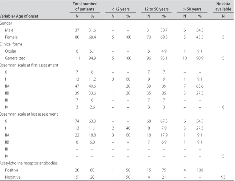
on-set younger than 12 years old, all patients were females and two were sisters. In the group older than 50 years, there was a greater proportion of males (1.2:1), and it was more evident over 60 years, with a ratio of 1.5:1 (Ta-ble 2). Age of onset ranged 0-74 years (0-64 years in fe-males and 15-74 years in fe-males). The mean age at on-set was 31.9±14.4 years. Age at presentation was greater in males (males 40.3±9.45, females 28.1±7.45; Student’s test; p<0.001). Modal ages at onset varied with gender: 16 years for females and 41 years for males. Patients with thymoma were older at irst symptoms compared to those without this neoplasm; only one patient was older than 50 years old, all the others were in the third and fourth de-cades of their lives (Table 3).
Disease duration ranged from ive months to 50 years, with a mean of 8.9±8.1 years, and mean duration of fol-low-up was 5.8±5.9 years.
Clinical characteristics
Ocular symptoms (ptosis and diplopia) and proxi-mal and distal limb weakness were the irst main
symp-Table 2. Sample characteristics according to age of onset of disease.
Variable/ Age of onset
Total number
of patients < 12 years 12 to 50 years > 50 years availableNo data
N % N % N % N % N
Gender
Male 37 31.6 – – 31 30.7 6 54.5
Female 80 68.4 5 100 70 69.3 5 45.5 5
Clinical forms
Ocular 6 5.1 – – 5 4.9 1 9.1
Generalized 111 94.9 5 100 96 95.1 10 90.9 5
Osserman scale at irst assessment
0 7 6 – – 7 7 – –
I 13 11.2 3 60 9 9 1 9.1
IIA 47 40.6 1 20 39 39 7 63.6
IIB 39 33.6 1 20 35 35 3 27.3
III 7 6 – – 7 7 – –
IV 3 2.6 – – 3 3 – – 6
Osserman scale at last assessment
0 74 63.3 – – 68 67.3 6 54.5
I 13 11.1 2 40 8 7.9 3 27.3
IIA 22 18.8 3 60 18 17.9 1 9.1
IIB 8 6.8 – – 7 6.9 1 9.1
III – – – – – – – –
IV – – – – – – – – 5
Acetylcholine receptor antibodies
Positive 20 80 1 50 15 79 4 100
toms in all age groups, but, from 12-50 years, inability to swallow and dysphonia were frequent. In those over 50 years old, dysphonia was as frequent as diplopia. Pa-tients with thymoma had limb weakness as the most im-portant symptom.
At irst neurological examination, oculobulbar palsies and proximal weakness of limbs were the main features in all age groups. In those patients over twelve years old, facial weakness was an important sign. Impairment of tendon relexes, muscle atrophy and hypotonia were less common. Few patients presented with myasthenic (n=12) or cholinergic (n=1) crisis at irst medical assessment.
Generalized myasthenia was the most common clin-ical presentation, but 5.1% (n=6) persisted as ocular my-asthenia (Table 2). According to the Osserman and Gen-kins18 classiication, the 122 cases were distributed in all
groups at irst evaluation. Most of them were classiied as IIA. Patients developed in a relatively benign manner, with no patients in group III or IV at last medical visit (Table 2). Patients with thymoma were classiied mostly as type IIB at irst assessment, but they also improved.
Serological studies were performed on 25 patients for AChR antibodies. Of those, 20 (80%) had detectable anti-bodies (Table 2). Repetitive nerve stimulation results were documented in 81 patients: 64 (79%) showed a
decremen-tal response, 16 (19.7%) had normal response, and one pa-tient had a decremental response at accessory nerve and incremental response at median nerve. Fourteen (11.5%) patients were known to have undergone a Tensilon test at presentation, and in 12 (85.7%) there was a response con-sistent with MG diagnosis. A prostigmine test was done in 20 (16.4%) patients and found to be positive in 18 (90%).
he main diseases that were associated with myas-thenia were arterial hypertension (18.8%; n=23) and di-abetes (14.7%; n=18). Two patients were under investi-gation for systemic lupus erythematosus (SLE), six had thyroid disorders, seven had osteoporosis and two were on treatment for rheumatoid arthritis. One patient had scleroderma.
Treatment procedures
All patients were taking pyridostigmine (acetylcholin-esterase inhibitor). Most of them, 109 (89.3%), used cor-ticosteroids during phases of the disease in order to im-prove therapeutic response. Azathioprine was used as an adjunct to steroids in forty (32.7%) patients. Plasmaphere-sis and immunoglobulin was necessary in 40 (32.7%) and nine (7.4%) patients, respectively. Other immunosuppres-sive drugs were used for associated diseases in two cases, one being cyclosporine and the other methotrexate.
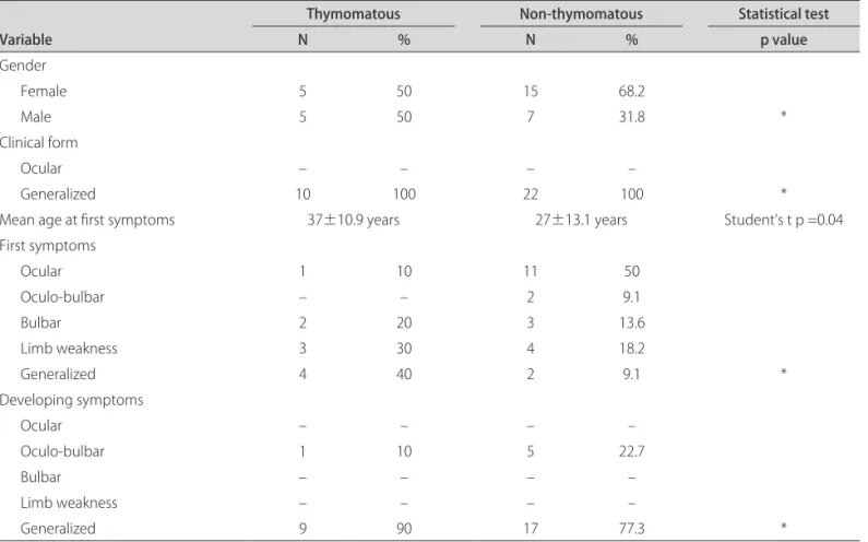
Table 3. Comparison of clinical and epidemiologic characteristics of thymomatous and non-thymomatous MG patients.
Variable
Thymomatous Non-thymomatous Statistical test
N % N % p value
Gender
Female 5 50 15 68.2
Male 5 50 7 31.8 *
Clinical form
Ocular – – – –
Generalized 10 100 22 100 *
Mean age at irst symptoms 37±10.9 years 27±13.1 years Student’s t p =0.04
First symptoms
Ocular 1 10 11 50
Oculo-bulbar – – 2 9.1
Bulbar 2 20 3 13.6
Limb weakness 3 30 4 18.2
Generalized 4 40 2 9.1 *
Developing symptoms
Ocular – – – –
Oculo-bulbar 1 10 5 22.7
Bulbar – – – –
Limb weakness – – – –
Generalized 9 90 17 77.3 *
Fifty-two patients (42.6%) underwent therapeutic thymectomy. Histopathology indings were available in 32 (61.5%) cases. he most common reported histopa-thology indings were hyperplasia (n=16; 50%; ive males and 11 females), and thymoma (n=10; 31.2%; ive males and ive females). Six specimens (18.8%) showed an atro-phic thymus.
DISCUSSION
General socio-demographic indings
In the present study, the majority of patients were fe-male and young. Male patients had a greater mean age of onset compared to females. Accordingly, we found a sin-gle modal age of onset of 41 years for males and 16 years for females. hese data are similar to those reported by Cunha et al. who found a single peak age of onset in males between 20 and 35 years old (44.8%) and in females be-tween 15 and 30 years old (45.2%)5. Christensen et al.
found a greater incidence in men older than 40 years, but demonstrated a bimodal pattern with one peak consist-ing of females at child-bearconsist-ing age and a second peak of older women19. Conversely, Singhal et al., in India,
stud-ied 836 patients and showed male preponderance with a ratio of 2.7:1 and a single (later) peak of males in the sixth and seventh decades3. Diferences across studies may be
in part explained by the fact that the samples vary in ge-netic and cultural aspects, which may afect disease ex-pression and the access to tertiary health services, as sug-gested by Sighal et al 3.
It has been suggested that the prevalence of MG falls after 70 years of age20. Such inding is consistent with this
research, where only one patient (male) was found to be older than 70. Recent population-based studies have sug-gested that MG has a relatively high incidence, but is un-der-diagnosed among the elderly21,22. hus, sampling
char-acteristics of the studies may impact such discrepant epi-demiologic indings.
Childhood MG is uncommon in Europe and North America, comprising 10-15% of MG cases23. he onset
of symptoms before 12 years of age was demonstrated in five (4.1%) patients. The lower prevalence reported in this study could be due to the fact that such patients were recruited from services that primarily deliver care to adults. his inding is in agreement with Evoli et al. who found a prevalence of juvenile myasthenia of 2.3% in the whole myasthenic population13. All patients in the group
with such early age of onset in the present study were fe-males. Such inding relects that of a previous analysis of 18 patients under 12 years old, where there was a fe-male predominance in pre-pubertal patients14, but
anoth-er study failed to show difanoth-erences in gendanoth-er distribution in prepubertal MG13. Two patients had ocular
myasthe-nia and three the generalized form of the disease. As for
disease severity in early childhood, this inding is similar to data from Evoli et al. and Morita et al. who found not only a high frequency of ocular myasthenia but also an even higher incidence of severe disease with respiratory involvement13,14.
Clinical presentation
The first main symptoms and signs were ocular, in agreement with Robertson et al. who observed 52% of patients with ocular involvement at the beginning of dis-ease and the more common signs and symptoms were ptosis and diplopia17. External ocular muscles are afected
initially in around 50% of cases and eventually in 90%; in 10% of patients, these muscles are the only afected ones (ocular myasthenia)20,24.
According to the Osserman and Genkins classiica-tion, patients were categorized into all groups at irst as-sessment, with the majority in group IIa.18 Coincident
with Christensen et al., 43% of the patients were into this group19. At inal neurological examination, 63.8% of all
patients were asymptomatic and none were in group III or IV. hus, the present study suggests that MG has a rel-atively benign course following adequate treatment.
Serological studies for AchR antibodies is speciic for MG and is positive in 80-85% in generalized myasthenia and 50-60% in ocular myasthenia20.hese data are
consis-tent with this sample and an Indian case series3.
Repetitive nerve stimulation is positive in virtually all cases of generalized MG but may be negative in nearly 50% of cases of the ocular form25. Overall, sensitivity is
around 60-75%20. his test was employed for diagnosis in
81 patients. he majority (79.01%) displayed a character-istic decremental response.
he sensitivity of the edrophonium test is around 86% for ocular myasthenia and 95% for the generalized form26.
It was positive in 12 (85.7%) in this series. Robertson et al. performed this test in 82 patients during clinical as-sessment, and 93% were found to be positive, in agree-ment with this study17.
Treatment procedures
Currently, many treatment modalities for myasthenia are available: acetylcholinesterase inhibitors, corticoster-oids, immunosuppressants, plasmapheresis, intravenous immunoglobulins and thymectomy1. Initial treatment
usually involves the use of acetylcholinesterase inhibi-tors, but adjuvant therapy is often needed for adequate disease control. Intravenous immunoglobulin or plasma-pheresis is generally used in early stages of treatment be-fore thymectomy, or later during an exacerbation20.
of eicacy27. However, to date, there have been no
pro-spective, randomized studies to assess the efectiveness of thymectomy for non-thymomatous MG patients. In a retrospective study, Werneck et al. concluded that there was no statistical diference between conservative treat-ment and thymectomy groups, regarding rates of remis-sion and/or improvement28.
Thymic hyperplasia was the most common histo-pathological inding of the present study. Several reports support these indings20,29. hymoma was the next most
common histopathology inding. Patients with thymoma had an older age of onset of symptoms, but there were no gender diferences. hese data are similar to those report-ed by Lavrnic et al.30.
In conclusion, the present paper reports the clinical and epidemiologic characteristics of MG in a sample from Ceará, Northeast Brazil. he indings presented here are similar to those reported in other surveys. In this sample, MG was more prevalent among young women and had a relatively benign course. Variations in indings among diferent studies may be explained by methodological and cultural/ethnic aspects. Further studies with diferent ep-idemiological designs (e.g., prospective and community-based) are needed to enhance our current knowledge of this complex disease.
REFERENCES
1. Meriggioli MN, Sanders DB. Autoimmune myasthenia gravis: emerging clini-cal and biologiclini-cal heterogeneity. Lancet Neurol 2009;8:475-490.
2. Tzartos SJ, Barkas T, Cung MT, et al. Anatomy of the antigenic structure of a large membrane autoantigen, the muscle-type nicotinic acetylcholine re-ceptor. Immunol Rev 1998;163:89-120.
3. Singhal BS, Bhatia NS, Umesh T, Menon S. Myasthenia gravis: a study from In-dia. Neurol India 2008;56: 352-355.
4. Drachman DB. Myasthenia gravis. N Engl J Med 1994;330:1797- 1810. 5. Cunha FM, Scola RH, Werneck LC. Myasthenia gravis: clinical evaluation of
153 patients. Arq Neuropsiquiatr 1999;57:457-464.
6. Sanchez JL, Uribe CS, Franco AF, Jimenez ME, Arcos-Burgos OM, Palacio LG. Prevalence of myasthenia gravis in Antioquia, Colombia. Rev Neurol 2002; 34:1010-1012.
7. Hantai D, Richard P, Koenig J, Eymard B. Congenital myasthenic syndromes. Curr Opin Neurol 2004;17:539-551.
8. Oosterhuis HJ. The natural course of myasthenia gravis: a long term follow up study. J Neurol Neurosurg Psychiatry 1989;52:1121-1127.
9. Heckmann JM, Owen EP, Little F. Myasthenia gravis in South Africans: racial diferences in clinical manifestations. Neuromuscul Disord 2007;17:929-934. 10. Alshekhlee A, Miles JD, Katirji B, Preston DC, Kaminski HJ. Incidence and mor-tality rates of myasthenia gravis and myasthenic crisis in US hospitals. Neu-rology 2009;72:1548-1554.
11. Phillips LH, Torner JC. Epidemiologic evidence for a changing natural histo-ry of myasthenia gravis. Neurology 1996;47:1233-1238.
12. Jacobson DL, Gange SJ, Rose NR, Graham NM. Epidemiology and estimated population burden of selected autoimmune diseases in the United States. Clin Immunol Immunopathol 1997;84:223-243.
13. Evoli A, Batocchi AP, Bartoccioni E, Lino MM, Minisci C, Tonali P. Juvenile myas-thenia gravis with prepubertal onset. Neuromuscul Disord 1998;8:561-567. 14. Morita MP, Gabbai AA, Oliveira AS, Penn AS. Myasthenia gravis in children:
analysis of 18 patients. Arq Neuropsiquiatr 2001;59:681-685.
15. Andrews PI, Massey JM, Sanders DB. Acetylcholine receptor antibodies in ju-venile myasthenia gravis. Neurology 1993;43:977-982.
16. De Assis JL, Zambon AA, Souza PS, Marchiori PE. Myasthenia gravis and thy-moma. Evaluation of 41 patients. Arq Neuropsiquiatr 1999;57:6-13. 17. Robertson NP, Deans J, Compston DA. Myasthenia gravis: a population based
epidemiological study in Cambridgeshire, England. J Neurol Neurosurg Psy-chiatry 1998;65:492-496.
18. Osserman KE, Genkins G. Studies in myasthenia gravis: review of a twenty-year experience in over 1200 patients. Mt Sinai J Med 1971;38:497-537. 19. Christensen PB, Jensen TS, Tsiropoulos I, et al. Incidence and prevalence of
myasthenia gravis in western Denmark: 1975 to 1989. Neurology 1993;43: 1779-1783.
20. Thanvi BR, Lo TC. Update on myasthenia gravis. Postgrad Med J 2004;80:690-700. 21. Aragones JM, Bolibar I, Bonill X, et al. Myasthenia gravis: a higher than
ex-pected incidence in the elderly. Neurology 2003;60:1024-1026.
22. Vincent A, Clover L, Buckley C, Grimley Evans J, Rothwell PM. Evidence of un-derdiagnosis of myasthenia gravis in older people. J Neurol Neurosurg Psy-chiatry 2003;74:1105-1108.
23. Phillips LH. The epidemiology of myasthenia gravis. Ann N Y Acad Sci 2003; 998:407-412.
24. Conti-Fine BM, Milani M, Kaminski HJ. Myasthenia gravis: past, present, and future. J Clin Invest 2006;116:2843-2854.
25. Oh SJ, Kim DE, Kuruoglu R, Bradley RJ, Dwyer D. Diagnostic sensitivity of the laboratory tests in myasthenia gravis. Muscle Nerve 1992;15:720-724. 26. Phillips LH, Melnick PA. Diagnosis of myasthenia gravis in the 1990s. Semin
Neurol 1990;10:62-69.
27. Rowland LP. Controversies about the treatment of myasthenia gravis. J Neu-rol Neurosurg Psychiatry 1980;43:644-659.
28. Werneck LC, Cunha FM, Scola RH. Myasthenia gravis: a retrospective study comparing thymectomy to conservative treatment. Acta Neurol Scand 2000; 101:41-46.
29. Tsinzerling N, Lefvert AK, Matell G, Pirskanen-Matell R. Myasthenia Gravis: a long term follow-up study of Swedish patients with speciic reference to thy-mic histology. J Neurol Neurosurg Psychiatry 2007;78:1109-1112. 30. Lavrnic D, Jarebinski M, Rakocevic-Stojanovic V, et al. Epidemiological and