SYSTEMATICS, MORPHOLOGY AND PHYSIOLOGY
Morphology of Male Reproductive System in Three Species of
Trypoxylon
(
Trypargilum
) Richards (Hymenoptera: Crabronidae)
P
OLYANAA. M
OREIRA1, V
INÍCIUSA. A
RAÚJO1, U
YRÁZ
AMA2ANDJ
OSÉL
INO-N
ETO31Depto. Biologia Animal, Univ. Federal de Viçosa, 36570-000, Viçosa, MG; [email protected] [email protected]
2Depto. Ciências Biológicas, Univ. Federal de Ouro Preto, 35400-000, Ouro Preto, MG; [email protected] 3Depto. Biologia Geral, Universidade Federal de Viçosa, 36570-000, Viçosa, MG; [email protected]
Neotropical Entomology 37(4):429-435 (2008)
Morfologia do Sistema Reprodutor Masculino em Três Espécies de Trypoxylon (Trypargilum) Richards (Hymenoptera: Crabronidae)
ABSTRACT - Variations in the adult male reproductive system among different groups of Hymenoptera offer characteristics that help studies on behavior and phylogenetics. The objective of this study was to describe the adult male reproductive system of three Trypoxylon (Trypargilum) species. For that, tissues were disseced, fi xed in 2.5% glutaraldehyde in 0.1 M sodium cacodylate buffer, pH 7.2 and postfi xed in 1% osmium tetroxide. The material was dehydratated and embedded for light and electron transmission microscopes. The species have similar reproductive systems, which are formed by a pair of testes, each one with three fusiforme follicles, from which emerges an efferent duct that later joins forming a deferent duct. The deferent duct opens into an ejaculatory duct. The fi rst half of the deferent duct is enlarged and differentiated in a region specialized in sperm storage, the seminal vesicle. The accessory gland fl ows in the post-vesicular region of the deferent duct. The testes and vesicles are both covered with a conjunctive capsule. Sexually mature individuals have all spermatogenesis stages in their follicles. Sperms are released from testes in bundles which are disorganized inside seminal vesicles.
KEY WORDS: Histology, testes, spheciform wasp
RESUMO - Variações no sistema reprodutor entre os diferentes grupos de Hymenoptera oferecem caracteres que auxiliam nos estudos de comportamento e fi logenia. O objetivo deste trabalho foi descrever o sistema reprodutor masculino de três espécies de Trypoxylon (Trypargilum). Para isso, os tecidos foram dissecados, fi xados em glutaraldeído 2,5% em tampão cacodilato de sódio 0,1 M, pH 7,2 e pós-fi xados em tetróxido de ósmio a 1%. O material foi desidratado e incluído para microscopias de luz e eletrônica de transmissão. As espécies possuem os sistemas reprodutores muito semelhantes, formados por um par de testículos, cada um com três folículos fusiformes, a partir dos quais emerge um ducto eferente que depois se juntam formando o ducto deferente. O ducto deferente termina no ducto ejaculatório. A primeira metade dos ductos deferentes é dilatada e diferenciada em uma região especializada no armazenamento de espermatozóides, a vesícula seminal. A glândula acessória desemboca na região pós-vesicular do ducto deferente. Testículos e vesículas seminais são envoltos por uma única cápsula conjuntiva. Indivíduos maduros sexualmente apresentam todos os estágios da espermatogênese em seus folículos. Os espermatozóides são liberados dos testículos em feixes, os quais estão desorganizados na vesícula seminal.
PALAVRAS-CHAVE: Histologia, testículo, vespa esfeciforme
The Trypoxylon Latreille genus has wide geographic distribution (Bohart & Menke 1976) and more than 660 species have been reported (Hanson & Menke 1995) in the Trypoxylon and Trypargilum Richards subgenera. The female Trypoxylon supply their nests with spiders; the nests are built completely with mud (Bohart & Menke 1976) or in pre-existing cavities, such as those made in wood by other insects.
1981, Wheeler & Krutzsch 1992, Duvoisin et al. 1999, Baer & Schmid-Hempel 2000, Cruz-Landim 2001, Cruz-Landim & Dallacqua 2002, Baer 2003, Dallacqua & Cruz-Landim 2003, Tavares et al. 2003, Ferreira et al. 2004).
Ferreira et al. (2004) in a study on 51 bee species from six families (according to the Michener classification, 1965) divided the male reproductive systems into four types according to the number of testicular follicles and the portion of the deferent duct covered by the scrotal membrane. In addition to this study, there are only two other studies on the male reproductive system of Apoidea, one by Dallacqua & Cruz Landim (2003) and another by Araújo etal. (2005) that both report bees. The present study describes the male reproductive system of three species of Trypoxylon (Trypargilum) and is the fi rst study on the subject in spheciform wasp.
Materials and Methods
Adult male Trypoxylon lactitarse (Saussure), T. rogenhofrei (Kohl) and T. aurifrons (Shuckard) were obtained from trap-nests placed in the Central Apiary of the Federal University of Viçosa, Viçosa, MG and on the farm Bela Vista, Conceição do Castelo, ES, Brazil. The trap-nests were made of bamboo were 10 to 18 cm long and 6 to 12 mm in diameter. Those occupied by Trypargilum were taken to the laboratory where they were kept until the emergence of the individuals.
Light microscopy.For the histological analysis, reproductive systems from male adults were fixed for 12h in 2.5% glutaraldehyde in 0.1 M sodium cacodylate buffer, pH 7.2 and post fi xed in 1% osmium tetroxide. They were then dehydrated in increasing alcohol concentration and embedded in Historesin® (GMA, Leica). Semithin sections were stained
with 1% sodium toluidine borate and amounted in Entelan®
(Merck). The analysis and photographic records were made in an Olympus CX-31 microscope. For the anatomical analyses, shortly after fi xing, some reproductive systems were photographed under an Olympus CX-31 microscope and an Olympus SZ40 stereoscopic microscope.
Transmission electron microscopy. Seminal vesicles were
fi xed for 3h in a solution of 2.5% glutaraldehyde, in 0.1 M cacodylate buffer, pH 7.2, 0.2% picric acid, 3% sucrose and 5 mM CaCl2. They were post fi xed in 1% osmium tetroxide in the same buffer and then dehydrated in acetone and embedded in Epon 812. Ultrathin sections were stained with uranyl acetate and lead citrate and observed with the Zeiss LEO 906 transmission electron microscope.
Results
The reproductive systems of these three Trypargilum species are similar (Fig. 1A). They have two testicles, each one with three fusiform follicles or testicular tubules (Figs. 1B, C). An efferent duct emerges from each follicle (Figs.
1B, C); three efferent ducts join forming the deferent duct (Fig. 1B). The anterior half of the deferent duct is dilated and differentiated in a seminal vesicle, where the spermatozoa are stored until copulation (Figs. 1B, 2D). The seminal vesicle is tubular and presents a fold in the middle region that divides it into two regions, which are parallel to each other and with the testicle (Figs. 1B, D). The seminal vesicles and the testicles are covered by a single conjunctive tissue capsule (Figs. 1A, D). In the deferent duct, shortly after the seminal vesicles region, the accessory gland opens (Figs. 1A, B) and, later, the deferent duct opens into the ejaculatory duct (Fig. 1B).
The follicles are covered by a layer of conjunctive tissue and entirely fi lled with cysts, which consist of germinative cells covered by somatic cells (Figs. 1C, E). Even in the sexually mature individuals, the follicles are completely fi lled by cysts at different phases of spermatogenesis. In each cyst, the spermatogenetic process is synchronized with all of the germinative cells in the same differentiation phase. During this process, the cysts migrate from the more anterior region of the follicle to the region close to the efferent duct. Thus, the follicles have differentiated regions along their length, with the youngest cells situated at the tip (Fig. 1C). Each cyst has up to 32 cells in the spermatid phase, a number that is maintained in the spermatozoa phase (Figs. 1E, 2F). At the end of the spermatogenesis a substance is secreted that assumes the shape of a hood, in which the anterior portion of the sperm heads are embedded (Figs. 2E, F). This substance keeps the spermatozoa together in bundles and thus they are transferred to the seminal vesicles (Fig. 2A) where this bundles will become disorganized (Figs. 2D, E). All the ducts present single epithelium, with spherical and basal cellular nuclei. A thin basal membrane separates the epithelial cells from a tunic formed by longitudinal and transversal bundles of muscular fi bers (Figs. 2A-E, 3A-C, 4D, F, H). The efferent duct and the pre-vesicle region of the deferent duct have epithelium with cubic cells with cytoplasmatic inclusions, heavily stained with toluidin blue in the tip domain (Figs. 2A-C). The seminal vesicles have histological differences between the anterior and posterior regions of the fold. The anterior region has a homogeneous secretion and the muscular tunic is thin (Figs. 3A, D), while in the posterior region the secretion is dense and heterogeneous and the muscular tunic is thick (Figs. 3C, E). The epithelium is formed by prismatic cells with spherical and basal nucleus. Above the nucleus, there are myelin fi gures and secretion vesicles with varied contents. At the tip, a large quantity of mitochondria is observed and the plasmatic membrane has microvilli (Figs. 3A, B). After the seminal vesicle, the deferent duct becomes narrow, almost obliterated, and projects into the region of the accessory gland insertion.
The accessory glands are oval (Fig. 4A) with epithelium with prismatic cells with small nucleus located in the basal third, and many secretion granules throughout the cytoplasm (Figs. 4B, C, E).
Discussion
The reproductive system of the species studied here is similar to that of Type I reported for bees of the Colletidae, Andrenidae, Halictidae and Megachilidae families (Ferreira et al. 2004). It is particularly similar to that of the Colletidae because of the presence of three follicles in the testicle, a single capsule covering the testicles and seminal vesicles, whose diameter is similar to those of the deferent duct. Furthermore, in both cases the post-vesicles deferent duct is short and opens in the fi rst part of the accessory gland. The similarity observed among the bees and in these three wasp species coincide with the morphological structural and molecular data that indicated the Crabronidae as a brother group of the bees (Lomholdt 1982, Melo 1999, Michener 2000). Further, Colletidae is considered a more basal group of bees (Michener 2000)
what is reinforced by the similarity between the Crabronidae reproductive system and that of these bees.
The number of spermatozoa per bundle may present interspecifi c variations that have been used for phylogenetic analyses (Cruz-Landim 2001, Schiff et al. 2001). In the species studied here, up to 32 spermatozoa were found per bundle. However, we observed up to 128 spermatozoa per bundle in Sceliphron laetum Smith (Sphecidae stricto sensu). In most of the corbiculada bees, the bundles have up to 64 spermatozoa, except in Meliponi where up to 128 spermatozoa are observed in the bundle (Cruz-Landim 2001). According to Virkki (1970, 1973), more derived insects tend to have fewer spermatozoa per bundle than the more basal insects. This coincides with that observed for Trypoxylon and Sceliphron, because the Sphecidae are considered more basal than the Crabronidae (Lomholdt 1982, Melo 1999). c
g dd
ad
f
ed
sv
dd
ej
t
g
c
A B
C
D E
ed
f f
sv
sv f
f
sv
sv f
f f
f
The continuous spermatozoa production in these three species, shown by the presence of cysts in difference phases of spermatogenesis in their testicles, is in line with the observation that these individuals mate throughout the
adult phase, which lasts for about two months in Trypoxylon lactitarse (Buschini 2007). This behavior was observed in other Trypargilum such as T. monteverdeae (Brockmann 1992), T. rogenhoferi (Garcia & Adis 1995) and T. vagulum f
m
ed
ep
A
ep
B
C
D
F E
ed
sv
(Coville et al. 2000). The Trypargilum males, in addition to copulating throughout the adult phase, also copulate several times in a short period of time. This behavior has also been observed in other Hymenoptera, in which the seminal vesicles have a valve or constriction that can regulate the quantity of spermatozoa in the copulation (Baer & Boomsma 2003, Damiens & Boivin 2005). Thus, it can be supposed that, in this species of Trypargilum, the fold that divides the seminal vesicle into two regions has the function of regulating the quantity of spermatozoa eliminated during copulation. Furthermore, the thicker muscular tunic in the posterior region of the seminal vesicles in the Trypargilum species also contributes to the assumption that only the spermatozoa in
this region are transferred to the female in the copulation. The tip portion of the seminal vesicles epithelium has many mitochondria, that has also been observed in other Aculeata such as Apis mellifera Lepeletier (Cruz-Landim & Cruz-Hofl ing 1969), Camponotus spp. (Wheeler & Krutzsch 1992), Melipona bicolor bicolor Lepeletier (Dallacqua & Cruz Landim 2003) and Scaptotrigona xanthotricha Moure (Araújo et al. 2005). It probably indicates high metabolic activity in these cells, involving mainly the regulation of the lumen pH (Wheeler & Krutzsch 1992).
As in most insects, the ejaculatory duct in these species is single, median and presents a cuticle, showing its ectodermic origin. Bushrow et al. (2006) reported the presence of two
A B
C
D
E L
ep
L
sb
mt
L m
m
ejaculatory ducts in Ancistrocerus antilope Panzer (Vespidae) that began at the base of the anterior accessory glands and joined later forming the common ejaculatory duct later.
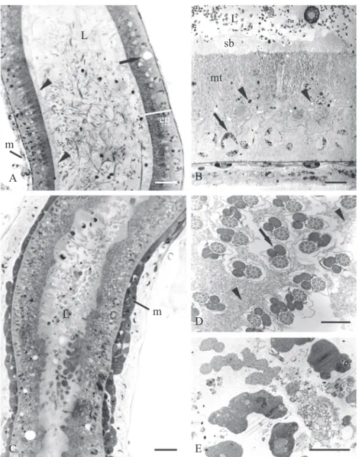
However, as these authors did not examine the wall of these ducts, we cannot know whether the two ducts that leave the accessory glands have cuticle and, therefore, whether they Fig. 4. A - Light micrograph of part of the accessory gland (g) showing its insertion in the deferent duct (dd) and the papilla formed in this region (arrow). B - Transversal section in the traced region of the anterior fi gure, showing the papilla formed (arrow) and the presence of secretion vesicle in glandular epithelium (arrowheads). C - Longitudinal section in the insertion region of the accessory gland (g) in the deferent duct (dd) where the papilla is again observed (arrow) and the glandular epithelium cells with basal nucleus (arrowhead). D - Transversal section of the post-vesicle deferent duct showing the reduced lumen (arrowhead). E - Cross section of the accessory gland showing epithelial cells (ep) with several secretion vesicles (arrowhead) and apocrine secretion release (arrow). F - Cross section of deferent duct in a region after the insertion of the accessory gland showing epithelial cells with basal nucleus (arrowhead) and the striated border (arrow). G - Longitudinal section of the transition between the deferent duct (dd) and the ejaculatory duct (ej). Note the difference between the epithelium of these regions, with the presence of striated border on the deferent duct. H - Cross section of the ejaculatory duct showing the cuticle at the tip of the epithelium (arrow), the spherical cellular nuclei (arrowhead) and the muscular layer (m). Accessory gland = g; deferent duct = dd; epithelium = ep; ejaculatory duct = ej; muscular layer = m. Bars: A = 0.2 mm; B-E = 50 μm; G and H = 15 μm.
g
dd
dd
A
g
dd
dd
g
ep
dd
ej
m B
C D
E F
are ejaculatory ducts or the posterior region of the deferent duct as we have reported for the three Trypargilum species.
Ackonowledgments
The authors thank Dr. Lucio A. O. Campos (UFV) for technical support, the two anonymous reviewers for their careful reading of the manuscripts and suggestions and the CNPq, CAPES and FAPEMIG for funding.
References
Araújo, V.A., U. Zama, C.A. Neves, H. Dolder & J. Lino-Neto. 2005. Ultrastructural, histological and histochemical characteristics of the epithelial wall of the seminal vesicle of mature Scaptotrigona xanthotricha Moure (Hymenoptera, Apidae, Meliponini). Braz. J. Morphol. Sci. 22: 193-201.
Baer, B. 2003. Bumblebees as model organisms to study male sexual selection in social insectes. Behav. Ecol. Sociobiol. 54: 521-534.
Baer, B. & H. Schmid-Hempel. 2000. The artifi cial insemination of bumblebee queens. Insectes Soc. 47: 183-187.
Baer, B. & J.J. Boomsma. 2003. Male reproductive investment and polyandry in fungus-growing ants. Behav. Ecol. 15: 426-432.
Bohart, R.M. & A.S. Menke. 1976. Sphecidae wasps of the world - a generic revision. University of California Press, Berkeley, 695p.
Brockmann, H.J. 1992. Male behavior, coustship and nesting in Trypoxylon (Trypargilum) monteverdeae (Hymenoptera: Sphecidae). J. Kans. Entomol. Soc. 65: 66-84.
Buschini, M.L.T. 2007. Life-history and sex allocation in Trypoxylon (syn. Trypargilum) lactitarse (Hymenoptera; Crabronidae). J. Zool. Syst. Evol. Res. 45: 206-213.
Bushrow, E.S., C.L. Fuller, D.P. Cowan & C.A. Byrd. 2006. Anatomy of the male reproductive system and sperm morphology in the caterpillar-hunting wasp Ancistrocerus antilope (Hymenoptera, Vespidae). Invertebr. Biol. 125: 354-362.
Coville, R.E., C. Griswold & P.L. Coville. 2000. Observations on the nesting biology and behavior of Trypoxylon (Trypargilum) vagulum (Hymenoptera: Sphecidae) in Costa Rica. Pan-Pac. Entomol. 76: 28-48.
Cruz-Landim, C. 2001. Organization of the cysts in bee (Hymenoptera, Apidae) testis: Number of spermatozoa per cyst. Iheringia Ser. Zool. 91: 183-189.
Cruz-Landim, C. & M.A. Cruz-Hofl ing. 1969. Electron microscope observations on honeybee seminal vesicle (Apis mellifera adansonii, Hymenoptera, Apidae). Pap. Avulsos Zool. (Sao Paulo) 14:145-151.
Cruz-Landim, C. & R.P. Dallacqua. 2002. Testicular reabsorption in adult males of Melipona bicolor bicolor Lepeletier (Hymenoptera, Apidae, Meliponini). Cytologia 67: 145-151.
Dallacqua, R.P. & C. Cruz-Landim. 2003. Ultrastructure of the ducts of the reproductive tract of males of Melipona bicolor bicolor Lepeletier (Hymenoptera, Apinae, Meliponini). Anat. Histol. Embryol. 32: 276-281.
Damiens D. & G. Boivin. 2005. Male reproductive strategy in Trichogramma evanescens: Sperm production and allocation to females. Physiol. Entomol. 30: 241-247.
Duvoisin, N., B. Baer & H. Schmid-Hempel. 1999. Sperm transfer and male competition in a bumblebee. Anim. Behav. 58: 743-749.
Ferreira A., F.C. Abdalla, W.E. Kerr & C. Cruz-Landim. 2004. Comparative anatomy of the male reproductive internal organs of 51 species of bees. Neotrop. Entomol. 33: 569-576.
Garcia, M.V.B. & J. Adis. 1995. Comportamento de nidifi cação de Trypoxylon (Trypargilum) rogenhoferi Kohl (Hymenoptera, Sphecidae) em uma fl oresta inundável de várzea na Amazônia Central. Amazoniana 13: 259-282.
Gotwald, W.H. & W. Budette. 1981. Morphology of the internal reproductive system in army ants: Phylogenetic implications (Hymenoptera: Formicidade). Proc. Entomol. Soc. Wash. 83: 72-92.
Hanson, P.E. & A.S. Menke. 1995. The sphecid wasps (Sphecidae). In: The Hymenoptera of Costa Rica, (Hanson PE, Gauld IA, eds). pp. 621-649. Oxford University Press: New York.
Lomholdt, O. 1982. On the origin of the bees (Hymenoptera: Apidae, Sphecidae). Entomologica Scand. 13: 185-190.
Melo, G.A.R. 1999. Phylogenetic relationships and classifi cation of the major lineages of Apoidea (Hymenoptera), with emphasis on the crabronid wasps. Sci. Pap. Nat. Hist. Mus. Univ. Kans. 14: 1-55.
Michener, C.D. 1965. A classifi cation of the bees of the Australian and South Pacifi c regions. Bull. Am. Mus. Nat. Hist. 130: 1-362.
Michener, C.D. 2000. The bees of the world. The Johns Hopkins University Press, Baltimore, 913p.
Schiff, N., A.J. Flemming & D.L.J. Quicke. 2001. Spermatodesmata of the sawfl ies (Hymenoptera: Symphyta): Evidence for multiple increases in sperm bundle size. J. Hym. Res. 10: 119-125.
Tavares, M.G., A.S.T. Irsigler & L.A.O. Campos. 2003. Testis length distinguishes haploid from diploid drones in Melipona quadrifasciata (Hymenoptera: Meliponinae). Apidologie 34: 449-455.
Virkki, N. 1970. Alticial beetles with 128 sperm cell per bumdle. J. Agric. Univ. P. Rico 54: 586-587.
Virkki, N. 1973. Evolution of sperm cell number per bundle in insects. An. Esc. Nac. Cienc. Biol. Mex. 20: 23-54.
Wheeler, D.E. & P.H. Krutzsch. 1992. Internal reproductive system in adult males of the genus Camponotus (Hymenoptera: Formicidae: Formicinae). J. Morphol. 211: 307-317.