ONTOGENETIC PATTERNS AND GENETIC VARIATION IN
Anopheles
(
Anopheles
)
intermedius
CHAGAS, 1908 AND
Anopheles (Anopheles) mattogrossensis
LUTZ & NEIVA, 1911
(DIPTERA: CULICIDAE) IN THE BRAZILIAN AMAZON
RODRIGUEZ, G. A. D., SANTOS, J. M. M. dos and MAIA, J. de F. INPA/CPCS, C.P. 478, CEP 69011-970, Manaus, AM, Brazil
Correspondence to: Joselita M. M. dos Santos, INPA/CPCS, C.P. 478, CEP 69011-970, Manaus, AM, Brazil Received February 18, 1999 – Accepted June 2, 1999 – Distributed May 31, 2000
(With 5 figures)
ABSTRACT
Changes in the expression of genes were observed during development in populations of Anopheles (Anopheles) intermedius and Anopheles (Anopheles) mattogrossensis. Esterase showed seven zones of activity: EST1 was present in all developmental stages of both species; EST2 was observed only in larvae of A. intermedius and larvae and pupae of A. mattogrossensis, with greater activity in pupae; EST3 and EST5 were present in all developmental stages, with greater intensity in larvae; EST4 and EST6 showed weak activity in larvae of A. mattogrossensis and was not found in A. intermedius.
Leucine aminopeptidase showed four zones of activity, of which LAP1 and LAP2 were found in all stages of A. intermedius, with highest activity in larvae, and in larvae only of A. mattogrossensis. LAP3 was detected in all stages of A. mattogrossensis and in larvae only of A. intermedius. LAP4 was detected only in larvae and pupae of A. mattogrossensis, with greater intensity in pupae.α−Glycerophosphate dehydrogenase showed a single zone of activity, detected in older fourth-instar larvae and becoming more intense from the pupal stage onwards.
Key words: Anopheles intermedius, Anopheles mattogrossensis, ontogeny, isozymes, malaria.
RESUMO
Padrões ontogenéticos e variação genética em Anopheles (Anopheles) intermedius Chagas, 1908 e Anopheles (Anopheles) mattogrossensis Lutz & Neiva, 1911 (Diptera: Culicidae) da
Amazônia brasileira
Foram observadas modificações na expressão gênica durante o desenvolvimento de Anopheles inter-medius e Anopheles mattogrossensis do subgênero Anopheles. A esterase revelou sete regiões de ativi-dade, sendo a EST1 presente durante todo o desenvolvimento das duas espécies; a EST2 observou-se apenas em larvas de A. intermedius e em larvas e pupas de A. mattogrossensis, com maior atividade em pupas; as EST3 e EST5 revelaram-se em todos os estágios, mostrando maior intensidade em larvas; as EST4 e EST6 foram detectadas com fraca atividade em A. intermedius durante todos os estágios, porém em A. mattogrossensis apresentaram forte atividade em pupas e fraca em larvas; a EST7 revelou-se apenas em pupas de A. mattogrossensis, não sendo observada em A. intermedius. A leucina aminopep-tidase revelou quatro regiões de atividade, das quais, LAP1 e LAP2 foram observadas durante todo o desenvolvimento de A. intermedius, com maior atividade em larvas, e em A. mattogrossensis reve-ladas apenas em larvas; a LAP3 detectou-se em todos os estágios de A. mattogrossensis e somente em larvas de A. intermedius; a LAP4 foi somente detectada em larvas e pupas de A. mattogrossensis, com maior intensidade em pupas. A α−GPDH apresentou uma única região de atividade, revelada em larvas de 4o estágio velhas e a atividade é intensificada a partir do estágio de pupa, em ambas espécies.
INTRODUCTION
Studies of the electrophoretic patterns of proteins and enzymes during ontogeny of different organisms provide data about differential gene action, enabling researchers to determine the exact moment when a specific gene becomes active in the synthesis of the corresponding enzyme (Wagner & Selander, 1974).
Several studies have pointed out that some enzymes are modified during the development either in terms of band intensity or in terms of the appearence of new forms and the desappearence of others (Wright & Shaw, 1969; O’Brien & MacIntire, 1972; Vedbrat & Whitt, 1975; Santos, 1979; Santos et al., 1985; Santos et al., 1996a,b; Scarpassa, 1988; Scarpassa et al., 1992; Maia & Santos, 1999). These studies, when taken as whole, enable a deeper understanding of how the genes in mosquitoes act and are expressed. The ontogeny changes provide a special insight that can be useful if accompained with the study of the larvicidal effects in nature. Besides, conventional morphology seldom predicts discriminating cha-racters in the first development stages, thus the enzymatic variants are very useful.
According to Vedbrat & Whitt (1975) the appearence and disappearence of a specific enzyme or isozyme may be due to: 1) the substitution of one type of cellular pathway for another; 2) the triggering or stopping of the activity of already existing enzymes; 3) or suppression or expression of a specific gene locus.
In this study we present the electrophoretic patterns of gene manifestation of esterase, leucine aminopeptidase and α-glycerophosphate dehy-drogenase during the development of Anopheles intermedius and Anopheles mattogrossensis of the
Anopheles subgenus
MATERIAL AND METHODS
The mosquitoes used in this study were obtained from natural populations of Janauarilândia in the Amazon State, and in Macapá in the State of Amapá. Individuals were studied by electropho-resis in three different stages of development, i. e., 4 th-instar larvae (young: 12 hours; after young: 24 hours and old: after 30 hours), pupae and adults.
Larvae were reared by the method of Santos et al.
(1981), slightly modified. For isozymes analysis, the material was collected into cuvettes and stored frozen at –70oC. Samples were homogenized with the aid of a glass stick in 15 ul of 0.5% 2-beta-mercaptoetanol (v/v) for larvae and 20 ul for pupae and adults, on concave porcelain plates kept on ice. After the homogenization, thin paper pieces measuring 2 x 2 cm were placed on the plates containing the samples, which were soaked up with Whatman n. 3 filter paper. These papers pieces were inserted vertically into the gel.
Three enzimatic systems were studied: esterases, leucine aminopeptidase and α -glycerophosphate dehydrogenase, at 12.5% starch gel. Buffer solutions and reaction mixtures were prepared by the methods of Santos et al. (1985) and Santos et al. (1996a).
RESULTS
Esterases
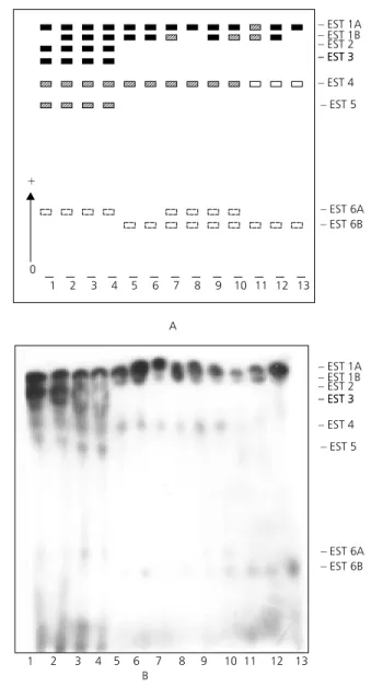
Analysis of esterases during the ontogenetic development showed the occurrence of seven activity bands changing in staining intensity according to each stage (Figs. 1 and 2).
Esterase 1 was detected in all development stages in both species. EST2 was only detected in larvae and EST4 and EST6 were detected in all phases of development, in A. intermedius EST2, EST4 and EST6 were detected in larvaeand pupae with higher activity in pupae in A. mattogrossensis. EST3 and EST5 showed a similar profile in both species during all phases of development with high activity in larvae and low activity in pupae and adults. A more cathodical band in A. mattogrossensis population denominated EST7 only detected in pupae, was also observed.
Most regions observed during development presented variations in the two species even though EST2 was only monomorphic for A. mattogrossensis and EST3 for A. intermedius.
EST1 showed two codominant alleles: ESTI*A
EST2 presented variation of more than two alleles in A. intermedius. EST3 showed two codominant alleles – EST3*A and EST3*B for the
A. mattogrossensis population, yet it was monomorphic for EST3*A allele in A. intermedius. The heterozygote profile presented two intense bands suggesting a monomeric structure for this protein. For EST4, EST5, EST6 and EST7 variation was observed in two alleles, whose heterozygotes showed two bands with the same staining intensity.
Leucine aminopeptidase
The electrophoretic patterns of leucine aminopeptidase isozymes during the ontogeny of
A. intermedius and A. mattogrossensis showed four activity bands (Figs. 3 and 4). LAP1 and LAP2 were observed in all development stages in A. intermedius, with greater activity in larvae. In A. mattogrossensis it was present only in larvae. LAP3 was observed only in A. intermedius larvae, though it was present in all development phases of A. mattogrossensis.
+
+ 0
0
1
1 2
2 3
3 4
4 5
5 6
6 7
7 8
8 9
9 10
10 11
11 12
12 13
13 – EST 1A
– EST 1A – EST 1B
– EST 1B – EST 2
– EST 2 – EST 3
– EST 3 – EST 3
– EST 3 – EST 4
– EST 4 – EST 5
– EST 5 – EST 6A
– EST 6A – EST 6B
– EST 6B A
B
LAP4 it was detected in larvae and pupae of
A. mattogrossensis with larger intensity in the latter. This band was not detected in A. intermedius,
possibly due to the small number of analyzed individuals. A more cathodic band, very close to the origin, with moderate to weak intensity, was observed in both species.
LAP1, which was more anodic, presented greater variation and intensity and stained with three distinct phenotypes – LAP1 A, LAP1 AB
and LAP1 B, in the population of A. intermedius,
and only LAP1 A and LAP1 AB in A. mattto-grossensis.
These phenotypes result from the com-bination of two codominant alleles – LAP1*A and
LAP1*B, the first one being the most frequent. LAP2, which also varied in both species, showed two codominant alleles – LAP2*A and LAP2*B. Heterozygotes profiles show two bands suggesting the enzyme having a monomeric structure. +
+ 0
0 1
1 2
2 3
3 4
4 5
5 6
6 7
7 8
8 9
9 10
10 11
11 12
12 13
13
– EST 1
– EST 2
– EST 3B – EST 3A
– EST 4 – EST 5
– EST 6 – EST 5 – EST 4 – EST 3B – EST 3A – EST 2 – EST 1
– EST 6
– EST 7 – EST 7
A
B
LAP3 also showed polymorphism, with two alleles – LAP3*A and LAP3*B. In the population of A. intermedius the phenotypes LAP3 A, LAP3 AB and LAP3 B were observed, whereas only LAP3 A and LAP3 AB were detected in A. matto-grossensis. The isozyme profile of the hetero-zygote, two equally stained bands, suggest that protein has a monomeric structure.
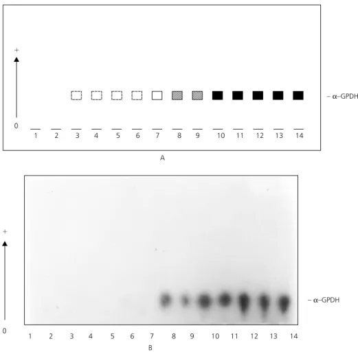
α-Glycerophosphate dehydrogenase
The electrophoretic patterns of α− glycero-phosphate dehydrogenase during ontogenetic development, start to be detected at weak intensity in strongly pigmented 4th-instar. Staining intensity increased in the pupal phase and continued during the adult stage. The same pattern was observed in both species (Fig. 5).
The α−GPDH profile consisted of a single electronegative activity band close to the origin. These data suggest that the genetic control of this enzyme occurs through one monomorphic locus
α−GPDH.
Table I shows comparative study of activity of enzyme analised during ontogenesis in mosquito species – Esterases, LAP and α–GPDH.
DISCUSSION
Changes in gene expression during onto-genetic development of A intermedius and A.
mattogrossensis were observed in the three enzymatic systems. The profile of EST3 and EST5 with the higher larvae activity appear to correspond with a decrease in the metabolic activity rate, metabolism being intensive in larvae, and decreasing for the following stages. These results are in agreement with Scarpassa (1988), who described seven activity bands for Anopheles nuneztovari of Tucurui, noting that esterases of this pattern are related to metabolic activity during development. EST2, present in the larvae of A. intermedius,
appears to be associated with the larval digestive system and metabolism, differing from that of adult. Larvae feeding on micro-organisms, algae, protozoa and similar foods may require these esterases, while adults feeding on sugary substances do not (VedBrat & Whitt, 1975).
EST4 and EST6 of A. intermedius present during all the development stages, could be related to more generalized functions than those recorded in a specific stage of development. It is thought that these esterases are related to the break down of lipids, used as an energy source. Freyvogel et al.
(1968), showed that lipids probably play an important role in blooddigestion for Aedes aegipty
and Anopheles stephensi. The fact that esterases are non-specific in their activity implies that they might function in the regulation of lipids, as suggested by the high levels of esterase found in the stomach epithelium of these mosquitoes. +
0
1 2 3 4 5 6 7 8 9 10 11 12 13 –LAP3 –LAP2 –LAP1
The appearence of EST2, EST4 and EST6 with a higher activity in A. mattogrossensis pupae suggest they may serve a regulatory function on ecdysone levels, postulating that this isozyme form in the pupal stage participates in the metabolism of this hormone. It was suggested by Whitmore et al. (1972) that carboxilesterase may play an important role in regulating the levels of juvenile hormone.
This hormone functions by favoring the expression of larval characteristics and is normally
present in the pupae. According to these authors, the induction of the enzymes is capable of degrading the hormone, a mechanism whereby insects can assure normal metamorphosis. Study of these enzymes may have important implications for those interested in the use of juvenile hormone and its analogs as insect-controlling agents, and suggests that these organisms have a “biochemical pool” that permit the degradation of foreign molecules (such as DDT) and the regulation of endogenous hormones when necessary. +
+ 0
0 1
1 2
2 3
3 4
4 5
5 6
6 7
7 8
8 9
9 10
10 11
11 12
12 13
13
–LAP3A
–LAP3A –LAP3B
–LAP3B –LAP4
–LAP4 –LAP2
–LAP2 –LAP1
–LAP1 A
B
Maia (1997) and Maia & Santos (1999) detected four activity regions for esterases during ontogeny of Anopheles albitarsis; EST1 was seen in the 4th-instar old larvae and pupae, EST2 and EST4 during all developmental stages, and EST3 was only detected in 4th-instar larvae. Similar results were obtained by Santos (1992) and Santos
et al. (1996a,b) who detected five activity regions during ontogeny in Anopheles darlingi: EST1 and EST2 showed the highest staining intensity in the larval stages, whereas EST3 and EST4 were most deeply stained in pupae and adults; EST5 was detected during all developmental stages.
Fig. 5 — Electrophoretic profiles of a-glycerophosphate dehydrogenase during the ontogenetic development of Anopheles mattogrossensis. Starch gel electrophoresis. Tris-phosphate buffer system, pH 7.4. (A) Scheme. (B) Photography. Samples 1 to 5 = 4 th-instar larvae; 6 to 9 = pupae; 10 to 14 = adults.
A
B +
+ 0
0 1
1 2
2 3
3 4
4 5
5 6
6 7
7 8
8 9
9 10
10 11
11 12
12 13
13 14
14
–α–GPDH
Several authors reported multiple bands of esterase activity in mosquitoes and they concluded that these bands are the result of genetic control of more than one locus. Among others we can mention Freyvogel et al. (1968) for Anopheles freeborni, A. stephensi, Ae. aegypti and Culex tarsalis; Vedbrat & Whitt (1975) for Anopheles albimanus and Narang et al. (1979) for Anopheles aquasalis.
The results concerning ontogeny of leucine aminopeptidase in A. intermedius and A. matto-grossensis are similar to those obtained by Maia & Santos (1999) in A. albitarsis. This author detected four activity regions: LAP1 and LAP2 was observed in the 4th-instar larvae; LAP3, was found only in pupae and adults; LAP4 was detected throughout development. However, six activity regions for this enzyme were observed in A. nuneztovari and A. darlingi. For the former species, Scarpassa (1988) and Scarpassa et al. (1992) reported for the population of Tucuruí (Pará) that LAP1 and LAP5 were detected at all developmental stages, LAP2 and LAP4 showed highest activity in larvae and reduced activity in pupae and adults, while LAP3 and LAP6 were characteristic of the last two stages. For A. darlingi Santos (1992) and Santos et al. (1996b) observed more activity of LAP1, LAP2 and LAP5 in larvae, while that for LAP3 was detected in pupae and adults, and LAP4 was restricted to pupae. The LAP4 of A. mattogrossensis detected in this study, may correspond with the LAP4 of A. darlingi revealed in the pupae stage, suggesting it may play a function in larval tissue histolysis at the time when the differentiation of adult tissues occurs.This hypothesis for the greater activity in pupae agrees with Sakai et al. (1969) and Pataryas et al. (1971) who reported the participation of the exopeptidases controlled by the LAP-D locus in the histolysis of larval tissues in the pupal stages of Drosophila melanogaster.
The electrophoretic patterns of α− glycero-phosphate dehydrogenase isozymes during the ontogeny of A. intermedius and A. mattogros-sensis – showing weak staining intensity in 4th-instar larvae with increasing activity in pupae and adults – are similar to those obtained by Narang
et al. (1979), Santos et al. (1996b) and Maia & San-tos (1999). These authors detected only one region, for this enzyme, whose activity increased until the
adult stage of A. aquasalis, A. darlingi and A. albitarsis. However Scarpassa (1988) reported two forms of the enzyme in A. nuneztovari: α–GPDH1 appear in old pupae and adults with more staining intensity in the latter, and is controlled by two codominant alleles whose heterozygotes suggest that the enzyme has a dimeric structure; α–GPDH2 was observed only in larvae, without allelic variation. Similar results were found by Mukiama (1980) who detected two loci of α–GPDH during ontogenesis of Ae. aegypti: α–GPDH1 in larvae and pupae, and α–GPDH2 only in the adult stage. Tadano (1984) reported polymorphism in
Aedes albopictus with one locus that showed activity only in adult phase. Another band less thick than usual was occasionally detected in larvae and pupae. Palabost-Charles (1980) also observed one polymorphic locus with two alleles in D. melanogaster. In this same specie, Wright & Shaw (1969) described three loci in the adult phase and verified that α–GPDH1 was concentrated in the thorax, while α–GPDH3 was found in the head and abdomen. According to these authors, the occurrence of α–GPDH1 in the adult’s thorax, where the flight muscles are located, suggests that the enzyme carries out two functions: 1) regeneration of NAD for the continuous glycolisis, and 2) production of energy needed for flight. The latter hypothesis is supported strongly by the fact that mutants defficient in the activity of α–GPDH cannot begin or maintain the flight (O’Brien & MacIntire, 1972). It is possible that the α–GPDH locus of the species treated in this paper corresponds to the α–GPDH1 locus of D. mela-nogaster, because the greatest activity of the enzyme was observed in the adult stage. On the other hand, this enzyme was polymorphic in some organisms, suggesting that α–GPDH it is not involved in the production of energy. Likewise, Zera (1981) reported high levels of polymorphism of this enzyme in aquatic hemipterous, noting that this may be the result of reductive selection pressure about this locus.
TABLE 1
Comparative study of activity of Esterases, LAP and ααααα–GPDH during ontogenesis in mosquito species.
Enzyme activity in ontogenesis Enzym Isozyme
L P A
Species References
+ + +
A. nuneztovari, A. intermedius and A. mattogrossensis
Scarpassa (1988) and Diaz Rodriguez (1998). 1
+ + − A. albimanus, A. darlingi and A. albitarsis
Vedbrat & Whitt (1975), Santos et al. (1996a, b) and Maia & Santos (1999).
+ − − A. intermedius Diaz Rodriguez (1998) + + − A. nuneztovari, A. darlingi,
and A. mattogrossensis
Scarpassa (1988), Santos et al.
(1996a, b) and Diaz Rodriguez (1998).
2
+ + + A. albimanus and A.
albitarsis
Vedbrat & Whitt (1975) and Maia Santos (1999).
+ − − A. nuneztovari Scarpassa (1988).
+ + − A. albitarsis Maia & Santos (1999). + + + A. intermedius and A.
mattogrossensis Diaz Rodriguez (1998)
− + − A. albimanus Vedbrat & Whitt (1975). 3
− + + A. darlingi Santos et al. (1996a, b). + + − A. nuneztovari and A.
mattogrossensis
Scarpassa (1988) and Diaz Rodriguez (1998). + + +
A. albimanus, A. intermedius and A. albitarsis
Vedbrat & Whitt (1975), Diaz Rodriguez (1998) and Maia Santos (1999).
4
− + + A. darlingi Santos et al. (1996b)
+ − − A. albimanus Vedbrat & Whitt (1975). 5
+ + +
A. nuneztovari, A. darlingi,
A. intermedius and A. mattogrossensis
Scarpassa (1988), Santos et al.
(1996a, b) and Diaz Rodriguez (1998).
+ + − A. nuneztovari and A.
mattogrossensis
Scarpassa (1988) and Diaz Rodriguez (1998). 6
+ + + A. albimanus and A.
intermedius
Vedbrat & Whitt (1975) and Diaz Rodriguez (1998).
+ − − A. albimanus Vedbrat & Whitt (1975)
+ + + A. nuneztovari Scarpassa (1988)
7
− + − A. mattogrossensis Diaz Rodriguez (1998) 8 + + + A. albimanus Vedbrat & Whitt (1975). Esterase
9 − + − A. albimanus Vedbrat & Whitt (1975). + − − A. mattogrossensis and A.
albitarsis
Diaz Rodriguez (1998) and Maia & Santos (1999).
LAP 1
+ + + A. nuneztovari, A. darlingi, and A. intermedius
Scarpassa et al. (1992), Santos et al.
REFERENCES
DIAZ RODRIGUEZ, G. A., 1998, Padrões Isoenzimáticos e Variabilidade genética em Anopheles (Anopheles) intermedius chagas, 1908 e Anopheles (Anopheles) mattogrossesnsis Lutz & Neiva, 1911 (Deptera: Culicidae) da Amazônia Brasileira. Dissertação de Mestrado. INPA/VA, Manaus, AM, 107p.
FREYVOGEL, T. A., HUNTER, R. L. & SMITH, E. M., 1968, Nonspecific Esterases in mosquitos. J. Histochem. Cytochem, 16(12): 765-790.
MAIA, J. F., 1997, Variabilidade Genética em populações naturais de Anopheles (Nyssorhynchus) albitarsis Lynch-Arribálzaga, 1878 (Diptera: Culicidae).
Dissertação de Mestrado, INPA, Manaus, AM, 118p.
MAIA, J. F. & SANTOS, J. M. M., 1999, Padrões onto-genéticos das esterases, leucina aminopeptidase e a-glicerofosfato desidrogenase em Anopheles (Nysso-rhynchus) albitarsis Lynch-Arribálzaga, 1878 (Dipte-ra: Culicidae). Acta Amazonica, 29(1): 135-144. MUKIAMA, T. K., 1980, Comparative dehydrogenase
activity during the ontogenesis of the yellow fever mosquito Aedes (Stegomyia) aegypti L. (Diptera: Culicidae). Comp. Biochem. Physiol., 67B: 6 5 9 .
NARANG, S. K., KITZMILLER, J. B., GALLER, R., RIOS, I. R. & NARANG, N., 1979, Genética de po-pulações de anofelinos. III. Análise eletroforética de
Anopheles aquasalis. Rev. Brasil. Pesq. Med. e Biol.,
12(4-5): 303-309.
TABLE 1 (continued)
Enzyme activity in ontogenesis Enzym Isozyme
L P A
Species References
+ − −
A. darlingi, A. mattogrossensis and A. albitarsis
Santos et al. (1996b), Diaz Rodriguez (1998) and Maia Santos (1999).
2
+ + + A. nuneztovari and A.
intermedius
Scarpassa et al. (1992) and Diaz Rodriguez (1998).
+ − − A. intermedius Diaz Rodriguez (1998). + + + A. mattogrossensis Diaz Rodriguez (1998). 3
− + + A. nuneztovari, A. darlingi, and A. albitarsis
Scarpassa et al. (1992), Santos et al.
(1996b) and Maia & Santos (1999). + + − A. mattogrossensis Diaz Rodriguez (1998). + + +
A. nuneztovari, A. intermedius and A. albitarsis
Scarpassa et al. (1992), Diaz Rodriguez (1998) and Maia Santos (1999).
4
− + − A. darlingi Santos et al. (1996b). + + + A. nuneztovari Scarpassa et al. (1992). 5
+ − − A. darlingi Santos et al. (1996b). + + + A. darlingi Santos et al. (1996b). LAP
6
− + + A. nuneztovari Scarpassa et al. (1992).
+ + +
A. darlingi, A. intermedius,
A. mattogrossensis and A. albitarsis
Santos et al. (1996b), Diaz Rodriguez (1998) and Maia Santos (1999).
+ + − Ae. aegypti Mukiama (1980)
− + + A.aquasalis and A.
nuneztovari
Narang et al. (1979) and Scarpassa (1988).
1
− − + Ae. albopictus Tadano (1984).
+ − − A. nuneztovari Scarpassa (1988)
αGPDH
2
− − + Ae. aegypti Mukiama (1980)
O‘BRIEN, S. J. & MACINTYRE, R. J., 1972, The α– Glycerophosphate Cycle in Drosophila melanogaster. I. Biochemical and Developmental Aspects. Biochem. Genet., 7: 141-161.
PALABOST-CHARLES, L., 1980, Maintenance Mecha-nism of Polymorphism at the α–Gpdh Locus in
Drosophila melanogaster. Biochem. Genet., 18(9-10): 905-913.
PATARYAS, H. A., HARITOS, A. A. & GELTI-DOUKA, H., 1971, Esterase and Leucine Aminopeptidase Zy-mograms in the Indian Meal Moth Plodia inter-punctella During Development. Experientia, 27(3): 344-345.
SAKAI, R. K., TUNG, D. A. & SCANDALIOS, J. G., 1969, Developmental Genetic Studies of Aminopep-tidase in Drosophila melanogaster. Molec. Gen. Ge-netics, 105: 24-29.
SANTOS, J. M. M., 1979, Aspectos biológicos e isoenzi-máticos de Anopheles (Nyssorhynchus) darlingi Root, 1926 (Diptera: Culicidae). Dissertação de Mestrado,
INPA/FUA, 87p.
SANTOS, J. M. M., 1992, Variabilidade Genética em Po-pulações Naturais de Anopheles (Nyssorhynchus) darlingi Root, 1926 (Diptera: Culicidae). Tese de Doutorado, INPA/FUA, 150p.
SANTOS, J. M. M., CONTEL, E. P. B. & KERR, W. E., 1981, Biologia de anofelinos amazônicos 1. Ciclo biológico, postura e estádios larvais de Anopheles darlingi Root, 1926 (Diptera: Culicidae) da Rodovia Manaus–Boa Vista. Acta Amazonica, 11(4): 789-797. SANTOS, J. M. M., CONTEL, E. P. B. & KERR, W. E., 1985, Biology of Amazonian Mosquitoes. III. Es-terase Isozymes in Anopheles darlingi. Acta Ama-zonica, 15(1-2): 167-177.
SANTOS, J. M. M., TADEI, W. P. & CONTEL, E. P. B., 1996a, Electrophoretic analysis of 11 enzymes in natural populations of Anopheles (N.) darlingi Root, 1926 (Diptera: Culicidae) in the amazon region. Acta Amazonica, 26(1/2): 96-114.
SANTOS, J. M. M., TADEI, W. P. & CONTEL, E. P. B., 1996b, Biology of Amazonian Anopheline. XX. Onto-geny of esterases, leucine aminopeptidase and a-glyce-rophosphate dehydrogenase in Anopheles ( Nysso-rhynchus) darlingi Root, 1926 (Diptera: Culicidae). Rev. Brasil. Biol., 56(3): 591-598.
SCARPASSA, V. M., 1988, Estudo do ciclo biológico e de isoenzimas na ontogênese de Anopheles (Nyssorhyn-chus) nuneztovari Gabaldón, 1940 (Diptera: Culcidae).
Dissertação de Mestrado, INPA/FUA, Manaus, AM, 1 7 2 p .
SCARPASSA, V. M., TADEI, W. P. & CONTEL, E. P. B., 1992, Biologia de Anofelinos amazônicos. XV. Leu-cina aminopeptidase em Anopheles (Nyssorhynchus) nuneztovari: ontogenia e variação genética. Acta Amazonica, 22(2): 229-238.
TADANO, T., 1984, Linkage Studies on a-Glycerophos-phate and Isocitrate Dehydrogenases in Aedes albo-pictus (Diptera: Culicidae). Biochem.Genet., 22(7-8): 587-595.
VEDBRAT, S. S. & WHITT, G. S., 1975, Isozyme ontog-eny of the mosquito, Anopheles albimanus. In: Iso-zymes, III. Developmental Biology. New York, Acade-mic Press., pp. 131-143.
WAGNER, R. P. & SELANDER, R. K., 1974, Isozymes in insects and their significance. Ann. Rev. Entomol.,
19: 117-138.
WHITMORE, D. JR., WHITMORE, E. & GILBERT, L. I., 1972, Juvenile Hormone Induction of Esterases: A Mechanism for the Regulation of Juvenile Hormone Titer. Proc. Nat. Acad. Sci., 69(6): 1592-1595. WRIGHT, D. A. & SHAW, C. R., 1969, Genetics and
Ontogeny of a-glicerophosphate dehydrogenase iso-zymes in Drosophila melanogaster. Biochem. Genet.,
3: 343.
ZERA, A. J., 1981, Extensive Variation at the α -Glycero-phosphate Dehydrogenase Locus in Species of Waterstriders (Gerridae: Hemiptera). Biochem. Genet.,