629 629 629 629 629 Mem Inst Oswaldo Cruz, Rio de Janeiro, Vol. 98(5): 629-635, July 2003
Location of Ribosomal Genes in the Chromosomes of
Anopheles
darlingi
and
Anopheles nuneztovari
(Diptera, Culicidae) from the
Brazilian Amazon
Míriam Silva Rafael/
+, Wanderli Pedro Tadei, Shirlei Maria Recco-Pimentel*
Coordenação de Pesquisas em Ciências da Saúde, Instituto Nacional de Pesquisas da Amazônia, Av. André Araújo 2936, 69083-000 Manaus, AM, Brasil *Departamento de Biologia Celular, Instituto de Biologia, Universidade Estadual de Campinas,
Campinas, SP, Brasil
Fluorescence in situ hybridization of Anopheles darlingi and A. nuneztovari demonstrated nucleolar organizer region activity at the end of the fourth larval instar, when the nucleolar organizer regions underwent gradual condensation. The heteromorphic sex chromosomes showed intraindividual size variation in the rDNA blocks located in the pericentromeric region and this coincided with the location of constitutive heterochromatin (C-banding).
Key words: Anopheles darlingi - Anopheles nuneztovari fluorescence in situ hybridization rDNA genes -nucleolar organizer region - sex chromosomes
Anopheles (Nyssorhynchus) darlingi Root, 1926and
A. (N.) nuneztovari Gabaldón, 1940 are important vectors of human malaria parasites. In Brazil, A. darlingi is the main vector of malaria, especially in the Amazon region, where more than 97% of all cases in the country occur (Tadei & Dutary-Thatcher 2000). In Colombia and Venezuela, the main vector is A. nuneztovari (Kitzmiller et al. 1973).
Intraspecific variation involving constitutive hetero-chromatin in mitotic chromosomes is a general phenom-enon in many groups of animals. Such structural changes result in the loss or gain of heterochromatin, as occurs in the chromosomes of Anopheles species in Thailand and Southeast Asia(Baimai et al. 1996, Baimai 1998). In A. nuneztovari and A. darlingi, which have karyotypes with 2n = 6 chromosomes, with a pair of submetacentric auto-somes (III), one pair of metacentric (II) and one pair of sex heteromorphic (XX/XY) chromosomes (Rafael & Tadei 1998), there is intraspecific variation in the heterochro-matic blocks located around the centromeric region in the sex chromosomes and autosomes (Rafael & Tadei 2000). Based on morphological variations (Faran & Linthicum 1981), geographic distribution, isoenzymes patterns (Rosa-Freitas et al. 1992, (Rosa-Freitas-Sibajev et al. 1995, Santos et al. 1999, Manguin et al. 1999), behavior, mitochondrial DNA sequences (Rosa-Freitas et al. 1992, Freitas-Sibajev et al. 1995, Conn et al.1999), random amplified polymorphic DNA (RAPD) patterns and internal transcribed spacer 2 (ITS2) region profiles from rDNA markers (Manguin et al. 1999) of different populations, A. darlingi is considered
This research was supported by PPG-7/Finep, Ministério da Ciência e Tecnologia, Brasília, DF, Brazil and Projeto Institucional do Inpa-PPI 3190, Manaus, AM, Brasil. +
Corresponding author. Fax: +55-92-643.3061. E-mail: msrafael@inpa.gov.br
Recived 8 March 2003 Accepted 7 May 2003
to be a single species. However, comparisons of chromo-somal variability among A. darlingi populations from Northern and Southern Brazil have revealed a higher fre-quency of heterozygous inversions in the northern popu-lations (Kreutzer et al.1972, Tadei et al.1982). In contrast, environmental and isoenzymatic variations (Fritz et al. 1995, Scarpassa et al. 1999), ITS2 (Fritz et al. 1994, Onyabe & Conn 1999) and mitochondrial DNA marker (Conn et al. 1998, Scarpassa et al.2000) analyses have shown impor-tant geographic interpopulational differences in A. nuneztovari, and it is still not clear whether A. nuneztovari
is a single variable species or a complex of species. The fixed inversion on the X-chromosome in Brazilian population of A. nuneztovari differed from that of Vene-zuelan and Colombian populations (Kitzmiller et al. 1973). This fixed inversion, the frequencies of inversions in au-tosome II, and the presence of a chromocenter were used by Conn (1990) and Conn et al.(1993) to identify the cytotypes A, B and C for A. nuneztovari. Cytotype A showed one fixed inversion on the X-chromosome (Bra-zilian Amazon), cytotype B had inversion 2La (Venezuela) and cytotype C had inversions 2b, 2Lc and 2Ld (Colom-bia and Western Venezuela).
630 630 630 630
630 rDNA of Two Neotropical Malaria Vectors • Míriam Silva Rafael et al.
(Parise-Maltempi & Avancini 2001). In mosquitoes, the rRNA genes are located within and around the hetero-chromatic regions, especially on the sex chromosomes in the subfamily Culicinae and in some species of the sub-family Anophelinae (Marchi & Pili 1994).However, there are no in situhybridization studies for A. darlingi and A. nuneztovari (both in the subgenus Nyssorhynchus).
In the present study of A. darlingi and A. nuneztovari
from Manaus (AM) and Macapá (AP) in the Brazilian Amazon, we investigated the NOR in the polytene chro-mosomes and the relationship of the NOR to the constitu-tive heterochromatin (C-banding) in the heteromorphic sex pair. We also compared the rDNA genes of both spe-cies using an rDNA probe (pDm 238 – D. melanogaster) in fluorescence in situ hybridization. The use of this method to map the ribosomal genes in the chromosomes of these mosquitoes should allow the identification of chromosomal signals which may be useful in differentiat-ing populations of both species. The FISH method should also be useful to separate the A. albitarsis complex, such as A. marajoara, which was incriminated as a primary malaria vector in Macapá (Conn et al. 2002).
MATERIALS AND METHODS
Mosquitoes rearing - From July 1998 to August 2000, individuals of A. darlingi and A. nuneztovari were col-lected at three locations in the Amazon region: Manaus (3o08’S, 60o01’W), and Novo Airão (1o56’S, 61o22’W) at Manairão locality, both in the state of Amazonas, and Macapá (0o02’S, 51o03’W) at Manuarum locality, in the state of Amapá. Adult females were captured from 18:30 to 20:30 h using oral aspirators while they were feeding on cattle, resting on stable walls or biting humans. Wild-caught adults were transported in moist chambers to the laboratory of malaria vectors at the Instituto Nacional de Pesquisas da Amazônia. Females were confined individu-ally in plastic cups for egg laying. The offspring were reared to the fourth instar larvae and prepupal stage. Mor-phological identification of the specimens was according to Forattini (1962) and Faran and Linthicum (1981).
Preparations of polytene chromosomes - Fourth in-star (early and middle stages) larvae and prepupae of fe-males A. darlingi and A. nuneztovari were used to pre-pare polytene chromosomes as described by French et al. (1962) and Kumar and Collins (1994). In some cases, during squashing of the salivary glands, the nucleolar contents and granular and fibrous compounds became dissociated and were lost. This loss of material, together with the fact that there were fewer specimens from Novo Airão than from Manaus, meant that there were fewer slide preparations from the former location. In all 71 slides con-taining well-spread polytene chromosomes were selected for in situ hybridization. In A. darlingi we analyzed 5, 21 and 18 slides from Novo Airão, Manaus and Macapá, respectively. In A. nuneztovari we analyzed 15 and 12 slides from Manaus and Macapá, respectively.
Preparations of mitotic chromosomes - Mitotic chro-mosomes in air-dried neuroblast preparations from fourth instar larvae (early and middle stages) and prepupae (F1) were examined using the techniques described by Imai et al. (1988). Thirty-nine treated and fixed slide preparations
containing 4 to 5 well-spread metaphase plates of A. darlingi and A. nuneztovari were used in the fluores-cence in situ hybridizationassays. We analyzed 2, 11 and 9 slides of A. darlingi from Novo Airão, Manaus and Macapá, respectively; and 7 and 10 slides of A. nuneztovari from Manaus and Macapá.
Fluorescence in situ hybridization - FISH of
poly-tene and mitotic chromosomes was done according to Viegas-Péquignot (1992), using a cloned rDNA probe (pDm
238) from D. melanogaster. Polytene and mitotic chromo-some preparations were pretreated with RNase (100 µg/ ml) at 37oC for 1 h. The slides were then dipped in 2X SSC for 2 min, dehydrated in an ethanol series for 2 min in each concentration, air-dried, denatured in 70% formamide (in 2X SSC) at 70oC for 2 min, and dehydrated in cold 50%, 75% and absolute ethanol. The rDNA probe was labelled with biotin-14-dATP using standard nick translation (GIBCO BRL) procedures. The labelled probe was dena-tured at 100oC for 10 min and added to the slides, which were then incubated in a humid chamber at 37o C for 36 h. Excess probe was removed by washing the slides twice in 50% formamide (in 2X SSC) and twice in 2X SSC (5 min per wash). The slides were subsequently blocked with the first antibody (antibiotin) in the presence of bovine se-rum albumin in a moist chamber at 37oC for 45 min. To detect the fluorescence signals, the slides were washed in PBT (0.4% of BSA 30% w/v, 0.1% of tween 20 and PBS buffer 1X) and then incubated with the second antibody (IgG-FITC - 1:100 v/v in PBT) in a moist chamber at 37oC for 45 min. After washing in PBT, the slides were counter-stained with propidium iodide (2 µg/ml), which was then removed by a quick wash. The slides were mounted with anti-fading (Vectashield) and examined using Zeiss Axioplan and Olympus fluorescence microscopes. Pho-tographs were obtained using 400-ASA color negative Kodak film.
RESULTS
Polytene chromosomes - The rDNA probe (pDm 238
-D. melanogaster) physically mapped the NOR at the proxi-mal end (5C region) of 58 salivary X-chromosomes of A. darlingi from Novo Airão, Manaus and Macapá. In A. nuneztovari, the genes were located in the 3C region of the right arm and the 4B region of the left arm in 67 X-chromosomes from Manaus and Macapá. In both spe-cies, the NOR was associated with the nucleolus through the presence of chromatin fibers and ribosomal cistrons protruding from the NOR, the activity of which revealed a gradual condensation on the X-chromosomes. The rDNA probe did not map to any other chromosome.
The early fourth instar larvae of A. darlingi from Novo Airão, Manaus and Macapá showed analogous, decondensed NOR with thin filaments of rDNA (Fig. 1A, B), whereas in the middle of fourth instar larvae, the NOR became less active (Fig. 1C). When the larvae reached the prepupal stage, the NOR became condensed (Fig. 1D).
631 631631 631631 Mem Inst Oswaldo Cruz, Rio de Janeiro, Vol. 98(5), July 2003
Mitotic chromosomes -The NOR was detected by FISH in the pericentromeric region of 63 sex chromosomes of A. darlingi from Novo Airão, Manaus and Macapá. Fifty-one nuclei of A. nuneztovari from Manaus and Macapá. The location of the rRNA genes showed similar intraindividual variation on the sex pairs and coincided with the centromeric heterochromatic blocks on these chromosomes. In both species, the fluorescence signals were greater in the X1 chromosome and smaller in the X2 chromosome. The X1 chromosome showed strong sig-nals in approximately 80% of the nuclei whereas 70% of the X2 chromosome had weak fluorescence signals (Figs 3, 4). The Y-chromosome was partially or almost totally
Fig. 1: polytene chromosomes of Anopheles darlingi after fluores-cence in situ hybridization with an rDNA (pDm 238) probe. Similar nucleolar organizer region (NOR) activity with different degrees of condensation was present in the 5C region (arrow) of the X chro-mosomes in samples from Manaus (A, D), Novo Airão (B) and Macapá (C). A, B: larvae at the beginning of the fourth instar showing decondensed NOR; C: larvae in the middle of the fourth stage, showing less NOR activity and rDNA filaments dispersed throughout the nucleolus; D: prepupae, with condensed NOR. Scale: 10 µm
Fig. 2: fluorescence in situ hybridization of Anopheles nuneztovari
632 632 632 632
632 rDNA of Two Neotropical Malaria Vectors • Míriam Silva Rafael et al.
mapped (Figs 3C, 4C). All interphase nuclei exhibited one or two nucleoli with fluorescence.
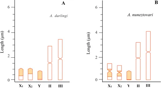
Fig. 5 shows the percentage of ribosomal cistrons mapped on the sex chromosomes of A. darlingi (A) from Novo Airão, Manaus and Macapá, and A. nuneztovari
(B) from Manaus and Macapá.
Fig. 3: fluorescence in situ hybridization of metaphase chromo-somes of Anopheles darlingi using an rDNA (pDm 238) probe. Analogous intraindividual rDNA gene markings were found in samples from Manaus (A), Novo Airão (B) and Macapá (C, D). A, B: fluorescence signals were larger in the X1 chromosomes and smaller in the X2 chromosomes (thin arrows); C: fluorescence sig-nals were weak in the X1 and X2 chromosomes (thin arrows); D: partially mapped Y chromosome (thin arrow). Scale: 10 µm
633 633633 633633 Mem Inst Oswaldo Cruz, Rio de Janeiro, Vol. 98(5), July 2003
DISCUSSION
Active NOR were detected on the polytene X-chromo-some of A. darlingi and A. nuneztovari in early fourth in-star larvae. In prepupae, the NOR became condensed and the nucleolus disappeared. The longest period in the de-velopment of both species, from eggs to adults, corre-sponds to the transformation of the fourth instar larvae into pupae (Santoset al.1981, Scarpassa & Tadei 1990). According to these authors, this period may be related to the levels of the insect hormone ecdysone. The effects of this moulting hormone on protein and rRNA synthesis in insects have been shown in cytological studies such as in
Rynchosciara (Amabis 1972). Salivary gland chromosomes from fourth instar larvae of S. ocellaris treated with ecdys-one showed very active NOR that were abruptly repressed when pupation was induced (Dessen & Perondini 1985). The structural regulatory mechanism of NOR may be in-fluenced by the functional stage of cells, and this can af-fect chromatin condensation and nucleolus size, as occurs in Drosophila (Bicudo 1982), Rynchosciara (Amabis 1972) and S. ocellaris (Dessen & Perondini 1991). A similar phe-nomenon probably occurs in Anopheles species, since the degree of NOR condensation in the salivary gland chro-mosomes of A. subpictus, the vector of malaria in India (Chaudhry 1986), varies in fourth instar larvae and pre-pupae. An analogous situation was reported by Tiepolo et al. (1974) who observed divergent DNA synthesis in hybrids of A. atroparvus and A. labranchiae. In A. darlingi and A. nuneztovari, the differences in the intensity of protein and RNA synthesis probably reflect the functional stage of the cells and the effect of hormones such as ecdysone on NOR transcriptional activity.
The presence of NOR in a single chromosome was detected using rRNA gene probes in Xenopus (Pardue et
al.1970), Drosophila of the mulleri/D. arizonensis com-plex (Bicudo 1982), S. occelaris (Dessen & Perondini 1991),
A. labranchiae and Orthopodomyia pulcripalpis (Marchi & Pilli 1994). Centromeric heterochromatin occurs at a single locus in X-chromosomes of A. gambiae and A. arabiensis (Collins et al. 1987). A. merus, A. melas,and A. quadrimaculatus have a second rDNA locus, probably located on the Y chromosome (Collins et al.1989). Like-wise, the D. melanogaster rDNAprobe mapped both the NOR in a single X chromosome (5C region) of A. darlingi
and the centromere (3C and 4B regions) of A. nuneztovari. In A. darlingi and A. nuneztovari, the NOR was puffed and exhibited chromatin fibers and fluorescence grains protruding from within the nucleolar mass.
The transcriptional activity of RNA genes and varia-tions in the intensity of NOR staining have been observed in the X chromosomes of D. hydei (Pardue et al. 1970), D. mulleri/D. arizonensis (Bicudo 1982) and the medfly C. capitata (Bedo & Webb 1989). In O. pulcripalpis, 18S and 28S rRNA probes showed the NOR in the 4C band of polytene chromosome I and in the nucleolus of polytene chromosomes of A. labranchiae. The 4C band is associ-ated with the nucleolus through fibers and grains that can be seen extending from the band into the nucleolus. This association may be related to the transcriptional ac-tivity in the NOR (Marchi & Pili 1994). A similar pattern of hybridization was observed in A. darlingi from Novo Airão, Manaus and Macapá, and A. nuneztovari from Manaus and Macapá.
The internal transcribed 2 (ITS2) variant, a spacer which separates the 18S from 5.8S rDNA sub-units, was ana-lyzed in A. nuneztovari mosquitoes by Onyabe and Conn (1999). According to these authors, multiple variants may be present on a single chromosome of this species, re-gardless of sex. These authors also suggested that the Fig. 5: map of the sex chromosomes of Anopheles darlingi (A) from Novo Airão, Manaus and Macapá and A. nuneztovari (B) from Manaus and Macapá based on the pDm 238 probe. Y-axis: average length of the chromosomes. X-axis: sex chromosomes (X1, X2, Y) and autosomes (II, III). The dashed area of the sex chromosomes of A. darlingi (A) corresponds to the average of the percentage of ribosomal cistrons mapped on the X1 (65.0% ± 13.3), X2 (48.1% ± 12.6) and Y (94.2% ± 6.4) chromosomes. The dashed area of the sex chromosomes of A. nuneztovari (B) corresponds to the average of the percentage of ribosomal cistrons of the long (64.8% ± 12) and short (25.7% ± 17.8) arms of the X1 chromosome, of the long (49.7% ± 20) and short (21% ± 15.1) arms of the X2 chromosome, and of the Y chromosomes (96.5% ± 4.2).
Y II III
X1 X2 X1 X2 Y II III
L
ength (
µ
m)
Le
ng
th
(µm)
A. nuneztovari
A B
A. darlingi
6
4
2
0
6
4
2
634 634 634 634
634 rDNA of Two Neotropical Malaria Vectors • Míriam Silva Rafael et al.
rDNA of A. nuneztovari may not be exclusively X-linked, thus resembling that of A. melas, A. merus, A. quadri-annulatus (Collins et al.1989) and A. quadrimaculatus
(Kumar & Rai 1990). These findings could be tested in the polytene chromosomes of A. nuneztovari by in situ hy-bridization, using rDNA-specific probes.
An rDNA (pDm 238 – D. melanogaster) probe was used to determine the relationship between the NOR and constitutive heterochromatin (C-banding) in A. darlingi
and A. nuneztovari. This probe mapped the X (X1 and X2) and Y chromosomes, whose gene sites coincided with the constitutive heterochromatin (C-banding) in the pericentromeric region and showed a conspicuous asso-ciation with the NOR of both species. This gene sites agree with the data for X1 acrocentric chromosomes of A. darlingi from Manaus and Macapá (Rafael & Tadei 2000). In these chromosomes, the constitutive heterochromatin was located in the centromeric region which extended to 1/3 of this chromosome whereas the X2 chromosomes showed fewer signals. In A. nuneztovari from Manaus, the intraspecific variations in the heterochromatic block signals in the submetacentric X1 (longer) and the X2 (shorter) chromosomes were the same as those of A. nuneztovari from Macapá.
Identical ribosomal gene locations were obtained in the centromeric X chromosome and in the Y chromosome of D. melanogaster, D. simulans and D. hydei (reviewed in Bicudo 1985). In 20 species of mosquitoes belonging to eight genera of the subfamily Culicinae, the rDNA was located on a single mitotic chromosome which had rRNA genes dispersed along the length of all three pairs of theese chromosomes (Kumar & Rai 1990, 1991). In contrast, in
A. petragnani, A. hispaniola (Marchi & Pili 1994), A. gambiae (Kumar & Collins 1994) and A. stephensi
(Redfern 1981), the rRNA genes were located on the sex pairs, mainly within heterochromatic regions (C-banding) or adjacent to them.
As in individuals with different rDNA amounts de-tected in populations of Anopheles, Culex, Aedes and
Orthopodomyia (Marchi & Pili 1994), the oriental Anoph-eles species are prone to variations in the content of con-stitutive heterochromatin in their mitotic chromosomes, especially in the sex pairs with consequent differences in size and shape (Baimai et al. 1996, Baimai 1998). In dipteran insects, constitutive heterochromatin may account for more than 60% of the length of the X chromosome (Baimai 1998). The fluorescence signals of rDNA around the cen-tromeric heterochromatic blocks of the sex pairs of A. darlingi and A. nuneztovari indicate that the rRNA genes are more dispersed in X1 than in X2 chromosomes. The differences of the fluorescence signals of the sex pairs of both species suggest intraindividual variation in the dis-tribution of rDNA copies associated with constitutive heterochromatin.
Intraspecific variation in the number of copies of rRNA genes has been reported in mosquitoes (Kumar & Rai 1990, 1991). This polymorphism probably results from re-combination and unequal crossing-over during the meio-sis. In Anopheles, the partially homologous X and Y chro-mosomes may undergo recombination and unequal cross-ing-over, and this may lead to differences in the arm length
and heterochromatin content of these chromosomes (Baimai & Traikpavasin 1987, Marchi & Mezzanotte 1990). The heterochromatin on eukaryotic chromosomes has a significant role in the regulation and concerted evolution of the genome and may serve similar functions in the chro-mosomes of A. darlingi and A. nuneztovari. It is uncer-tain whether the acrocentric X chromosome of A. darlingi
originated from the X metacentric chromosome. The meta-centric-acrocentric chromosomes may originate via heterochromatinization of the metacentric-acrocentric arm and their successive loss. In A. nuneztovari, which shows polymorphism in the size of the X1 (longer) and X2 (shorter) submetacentric chromosomes, similar hete-rochromatinization may occur. Detailed investigations of the dynamics of heterochromatin loss, particularly at the molecular level, and of its evolutionary significance in the genome of A. darlingi and A. nuneztovari, remain to be undertaken.
ACKNOWLEDGMENTS
To Klélia Aparecida de Carvalho and Luciana Bolsoni Lourenço (Departamento de Biologia Celular, Unicamp, SP, Brazil) for technical assistance.
REFERENCES
Amabis JM 1972. Indução e Repressão de “Puffs” em Rhynchosciara, Memórias de Mestrado, USP, São Paulo, 40 pp.
Baimai V 1998. Heterochromatin accumulation and karyotypic evolution in some dipteran insects. Zool Studies37: 75-88. Baimai V, Traipakvasin A 1987. Intraspecific variation in sex chromosomes of species B of the Anopheles dirus complex in Thailand. Genome 29: 401-404.
Baimai V, Treesucon A, Kijchalao U 1996. Heterochromatin variation in chromosome X in a natural population of Anoph-eles willmori (Diptera: Culicidae) of Thailand. Genetica 97: 235-239.
Bedo DG, Webb GC 1989. Conservation of nucleolar structure in polytene tissues of Ceratitis capitata (Diptera: Tephri-tidae). Chromosoma 98: 443-449.
Bicudo HEMC 1982. Silver staining and the nucleolar organiz-ing activity in Drosophila species of the mulleri complex and their hybrids. Rev Bras Genet5: 31-50.
Bicudo HEMC 1985. Organizadores de nucléolos em insetos.
Ciênc Cult 37: 430-439.
Chaudhry, S 1986. Sex chromosome in mosquitoes. I. NOR associated gene activation and asynapsis in the salivary X-chromosome of Anophelessubpictus (Culicidae: Diptera).
Caryologia39: 211-215.
Collins FH, Mendez MA, Rasmussen MO, Mehaffey PC, Besansky NJ, Finnerty V 1987. A ribosomal RNA gene probe differentiates member species of the Anopheles gambiae complex. Am J Trop Med Hyg37: 37-41. Collins FH, Paskewitz SM, Finnerty V 1989. Ribosomal RNA
genes of the Anopheles gambiae species complex. Adv Dis Vector Res 6: 1-28.
Conn J 1990. A genetic study of the malaria vector Anopheles nuneztovari from Western Venezuela. J Am Mosq Contr Assoc6: 400-405.
Conn J, Puertas YR, Seawright JA 1993. A new cytotype of
Anopheles nuneztovari from Western Venezuela and Co-lombia. J Am Mosq Contr Assoc9: 294-301.
635 635635 635635 Mem Inst Oswaldo Cruz, Rio de Janeiro, Vol. 98(5), July 2003
Mosq Contr Assoc15: 468-474.
Conn JE, Mitchell SE, Cockburn AF 1998. Mitochondrial DNA analysis of the neotropical malaria vector Anopheles nuneztovari. Genome 41: 313-327.
Conn JE, Wilkerson RC, Segura MNO, De Souza RTL, Schlichting CD, Wirtz RA, Póvoa MM 2002. Emergence of a new Neotropical malaria vector facilitated by human mi-gration and changes in land use. Am J Trop Med Hyg66: 18-22.
Dessen BEM, Perondini ALP 1985. Effects of ecdysterone on nucleolar activity in salivary glands of Sciara ocellaris.
Caryologia38: 309-317.
Dessen BEM, Perondini ALP 1991. Nucleolar subunits in the salivary gland nuclei of Sciara ocellaris (Diptera, Sciaridae).
Rev Bras Genet14: 673-683.
Faran ME, Linthicum KJ 1981. A handbook of the Amazonian species of Anopheles (Nyssorhynchus) (Diptera: Culicidae).
Mosq Syst 13: 1-81.
Forattini OP 1962. Entomologia Médica, Faculdade de Higiene e Saúde Pública, USP, São Paulo, Vol.1, 662 pp.
Freitas-Sibajev MGR, Conn J, Mitchell SE, Cockburn AF, Seawright JA, Momen H 1995. Mitochondrial DNA and morphological analyses of Anopheles darlingi populations from Brazil (Diptera: Culicidae). Mosq Syst 27: 78-99. French WL, Baker RH, Kitzmiller JB 1962. Preparation of
mosquito chromosomes. Mosq News 22: 377-383. Fritz GN, Bermudez H, Seawright JA 1995. Genetic
differen-tiation and diagnostic loci of Anopheles nuneztovari, An. trinkae and An. rangeli (Diptera: Culicidae). J Med Entomol 32: 663-672.
Fritz GN, Conn J, Cockburn A, Seawright J 1994. Sequence analysis of the ribosomal DNA internal transcribed spacer 2 from populations of Anopheles nuneztovari (Diptera: Culicidae). Mol Biol Evol11: 406-416.
Imai HT, Taylor RW, Crosland MWJ, Crozier RH 1988. Modes of spontaneous chromosomal mutation and karyotype evo-lution in ants with reference to the minimum interaction hypothesis. Jpn J Genet63: 159-185.
Kitzmiller JB, Kreutzer RD, Tallaferro E 1973. Chromosomal differences in populations of Anopheles nuneztovari. Bull WHO48: 435-445.
Kreutzer RD, Kitzmiller JB, Ferreira E 1972. Inversion poly-morphism in the salivary gland chromosomes of Anopheles darlingi Root. Mosq News 32: 555-565.
Kumar A, Rai KS 1990. Chromosomal localization and copy number of 18S + 28S ribosomal RNA genes in evolution-arily diverse mosquitoes (Diptera; Culicidae). Hereditas 113: 277-289.
Kumar V, Collins FH 1994. A technique for nucleic acid in situ
hybridizationtopolytene chromosomes of mosquitoes in the Anopheles gambiae complex. Insect Mol Biol3: 41-47. Kumar A, Rai KS 1991. Chromosomal localization and genomic organization of cloned repetitive DNA fragments in mos-quitoes (Diptera: Culicidae). J Genet 70: 189-202. Manguin S, Wilkerson RC, Conn JE, Rubio-Palis Y,
Danoff-Burg JA, Roberts DR 1999. Population structure of the primary malaria vector in South America, Anopheles darlingi, using isozyme, random amplified polymorphic DNA, internal transcribed spacer 2, and morphologic mark-ers. Am J Trop Med Hyg60: 364-376.
Marchi A, Mezzanotte R 1990. Inter- and intraspecific hetero-chromatin variation detected by restriction endonuclease digestion in two sibling species of the Anopheles maculipennis complex. Heredity65: 135-142.
Marchi A, Pili E 1994. Ribosomal RNA genes in mosquitoes: localization by fluorescence in situ hybridization (FISH).
Heredity 72: 599-605.
Onyabe DY, Conn JE 1999. Intragenomic heterogeneity of a ribosomal DNA spacer (ITS2) varies regionally in the neotropical malaria vector Anopheles nuneztovari (Diptera: Culicidae). Insect Mol Biol8: 435-442.
Pardue ML, Gerbi SA, Eckhardt RA, Gall JG 1970. Cytologi-cal loCytologi-calization of DNA complementary to ribosomal RNA in polytene chromosomes of Diptera. Chromosoma 29: 268-290.
Parise-Maltempi PP, Avancini RMP 2001. C-banding and FISH in chromosomes of the blow flies Chrysomya megacephala
and Chrysomya putoria (Diptera, Calliphoridae). Mem Inst Oswaldo Cruz96: 371-377.
Rafael MS, Tadei WP 1998. Metaphase karyotypes of Anoph-eles (Nyssorhynchus) darlingi Root and A. (N.) nuneztovari
Gabaldón (Diptera; Culicidae). Genet Mol Biol21: 351-354. Rafael MS, Tadei WP 2000. Heterochromatin variation in chro-mosomes of Anopheles (Nyssorhynchus) darlingi Root and
A. (N.) nuneztovari Gabaldón (Diptera: Culicidae). Genet Mol Biol 23: 67-70.
Redfern CPF 1981. Satellite DNA of Anopheles stephensi Liston (Diptera: Culicidae): chromosomal location and under-rep-lication in polytene nuclei. Chromosoma82: 561-581. Rosa-Freitas MG, Broomfield G, Priestman A, Milligan JJM,
Momen H, Molyneux DH 1992. Cuticular hydrocarbons, isoenzymes and behaviour of three populations of Anoph-eles darlingi from Brazil. J Am Mosq Control Assoc8: 357-366.
Santos JMM, Contel EPB, Kerr WE 1981. Biologia de ano-felinos amazônicos. I. Ciclo biológico, postura e estádios larvais de Anopheles darlingi Root, 1926 (Diptera: Culi-cidae) da Rodovia Manaus-BoaVista. Acta Amaz 11: 789-797.
Santos JMM, Lobo JA, Tadei WP, Contel EPB 1999. Intra-populational genetic differentiation in Anopheles (N.)
darlingi Root, 1926 (Diptera: Culicidae) in the Amazon region. Genet Mol Biol22: 325-331.
Scarpassa VM, Tadei WP 1990. Biologia de anofelinos amazônicos. XIII. Estudo do ciclo biológico de Anopheles nuneztovari Gabaldón 1940 (Diptera, Culicidae). Acta Amaz 20: 95-117.
Scarpassa VM, Geurgas S, Azeredo-Espin AML, Tadei WP 2000. Genetic divergence in mitochondrial DNA of Anoph-eles nuneztovari (Diptera: Culicidae) from Brazil and Co-lombia. Genet Mol Biol23: 71-78.
Scarpassa VM, Tadei WP, Suarez MF 1999. Population struc-ture and genetic divergence in Anopheles nuneztovari
(Diptera: Culicidae) from Brazil and Colombia. Am J Trop Med Hyg 60: 1010-1018.
Tadei WP, Dutary-Thatcher B 2000. Malaria vectors in the Brazilian Amazon: Anopheles of the subgenus Nys-sorhynchus. Rev Inst Med Trop São Paulo42: 87-94. Tadei WP, Santos JMM, Rabbani MG 1982. Biologia de
anofelinos amazônicos. V. Polimorfismo cromossômico de
Anopheles darlingi Root (Diptera, Culicidae). Acta Amaz 12: 353-369.
Tiepolo L, Diaz G, Laudani U 1974. Differential DNA synthe-sis in homologous regions of hybrid polytenic chromosomes.
Chromosoma 45: 81-89.