AR
TIGO ORIGINAL / ORIGINAL AR
TICLE
INTRODUCTION
Infection with Helicobacter pylori induces per-sistent inlammation in the gastric mucosa, causing gastritis in almost all infected individuals. Although most hosts remain asymptomatic, others develop peptic or duodenal ulcers, gastric adenocarcinoma and lymphomas, especially mucosa-associated lym-phoid tissue (MALT) lymphoma(26). The mechanism whereby the bacterium produces different pathological manifestations in the stomach and duodenum are not fully understood. In this respect, bacterial virulence factors, environmental factors and host genetic fac-tors probably contribute to the differences in clinical evolution(5).
Virulence factors play an important role in the pathogenesis of H. pylori infection, since virulent strains are more aggressive and increase the risk of
HELICOBACTER PYLORI
INFECTION
IN PATIENTS WITH DIFFERENT
GASTROINTESTINAL DISEASES FROM
NORTHERN BRAZIL
Igor Dias Ferreira
VINAGRE
1, André Lima de
QUEIROZ
1,
Mário Ribeiro da
SILVA JÚNIOR
2, Ruth Maria Dias Ferreira
VINAGRE
3and Luisa Caricio
MARTINS
4Received 21/4/2015 Accepted 18/6/2015
ABSTRACT - Background -The mechanisms whereby Helicobacter pylori produces different pathological manifestations in the
stomach and duodenum are not fully understood. Considering the geographic diversity in the prevalence of virulence factors of this microorganism and their association with the development of different diseases, the search for pathogenicity markers such as CagA and VacA alleles by molecular techniques has intensiied. Objectives - To investigate the presence of H. pylori infection and the frequency of different genotypes of this bacterium in patients with gastrointestinal diseases from Northern Brazil, and to establish their association with the histopathological indings. Methods - In a prospective study, samples were collected from 554 patients with
different gastrointestinal diseases (gastritis, duodenal ulcer, gastric ulcer, and gastric cancer) seen at a referral hospital attending the entire State of Pará, located in the metropolitan region of Belém. Data such as gender and age obtained with an epidemiological questionnaire were analyzed. The presence of H. pylori and the bacterial genotype were investigated by PCR. Gastric biopsies were
assessed histologically. Results - The prevalence of H. pylori infection was 91%. Infection was more frequent among patients with gastric ulcer and gastric cancer. In these groups, there was a predominance of men and older patients when compared to the other two groups studied. The predominant bacterial genotype was s1m1cagA+, which was more frequent among patients with gastric ulcer, duodenal ulcer and gastric cancer. A signiicant association was observed between s1m1cagA+ strains and a higher degree of inlammation, neutrophil activity and development of intestinal metaplasia. Conclusion - The present study demonstrates a high
incidence of H. pylori infection in the patients analyzed, especially among those with gastric ulcer and gastric cancer. Virulent
s1m-1cagA+ strains predominated and were associated with more severe lesions.
HEADINGS - Helicobacter pylori. Helicobacter infections. Gastrointestinal diseases. Genotype.
Declared conflict of interest of all authors: none Disclosure of funding: no funding received
1 Universidade Federal do Pará, Belém, PA, Brasil; 2 Departamento de Gastroenterologia, Hospital Santo Antonio Maria Zaccaria, Bragança, PA, Brasil; 3 Departamento de Clínica Médica, Hospital Ofir Loiola, Belém, PA, Brasil; 4 Laboratório de Patologia Clínica de Doenças Tropicais, Universidade Federal do Pará, Belém, PA, Brasil. Correspondence: Luisa Caricio Martins. Trav. Mauriti, 3269, ap. 704 B - Marco - CEP: 66095-360 - Belém, PA, Brasil. E-mail: [email protected]
developing severe clinical manifestations(6). The most important H. pylori virulence factor is the cytotoxin-associated gene pathogenicity island (cag PAI). Cytotoxin-associated gene A (CagA) is a protein with a molecular mass of approximately 125–140 kDa, which is encoded by the cagA gene. The protein is translocated into gastric epithelial cells by a type IV secretion system, encoded by the cag pathogenic-ity island (cag PAI)(23). Inside epithelial cells, CagA is phosphorylated on tyrosine residues by host cell Src kinases and stimulates cell signaling pathways(29) which, in turn, cause elongation of the cell(30) and activation of proto-oncogenes(19).
Speciic variants of the vacA allele exhibit different levels of toxin activity and are associated with different risks of gastrointestinal diseases. There are two types of signal regions (s1 and s2) and mid-regions (m1 and m2) that possess dif-ferent vacuolating activities. In general, genotypes vacA s1 and m1 produce large amounts of toxin and induce greater vacuolating activity in the gastric epithelium, while genotypes s2 and m2 produce little or no toxin. VacA s2-m2 strains are rarely associated with the development of peptic ulcer and gastric cancer(32).
Helicobacter pylori strains have been divided into two groups: type I (cag+/s1m1 VacA) that causes ulceration and cancer, and type II (cag-/s2m2 VacA) that induces less inlammation and tissue damage(26).
Since considerable geographic diversity in the prevalence of H. pylori virulence factors has been reported, the objec-tive of the present study was to investigate the presence of H. pylori infection and the frequency of association of dif-ferent bacterial genotypes with gastrointestinal diseases in 554 patients seen at a referral hospital in the Metropolitan Region of Belém, Pará, Brazil, as well as their association with different histopathological indings. The hospital re-ceives patients from across the State of Pará.
METHODS
Patients
Gastric biopsies were collected from 554 patients with dif-ferent gastrointestinal disorders seen at the endoscopy sector of the Oir Loiola Hospital from 2013 to 2014. This Hospital is a reference in cancer treatment and receives patients from throughout the region suspected of having this disease for diagnostic investigation.
A speciic questionnaire elaborated and applied by the authors and completed by the patients was used to obtain data such as gender, age, and symptoms. The patients agreed to participate in the study by signing a free informed consent form.
During endoscopy, four biopsy fragments were obtained from the stomach of each patient. Biopsies of the lesion and perilesional mucosal area were obtained from each patient for histological analysis and two antrum specimens were also analyzed using molecular methods. None of the patients had received antimicrobial drugs, H2-receptor antagonists, acid pump inhibitors, nonsteroidal anti-inlammatory drugs, or any medication for at least 60 days prior to endoscopy.
All patients included were of low socioeconomic level and had similar cultural habits. All patients were born in Pará State and had the same ethnic background, approximately 50% Portuguese, 40% Amerindian and 10% African(28). The study was approved by the Ethics Committee of the Center of Tropical Medicine, Federal University of Pará.
Extraction of DNA and PCR
Gastric fragments were collected from the antrum of each patient for histopathological analysis and extraction of bacterial DNA(17). Total DNA was extracted from frozen
gastric biopsy specimens using the following procedure: 10 mL proteinase K and 300 mL lysis buffer (200 mM Tris-HCl, 25 mM EDTA, 300 mM NaCl, 1.2% sodium dodecyl sulfate) were added to the biopsy specimens and the mixture was incubated at 55°C for 12 h. The lysate was extracted with an equal volume of phenol-chloroform, precipitated with isopropanol, and washed with 70% ethanol. The pellet was dried and suspended in 200 ml sterile distilled water. The extracted DNA was stored at -20°C.
A set of primers (p1 and p2)(13) that amplify a gene frag-ment of 298 bp found in all H. pylori strains was used to detect bacterial DNA. Only positive samples were used for ampliication of the cagA gene.
The protocol and primers described by Tummuru et al.(34) were used for ampliication of the cagA gene. Negative and positive controls were included in all reactions. The PCR products were separated by electrophoresis on 2% agarose gel stained with ethidium bromide and visualized under a UV transilluminator.
Histopathological analysis
Hematoxylin-eosin staining was used for histopatho-logical analysis of the gastric biopsies. The histopathohistopatho-logical parameters were scored from 0-3 according to criteria of the Sydney System for analysis of polymorphonuclear and mononuclear cell iniltration(20).
Statistical analysis
The chi-square test and G test were used to compare frequencies between groups. Risk factors were estimated by univariate analysis using odds ratios. Statistical signiicance was accepted at a level of 95%. The BioEstat 5.0 program was used for data analysis(4).
RESULTS
A total of 554 patients with different gastrointestinal diseases were analyzed; of these, 49.3% (273/554) had gastritis, 9.9% (55/554) had gastric ulcers, 13.4% (74/554) had duodenal ulcers, and 27.4% (152/554) had gastric adenocarcinoma. Of the 273 patients with gastritis, 73% (200/273) were supericial type, 22% (61/273) erosive and 5% (12/273) atrophic.
Epidemiological and histopathological data
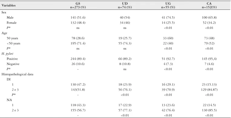
Table 1 shows the comparison of the frequency of gender, age, H. pylori infection and histopathological data between groups. Among the patients studied, 58.1% (322/554) were males and 41.9% (232/554) were females. There was a pre-dominance of male patients among those with gastric ulcer (χ2=8.81, P<0.01) and gastric cancer (χ2=7.38, P<0.01) when compared to patients with gastritis and duodenal ulcer.
In the histological analysis, 34% (187/554) of the biop-sies showed mild degree of inlammation, 37% (207/554) moderate, and 29% (160/554) intense. Neutrophil activity in 30% of biopsies (170/554) were mild, 39% (214/554) were moderate and 31% (170/554) intense. Comparing the histo-pathological data between different diseases studied, it was observed that patients with duodenal ulcer, gastric ulcer and gastric adenocarcinoma exhibited a higher degree of inlam-mation and neutrophil activity than patients with gastritis. Investigation of H. pylori infection by a molecular biology method (PCR) detected bacterial DNA in gastric biopsies of 91% (506/554) of the cases analyzed. Helicobacter pylori was detected in 89.4% (244/273) of patients with gastritis, 89.2% (66/74) of patients with duodenal ulcer, 92.7% (51/55) of patients with gastric ulcer, and 95.4% (145/152) of patients with gastric cancer.
Genotyping of H. pylori vacA and cagA genes
Characterization of the vacA alleles of the bacterial strains showed that all isolates carried the vacA gene. Among the patients analyzed, 97.6% (494/506) were colonized with only one strain, which exhibited only one vacA genotype (s1/ m1 or s1/m2 or s2m2), and 2.4% (12/506) were infected with more than one strain, with the detection of more than one vacA genotype (s1/m1 and s2/m2).
Analysis of the vacA gene showed that 88% (446/506) of the patients carried allele s1 of the signal region of the gene. There was a predominance of allele m1 of the mid-region in 87% (440/506) of the patients. In patients with monoinfec-tion, 86.6% (428/494) of the bacterial strains detected carried
the s1/m1 genotype, 1.2% (6/494) the s1/m2 genotype, and 12.2% (60/494) the s2/m2 genotype.
The cagA gene was detected in 85.6% (433/ 506) of the patients infected with H. pylori. With respect to gastrointestinal diseases, the presence of the cagA gene was observed in 80.3% (196/244) of patients with gastritis, in 98.5% (65/66) of patients with duodenal ulcer, in 88.2% (45/51) of patients with gastric ulcer, and in 88.3% (128/145) of patients with gastric cancer.
Comparison of the different vacA alleles and presence or absence of cagA showed a higher prevalence of virulent s1m1cagA+ strains in patients with monoinfection. A predominance of the s1m1/s2m2cagA+ genotype was observed in patients infected with multiple strains. Another genotype found in mixed infections was s1m1/s2m2cagA- (Table 2).
TABLE 1. Epidemiological and histopathological data of patients with different gastrointestinal diseases
Variables n=273 (%)GS n=74 (%)UD n=55 (%)UG n=152(%)CA
Sex
Male 141 (51.6) 40 (54) 41 (74.5) 100 (65.8)
Female 132 (48.4) 34 (46) 14 (25.5) 52 (34.2)
P* ns ns <0.01 <0.01
Age
50 years 78 (28.6) 19 (25.7) 33 (60) 73 (48)
<50 years 195 (71.4) 55 (74.3) 22 (40) 79 (52)
P* ns ns <0.01 <0.01
H. pylori
Positive 244 (89.4) 66 (89.2) 51 (92.7) 145 (95,4)
Negative 26 (10.6) 8 (10.8) 4 (7.3) 7 (4.6)
P* - ns <0.01 <0.01
Histopathological data DI
1 130 (47.2) 18 (23.9) 16 (29.1) 23 (15.13)
2 e 3 143(51.8) 56 (76.1) 39 (70.9) 129 (84.87)
P* - <0.01 <0.01 <0.01
NA
1 118 (43.3) 17 (22.9) 13 (23.6) 22 (14.5)
2 e 3 155 (56.7) 57 (77.1) 42 (76.4) 130 (85.5)
- <0.01 <0.01 <0.01
G test ns: not signiicant; DU: duodenal ulcer; GU: gastric ulcer; GC: gastric cancer; DI: degree of inlammation; NA: neutrophil activity. 1=mild, 2=moderate, 3=intense.
TABLE 2. Frequency of different bacterial genotypes detected in all patients studied
Genotype Frequency %
Monoinfection
s1m1cagA+ 422 83.4
s1m1cagA- 6 1.2
s1m2cagA- 6 1.2
s2m2cagA- 60 11.8
Mixed infection
s1m1/s2m2 cagA+ 11 2.2
s1m1/s2m2 cagA- 1 0.2
Association between H. pylori genotypes and histo-pathological findings
Only patients with monoinfection (n=494) were studied since the number of patients with mixed infection was not statistically signiicant for this analysis. Patients infected with s1m1cagA+ strains exhibited a higher degree of inlamma-tion and neutrophil activity than patients infected with other strains (Table 4).
TABLE 4. Association of the different vacA alleles and presence of the cagA gene with histopathological data (degree of inlammation and neutrophil activity) in monoinfected patients
vacA cagA DI OR/P-value NA OR/P-value
Alleles 1 2 and 3 (95% CI) 1 2 and 3 (95% CI)
s1m1 (+) 96 326 - 86 336
s1m1 (-) 1 5 8.24/0.01 3 3 11.72/0.01
s1m2 (-) 6 - (4.72-14.39) 6 - (6.53-21.01)
s1m2 (-) 44 16 45 15
OR: odds ratio; 95% CI: 95% conidence interval; DI: degree of inlammation; NA: neutrophil activity. 1=mild, 2=moderate, 3=intense.
TABLE 3. Prevalence of cytotoxin-associated gene (cagA) and allele combi-nations of the vacA gene in patients with different gastrointestinal diseases
Genotype Gastritis %n=244 GU %n=51 Cancer %n=145 DU %n=66
Monoinfection
s1-m1-cagA+ 188 (77) 44 (86.3) 126 (86.9) 64 (97)
NV 48 (19.7) 6 (11.8) 16 (11) 2 (3)
Mixed infection
s1 m1\s2 m2 cagA+ 8 (3.3) 1 (1.9) 2 (1.4)
-s1 m1\-s1 m2 cagA- - - 1(0.7)
-GU: gastric ulcer; DU: duodenal ulcer; NV: non-virulent strain (s1-m1-cagA-, s1-m2-cagA-, s2-m2-cagA-).
TABLE 5. Association of the different vacA alleles and presence of the cagA gene with intestinal metaplasia
vacA cagA Metaplasia OR/P-value
Alleles Present Absent (95% CI)
s1m1 (+) 117 305
s1m1 (-) 1 5 7.28/0.03
s1m2 (-) - 6 (2.23-23.72)
s1m2 (-) 3 57
OR: odds ratio; 95% CI=95% conidence interval.
DISCUSSION
Several authors have demonstrated differences in the geographical distribution of H. pylori genotypes, which di-rectly inluence the clinical relevance of the strains(35). This study was conducted to increase knowledge regarding the prevalence of H. pylori virulence factors in the State of Pará, Northern Brazil. Additionally, infection with this bacterium was related to different gastrointestinal diseases and to the histopathological alterations found in these cases.
The largest number of cases of gastric cancer was due to the study had been conducted at a cancer referral hospital endoscopy service in the region.
A predominance of males was observed among patients with gastric ulcer and gastric cancer. These patients were older than those of the other two groups studied. This higher frequency of gastric ulcers and cancer among men has been associated with lifestyle, since men are more frequently exposed to stress and environmental factors related to habits such as alcohol consumption and smoking(9). The higher in-cidence of gastric ulcers and cancer in individuals older than 50 years has been related to sequential changes characterized by metaplastic alterations that occur in the gastric mucosa secondary to infection with H. pylori(9, 16).
Detection of infection with H. pylori by a molecular biology method (PCR) revealed a prevalence of 91% among patients with different gastrointestinal diseases. A high inci-dence of gastric carcinoma and peptic ulcer, as well as a high prevalence of infection with H. pylori, is observed in Brazil, especially in the State of Pará(1, 18, 36, 37).
Some characteristics of H. pylori strains have been associated with the progression of infection to more severe disease. In this respect, some genotypes, such as alleles s1 and m1 of the vacA gene and presence of the cagA gene, are considered pathogenicity markers since they are associated with cytotoxin production and the induction of more intense epithelial lesions and inlammatory reactions. In many countries, these genotypes have been associated with the progression of infection to diseases such as peptic ulcer and gastric adenocarcinoma(1, 3, 10, 12, 14, 22, 31, 38).
Frequency of different genotypes in patients with gastrointestinal diseases
Analysis of the correlation between the endoscopic diag-nosis of gastrointestinal disease and H. pylori strains showed that virulent s1m1cagA+ strains were more frequent in pa-tients with gastric ulcer (G=34.18; P=0.01), duodenal ulcer (G=62.04; P=0.01), and gastric cancer (G=2.29; P=0.12)) compared to patients with gastritis. The comparison of strains between the group with gastric ulcer and the groups with duodenal ulcer (G=62.04; P=0.01) and gastric cancer (G=0.016; P=0.89) was not signiicant. Table 3 shows the frequency of the different genotypes in patients with gas-trointestinal diseases.
The cagA gene was identiied in 85.6% of the patients with gastrointestinal diseases studied. Similar results have been reported in other studies conducted in the State of Pará(31, 38) and in other Brazilian states, as well as in other countries(2, 11, 25, 40). When only H. pylori-positive patients were compared between the four groups studied, an even stronger association was observed between infection with cagA-positive H. pylori strains and gastrointestinal diseases. Analysis of the different diseases showed a higher frequency of the cagA gene in H. pylori-positive patients with duodenal ulcer compared to patients with gastritis, gastric ulcer and gastric cancer. Queiroz et al.(24) found that, in Brazil, 95.0% and 62.3% of strains isolated from children with and without duodenal ulcers are cagA-positive, respectively. Other studies have reported a higher frequency of the cagA gene in H. pylori-positive patients with peptic ulcer and gastric cancer(17, 33, 38). In the present study, 97.6% of the patients were infected with only one type of bacterial strain (monoinfection) and 2.4% were infected with more than one type of strain (mixed infection). In Brazil, the frequency of multiple infections ranges from 3.4% to 17% depending on the populations in-volved and the region where the study was conducted(2, 17, 33, 37). Characterization of the vacA alleles and investigation of the presence of the cagA gene demonstrated that the pre-dominant genotype in this study was s1m1cagA+ (83.4%). Previous studies conducted in the State of Pará reported a higher prevalence of this genotype in patients with different gastrointestinal diseases(17, 31, 37, 38). The same was observed in studies conducted in other Brazilian states such as Belo Horizonte (Minas Gerais)(2), Salvador (Bahia)(8), and Botucatu (São Paulo)(33).
Analysis of the genotypes of H. pylori contributes to explain the distribution of gastrointestinal diseases around the world, given the fact that, although the prevalence of H. pylori infection is higher in developing countries, some developed countries such as Japan, Portugal and Spain have high frequencies of severe gastrointestinal diseases such as peptic ulcers and gastric cancer. Studies conducted in these countries have associated this fact with the local predominance of H. pylori strains carrying genotype s1b/ m1/cagA+(15, 21, 35).
In the present study, virulent s1m1cagA+ strains were more frequent in patients with gastric ulcer, duodenal ulcer and gastric cancer compared to patients with gastritis. These data indicate a greater association between infection with bacterial strains carrying this genotype (s1m1cagA+) and an increased risk of developing more severe forms of infection. This association is supported by studies conducted in Brazil and in other countries(7, 8, 17, 33, 37, 39).
With respect to the association of different alleles of the vacA gene and presence of the cagA gene with histopatho-logical findings, we observed that patients infected with s1m1cagA+ strains exhibited a high degree of inlammation and neutrophil activity in the gastric mucosa, as well as an increased risk of developing intestinal metaplasia. These indings agree with the results reported in studies conducted in Brazil and in other countries(2, 31, 37, 40) and support the cor-relation of pathogenic vacA and cagA genotypes of H. pylori with chronic aggression to the mucosa, which is expressed at the morphological level by an inlammatory iniltrate rich in enzymes and free radicals that damage epithelial cells(33).
In conclusion, the results of the present study demon-strate the importance of bacterial factors, such as genotype of the infecting strain, for the pathogenesis of gastrointestinal diseases. These indings are supported by data indicating that patients infected with s1m1cagA+ strains have an increased risk of developing more severe forms of infection. These indings are useful to identify patients who are at a higher risk of developing more severe gastrointestinal diseases, and therefore contribute to the prognosis and treatment of these cases. Further studies establishing the relationship between bacterial, host and environmental factors are necessary to determine what makes a patient to develop duodenal ulcer, gastric ulcer or gastric cancer, since the distribution of the genotypes of infecting strains does not differ between patients with these diseases.
Authors’contributions
Vinagre IDF, Queiroz AL and Vinagre RMDF: responsible researcher. Silva Jr MR: examination of upper gastrointestinal endoscopy and biopsy. Martins LC: histopathological analysis and genotyping of H. pylori.
Vinagre IDF, Queiroz AL, Silva Jr MR, Vinagre RMDF, Martins LC. Infecção pelo Helicobacter pylori em pacientes com diferentes doenças gastroin-testinais do Norte do Brasil. Arq Gastroenterol. 2015,52(4):xxx.
RESUMO - Contexto - Os mecanismos pelos quais o H. pylori produz diferentes quadros patológicos no estômago e no duodeno não são totalmente
conhecidos. Considerando a diversidade geográica relacionada à prevalência dos fatores de virulência desse microrganismo e sua associação com o desenvolvimento de diferentes doenças, vem se intensiicando a pesquisa de marcadores de patogenicidade, como o CagA e os alelos do VacA por técnicas moleculares. Objetivos - O objetivo desse estudo foi investigar a presença da infecção por H. pylori, e a frequência dos diferentes genótipos dessa bactéria em pacientes com doenças gastrointestinais da nossa região, procurando estabelecer sua associação com os achados histopatológicos.
Métodos - Em estudo prospectivo, foram coletadas amostras de 554 pacientes com diferentes doenças gastrointestinais (gastrite, úlcera duodenal,
úlcera gástrica e câncer gástrico), atendidos em hospital de referência para todo o Estado do Pará, localizado na região metropolitana de Belém. Foram analisados dados obtidos através de questionário epidemiológico, relacionados ao sexo e faixa etária desses pacientes. A presença do H. pylori
e do genótipo bacteriano foi detectada utilizando a PCR. As biopsias gástricas foram avaliadas histologicamente. Resultados - Observou-se uma
prevalência de 91% da infecção pelo H. pylori, sendo mais frequente nos portadores de úlcera gástrica e câncer gástrico, nos quais houve predomínio do sexo masculino e a idade foi maior que a dos outros dois grupos estudados. O genótipo bacteriano predominante foi o s1m1cagA positivo, sendo mais frequentes entre os pacientes com úlcera gástrica, úlcera duodenal e câncer gástrico. Houve associação signiicante das cepas com o genótipo s1m1cagA positivo com maior grau de inlamação, atividade neutrofílica e desenvolvimento de metaplasia intestinal. Conclusão - Nosso estudo
demonstra a alta incidência da infecção pelo H. pylori nos pacientes analisados em nosso meio, especialmente em portadores de úlcera e câncer gástricos. As cepas virulentas s1m1cagA+ foram predominantes e estavam associadas a lesões mais graves.
REFERENCES
1. Araújo Filho I, Brandão Neto J, Pinheiro LAM, Azevedo IM, Freire FHMA, Medeiros AC. Prevalence of Helicobacter pylori infection in advanced gastric
carcinoma. Arq. Gastroenterol. 2006;43(4):288-92.
2. Ashour AAR, Magalhães PP, Mendes EN, Collares GB, Gusmão VR, Queiroz DMM, et al. Distribution of vacA genotypes in Helicobacter pylori strains isolated from Brazilian adult patients with gastritis, duodenal ulcer or gastric carcinoma. FEMS Immunol Med Microbiol. 2002;33:173-8.
3. Atherton JC, Peek RM, Tham KT, Cover TL, Blaser MJ. Clinical and patho-logical importance of heterogeneity in vacA the vacuolating cytotoxin gene of
Helicobacter pylori. Gastroenterology. 1997;112(1):92-9.
4. Ayres M, Ayres MJ, Ayres DL, Santos AS. Bioestat 5.0. Aplicações estatísticas nas áreas das ciências biológicas e médicas. Sociedade Civil Mamirauá MCT - CNPq, Belém, 2007:364p.
5. Blaser MJ, Atherton JC. Coadaptation of Helicobacter pylori and humans: ancient history, modern implications. J. Clin. Invest. 2009;1(119):2475–87.
6. Blaser MJ, Berg DE. Helicobacter pylori genetic diversity and risk of human disease. J Clin Invest. 2001;107(7):767–73.
7. Bolek BK, Salih BA, Sander E. Genotyping of Helicobacter pylori strains from gastric biopsies by multiplex polymerase chain reaction. How advantageous is it? Diagn Microbiol Infect Dis. 2007;58(1):67–70.
8. Brito CA, Silva LM, Jucá N, Leal NC, de Souza W, Queiroz D, Cordeiro F, Silva NL. Prevalence of cagA and vacA genes in isolates from patients with Helicobacter pylori-associated gastroduodenal diseases in Recife, Pernambuco, Brazil. Mem Inst Oswaldo Cruz. 2003;98(6):817-21.
9. Casali JJ, Franzon O, Kruel NF, Neves BD. Análise epidemiológica e emprego do teste rápido da urease em pacientes com úlcera péptica perfurada. Rev Col Bras Cir. 2012;39(2):93-8.
10. Figueiredo C, Van Doorn LJ, Nogueira C, Soares JM, Pinho C, Figueira P, Quint WG, Carneiro F. Helicobacter pylori genotypes are associated with clinical outcome in Portuguese patients and show a high prevalence of infections with multiple strains. Scand J Gastroenterol. 2001;36:128-35.
11. González-Vázquez R, Herrera-González S, Cordovs-Espinoza MG, Zúniga G, Giono-Cerezo S, Hernández- Hernández JM, León-Ávila G. Helicobacter pylori: detection of iceA1 and iceA2 genes in the same strain in Mexican isolates. Arc Med Res. 2012;43(5):339–46.
12. GwackJ, ShinA, KimCS, KoKP, KimY, JunJK, et al. CagA-producing Helico-bacter pylori and increased risk of gastric cancer: a nested case–control study in Korea. British Journal of Cancer. 2006;95(5):639–64.
13. Hammar M, Tysziewicz T, Wadstrom T, O’Toole PW. Rapid detection of Heli-cobacter pylori in gastric biopsy material by polymerase chain reaction. J Clin Microbiol. 1992;30(1):54–8.
14. Higashi H, Tsutsumi R, Fujita A, Yamazaki S, Asaka M, Azuma T, Hatakeya-ma M. Biological activity of the Helicobacter pylori virulence factors CagA is determined by variation in the tyrosine phosphorylation sites. Proc Natl Acad Sci USA. 2002;99(22):14428-33.
15. Kim N, LimI SH, Lee KH, Choi SE, Jung HC, Song IS, Kim CY. Long-term effects of Helicobacter pylori eradication on intestinal metaplasia in patients with duodenal and benign gastric ulcers. Dig Dis Sci. 2000;45(9):1754-62.
16. Komen NA, Bertleff MJ, Van Doorn LJ, Lange JF, de Graaf PW. Helicobacter
genotyping and detection in peropetative lavage luid in patients with perforated peptic ulcer. J Gastrintest Surg. 2008;12(3):555-60.
17. Martins LC, Corvelo TC, Demachki S, Araujo MT, Assumpcao MB, Vilar SC, Freitas FB, Barbosa HP, Fecury AA, do Amaral RK, Dos Santos SE. Clinical and pathological importance of vacA allele heterogeneity and cagA status in peptic ulcer disease in patients from North Brazil. Mem Inst Oswaldo Cruz. 2005;100(8):875-81.
18. Martins LC, Corvelo TCO, Oti HT, Barile KAS. Seroprevalence of antibodies against Helicobacter pylori CagA antigen in patients with gastric ulcer in the North region of Brazil. Rev Soc Bras Med Trop. 2002;35(4):307-10.
19. Meyer-ter-Vehn T, Covacci A, Kist M, Pahl HL. Helicobacter pylori activates mitogen-activated protein kinase cascades andinduces expression of the proto-oncogenes c-fos and c-jun. JBiol Chem.2000;275(21):16064-72. 20. Misiewicz JJ. The Sydney System: a new classiication of gastritis. Introduction.
J Gastroenterol Hepatol. 1991;6(3):207-8.
21. Nogueira C, Figueiredo C, Carneiro F,Gomes AT, Barreira R, Figueira P, et al.
Helicobacter pylori genotypes may determine gastric histopathology. Am. J. Pathol. 2001;158:647-54.
22. Nomura M, Lee J, Stemmermann GN, Nomura RY, Perez-Perez GI, Blaser MJ.
Helicobacter pylori CagA seropositivity and gastric carcinoma risk in a Japanese American population. J Infect Dis. 2002;186(8):1138-44.
23. Odenbreit S, Puls J, Sedlmaier B, Gerland E, Fischer W, Hass R. Translocation of
Helicobacter pylori CagA into gastric epithelial cells by type IV secretion. Science 2000;287(5457):1497-500.
24. Queiroz DM, Mendes, EN, Carvalho AS, Rocha GA, Oliveira AMR, Soares AST, et al. Factors associated with Helicobacter pylori by a cagA-positive strain in children. J Infect Dis. 2000;181(2):626-30.
25. Ribeiro ML, Godoy AP, Benvengo YH, Mendonca S, Pedrazzoli Jr J. Clinical relevance of the cagA, vacA and iceA genotypes of Helicobacter pylori in Brazilian clinical isolates. FEMS Immunol Med Microbiol. 2003;36:181-5.
26. Romero-Adrián TB, Leal-Montiel J, Monsalve-Castillo F, Mengual-Moreno E, McGregor EG, Perini L, Antúnez A. Helicobacter pylori: Bacterial Factors and the Role of Cytokines in the Immune Response. Curr. Microbiol. 2009;60:143–55. 27. Ricci V, Romano M, Boque P. Molecular cross-talk between Helicobacter pylori
and human gastric mucosa. World J Gastroenterol. 2011;17(11)1383-99. 28. Santos SEB, Guerreiro JF. The indigenous contribution to the formation the
population of the Brazilian Amazon region. Genetics and Molecular Biology. 1995;18:311-5.
29. Selbach M, Moese S, Hauck CR, Meyer TF, Backert S. Src is the kinase of the Heli-cobacter pylori CagA protein in vitro and in vivo. J Biol. Chem.2002;277(9):6775-8. 30. Segal ED, Cha J, Lo J, Falkow S, Tompkins LS. Altered states: involvement of
phosphorylated CagA in the induction of host cellular growth changes by Heli-cobacter pylori. Proc Natl Acad Sci. 1999;96(25):14559-64.
31. Silva Jr MR, Vinagre RMDF, Silva AV, Oliveira CSF, Santos KN, Costa RAA, et al. Difference in virulence markers between Helicobacter pylori strains from the Brazilian Amazon Region. Rev Soc Bras Med Trop. 2013;46(3):358-60. 32. Sugimoto M, Yamaoka Y. Virulence factor genotypes of Helicobacter pylori affect
cure rates of eradication therapy. Arch. Immunol Ther Exp. 2009;57(1):45–56. 33. Thomazini CM, Pinheiro NA, Pardini MI, Naresse LE, Rodrigues MAM.
Helicobacter pylori and gastric cancer: distribution of cagA and vacA genotypes in patients with gastric carcinoma. J Bras Patol Med Lab. 2006;42:25-30. 34. Tummuru MK, Cover TL, Blaser MJ. Cloning and expression of a high
Molec-ular mass major antigen of Helicobacter pylori: evidence of linkage to cytotoxin production. Infect Immun. 1993;61(5):1799–809.
35. Van Doorn LJ, Figueredo C, Mégraud F, Pena S, Midoo P, Queiroz DMM, et al. Geographic distribution of vacA allelic types of Helicobacter pylori. Gastroenterology. 1999;116(4):823-30.
36. Vinagre RMD, Campos BP, Sousa RMP. Case study of stomach adenocarcinoma conducted at a cancer referral hospital in northern Brazil. Arq Gastroenterol. 2012;49(2):125-9.
37. Vinagre RMDF, Corvelo TCO, Arnaud VC, Leite ACK, Martins LC. Determinação das cepas do Helicobacter pylori e do polimorismo do gene da interleucina-8 em pacientes com câncer gástrico. Arq Gastroenterol. 2011;48(1):46-51.
38. Vinagre RMDF, Vilar e Silva A, Fecury AA, Martins LC. Role of infection and lifestyle habits in the development of gastroduodenal diseases in a population from the Brazilian Amazon. Arq Gastroenterol. 2013;50(3):170-4.
39. Wada A, Yamasaki E, Hirayama T. Helicobacter pylori vacuolating cytotoxin, VacA, is responsible for gastric ulceration. J Biochem. 2004;136(6):741-6. 40. Yakoob J, Abid S, Jafri W, Ahmad Z, Ahmed R, Islam M. Distribution of