DOI: 10.1590/0004-282X20150032
ARTICLE
Alpha-synuclein A53T mutation is not
frequent on a sample of Brazilian Parkinson’s
disease patients
Mutação A53T da alfa-sinucleína não é frequente em amostra de população brasileira
com doença de Parkinson
Gabriela S. Longo1, Marcela A. S. Pinhel2, Michele L. Gregório3, Bruno A. P. Oliveira2, Driele C. G. Quinhoneiro2,
Waldir A. Tognola1, Fábio N. Oliveira1, Denise Poltronieri Martins1, Sabrina M. Cezario1, Caroline L. Sado4,
Marcelo A. Nakazone1, Maria C. J. Calastri1, Dorotéia R. S. Souza1
Parkinson’s disease (PD) is one of the major degenera-tive disorders, affecting 2% of the population over 65 years
and up to 4% in people over 85 years1. In Brazil, statistics
show that the incidence of PD amounts to 150/200 cases per 100,000 inhabitants2. Most cases of PD are idiopathic3.
However, in approximately 5%-10% of the cases, there is a genetic component with both dominant and recessive
inheritance patterns3.The chromosomal loci linked to the
familial forms of PD are termed PARK1-13. These loci in-clude six autosomal dominant genes [PARK 1or PARK 4 (alpha synuclein - SNCA), PARK 3 (Lewy bodies), PARK 5 (ubiquitin carboxyl-terminal esterase L1 - UCHL1), PARK 8 (leucine-rich repeat kinase 2 - LRRK2) and PARK13 (HTRA serine peptidase 2 – HTRA2)], 4 recessives genes [PARK 2
1Faculdade de Medicina de São José do Rio Preto, Sao José do Rio Preto SP, Brazil;
2Universidade de São Paulo, Faculdade de Medicina de Ribeirão Preto, Ribeirao Preto SP, Brazil;
3Universidade de Franca, Franca SP, Brazil;
4Universidade Federal de São Paulo, Sao Paulo SP, Brazil.
Correspondence: Marcela Augusta de Souza Pinhel; Faculdade de Medicina de Ribeirão Preto; Avenida dos Bandeirantes, 3900; 14090-900 Ribeirão Preto SP, Brasil; E-mail: [email protected]
Conflict of interest: There is no conlict of interest to declare.
Received 02 July 2014; Received in inal form 15 January 2015; Accepted 04 February 2015. ABSTRACT
Introduction: The pathogenesis of Parkinson’s disease (PD) involves both genetic susceptibility and environmental factors, with focus on the mutation in the alpha-synuclein gene (SNCA). Objective: To analyse the polymorphism SNCA-A53T in patients with familial PD (FPD) and sporadic PD (SPD). Method: A total of 294 individuals were studied, regardless of sex and with mixed ethnicity. The study group with 154 patients with PD, and the control group included 140 individuals without PD. The genotyping of SNCA-A53T was performed by PCR/RFLP. Signiicance level was p < 0.05. Results: Among all patients, 37 (24%) had FPD and 117 (75.9%) had SPD. The absence of SNCA-A53T mutation was observed in all individuals. Conclusion: SPD is notably observed in patients. However, the SNCA-A53T mutation was absent in all individuals, which does not differ controls from patients. This fact should be conirmed in a Brazilian study case with a more numerous and older population.
Keywords: Parkinson’s disease, alpha-synuclein, mutation.
RESUMO
Introdução: A patogênese da doença de Parkinson (DP) envolve fatores ambientais e suscetibilidade genética, destacando-se a mutação de alfa-sinucleína (SNCA.) Objetivos: Analisar a variante genética SNCA-A53T em pacientes com DP familiar (DPF) e DP esporádica (DPE).
Método: Foram estudados 294 indivíduos, independente de sexo, com etnia miscigenada, sendo 154 com DP e 140 sem a doença (grupo controle). A genotipagem de SNCA-A53T foi realizada por PCR/RFLP. Nível de signiicância para p < 0,05. Resultados: Entre os pacientes, 37(24%) tinham DPF e 117 (75,9%) DPE. A ausência da mutação SNCA-A53T em todos os indivíduos. Conclusão: DPE é destacada entre os pacientes, no entanto a mutação SNCA-A53T ausente em todos os indivíduos, não diferenciando os grupo controle e pacientes, o que deve ser conirmado em população brasileira, considerando uma ampla casuística, além da ancestralidade.
507
Gabriela S. Longo et al. Mutation of a-synuclein in Parkinson’s disease (parkin - PRKN), PARK 6 (PTEN-induced putative kinase
1 - PINK1), PARK 7 (oncogene DJ1), PARK 9 (ATPase
type 13A2 - ATP13A2)] and PARK11 (GIGYF2 - Grb10 Interacting GYF Protein-2),one gene linked to X chromo-some (PARK12) and a form with unknown inheritance
pattern (PARK10)4,5,6. Mutations identified in five major
genes were related with genetic etiology of familial PD,
such as a-synuclein, which mediates autosomal dominant
forms of the disease4.
he SNCA gene, located on human chromosome
4q21.3-q22, has 7 exons and encodes the a-synuclein pro-tein of 140 amino acids, which is divided into three regions:
N-terminal a-helical region (where mutations occur in the
event of PD); central region (hydrophobic NAC sequence,
known as non β-amyloid component), responsible for the
for-mation of ibrils that cause toxicity; and the acidic C-terminal
region5. he analysis of the sequence of exon 4 revealed that
a single change of G →A (Guanine→Adenine) in the position 209 of the nucleotide chain causes the amino acid alanine to be replaced by the amino acid threonine at position 53 of the
a-synucleinprotein6.
The a-synuclein protein is the major protein
com-ponent of intracellular neuron deposits observed in PD7.
Although the function and the underlying dysregulation
of SNCA mechanisms are not fully understood, the
for-mation and expression of oligomers of this protein play a
critical role in the development of the disease8. This
wild-type a-synuclein protein is the major component of LB, in
both familial and sporadic PD, which highlights the role
of the SNCA gene variant in all presentations of the
dis-ease9. Functionally, relevant genetic variants in and near
the SNCA gene are risk factors for sporadic PD (SPD). For example, the A53T mutation promotes greater aggrega-tion and fibrillaaggrega-tion in the LBs, and interferes with
neuro-transmission9, which confirms its role in the pathogenesis
of the disease.
herefore, this study aimed to analyse the frequency of
the genetic variant SNCA-A53T in a Brazilian population
with familial PD (FPD) or sporadic PD.
METHOD
A total of 294 individuals divided into two groups were studied: Study Group with 154 PD patients and Control Group formed by140 subjects, who had not been diag-nosed with PD. The study group included PD patients
older than 50 years, regardless of ethnicity10 and sex.
The patients were examined at the Outpatient Clinic for Movement Disorders of Hospital de Base, Medical School of São José do Rio Preto.
Diagnosis of PD followed the criteria recommended by Jankovic11, including bradykinesia, rigidity, resting tremor,
postural instability, unilateral onset, L-dopa response for
more than ive years, levodopa-induced dyskinesia, pro -gressive disorder, persistent asymmetry and clinical course of ten years or more, as well as complementary tests. he Control Group was selected in ambulatory clinics of that institution, namely, orthopedics, gynecology, among others. All subjects were informed about the study and conirmed their willingness to participate by signing an informed con-sent form. he study was approved by the Ethics Research Committee of institution.
he subjects were studied considering allele and geno
-typic frequencies for theSNCA-A53T mutation. Blood was
collected by venepuncture with EDTA, and the genomic DNA was extracted from whole blood (5 mL) following the salting-out method12. PCR ampliication of DNA was per
-formed in a thermocycler (Eppendorf - Mastercycler). Each tube included 0.5 mL of each deoxynucleotide (0.8 mM); 2.5mLof10 X PCR bufer; 2.5mL of 10% dimethyl sulfoxide; 2.5mL of each primer (2.5mM); 0.2mL of Taq polymerase (5U/mL); 11 mL of Milli Q water; 2mL of genomic DNA dilu-tion (0.2 mg). For the PCR, the following primer sequences were used: (exon 4): P1- 5’ GCT AAT CAG CAA TTT AAG GCT AG 3’; P2- 5’ GAT ATG TTC TTA GAT GCT CAG 3’. he initial DNA denaturation was achieved at 94ºC for 4 minutes. Next, the reaction mixture was subjected to 35 cycles of 94ºC for 20 seconds and of 55 ºC for 30 seconds, to an extension at 72°C for 45 seconds, and to a inal cycle
at 72°C for 10 minutes13. he post PCR product was digest
-ed with the restriction enzyme Tsp45I at 37°C for 4 hours and coloured by GelRed® (Uniscience). Electrophoresis was performed in 1% agarose gel at constant current of 100 V during 40 minutes, in order to separate fragments of 88pb, 128pb and 216pb in the presence of mutation (AG); 216pb for homozygous wild-type (GG) as well as 88pb and 128pb for homozygous mutant (AA). A sample of standard DNA (100bp Invitrogen) was used for comparison of the electro-phoretic bands (Figure).
he quantitative and qualitative analysis was performed by t-test and Chi-square test, respectively, with the use of the MiniTab14 and GraphPad3 software. he adopted level of sig -niicance was p < 0.05.
RESULTS
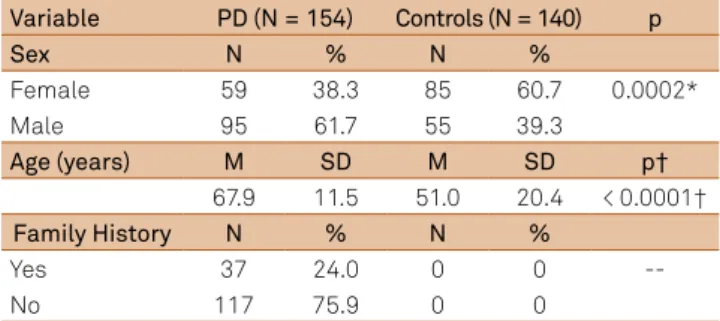
Table shows demographic data and history of PD pa-tients. he mean age was 67.9 ± 11.5 years in the study group, and 51.0 ± 20.4 years in the control group (p < 0.0001). he study included only those patients with disease duration longer than 10 years.
here was a prevalence of males in the study group (N = 95; 61.6%) compared with the control group (N = 55; 39.2%%; p = 0. 0002). In the study group, 37 patients (24.0%) showed his-tory of familial PD versus 117 patients (75.9%) with sporadic PD. Patients were qualiied as familial, when they had at least one relative of irst degree diagnosed with PD, regardless of Mendelian pattern of inheritance. Sporadic cases included individuals without any family history of the disease in the past three generations.
he genome analysis of all patients and controls showed homozygous wild-type genotype (GG), which demonstrates
the absence of SNCA-A53T mutation in both groups.
DISCUSSION
his study has not found any association of A53T muta
-tion in the SNCA gene with Parkinson’s disease. his corrob
-orates the data presented by Moura et al.14, who conducted
research on Brazilian populations and quantiied the SNCA
gene mutations without inding any alterations. SNCA mul -tiplications have been implicated in autosomal dominant
forms of PD since 200315. However, it became clear that
whole-gene multiplications in SNCA locus are a rare form of parkinsonism and may account for only a small fraction of the total number of PD patients16,17.
Remarkably, further Brazilian studies found no
SNCA-A53T mutation in PD patients14,18,19. Furthermore,
other genes re1ated to PD, such as APOE, GSTs and LRRK2
(PARK8), also showed no diference between patients and controls in this population20,21,22,23. On the other hand,
muta-tions in the genes GIGYF (PARK11), ATP13A2 (PARK9)and
GBA were associated with PD6.
Vaughan et al.13 evaluated 7 exons of SNCA gene in 30
European and American children with autosomal dom -inant PD, who did not show either A53T mutation or oth-er mutations. Anothoth-er study, also with PD patients, did not conirm the presence of this genetic variant in the SNCA
gene24. Moreover, Athanassiadou et al.25 assessed 19
fami-lies with no biological relations, where at least two relatives of irst/second degree were afected with PD. he authors
observed the presence of the mutation in several members of those 7 families, showing autosomal dominant inheritance pattern. Choi et al.26 identiied the A53T mutation in only
1 of 72 patients with early-onset PD, whose family history was consistent with autosomal dominant PD.
In this study, the selected population was of mixed
eth-nicity10, unlike the population in which the SNCA-A53T
mu-tation was found27. Furthermore, in this study, control
sub-jects, without a family history of PD, were selected from other speciality clinics, which made it diicult to perform match -ing pair analysis on sex and age with the group of patients. his may be a limitation of this study. Elderly individuals (> 60 years) have been treated in the several speciality clinics of the hospital and showed other neurodegenerative diseas-es, which were considered as exclusion criteria for this study. However, these discrepancies seem to have little rel-evance, considering the absence of said mutation in both groups, which seems to be diferent in the Brazilian popula -tion14,18,19. Additionally, individuals remained in the control
group, regardless of the matching pair analysis with patients, which enabled a larger number of individuals to perform comparative analysis.
Mutations in the SNCA gene enhance the production of
alpha-synuclein protein, which contributes to the
pathogen-esis of PD28. SNCA is expressed throughout the brain,
spe-cially in presynaptic nerve terminals, and SNCA-A53T rather
tends to form aggregates there are critical to Lewy bodies for-mation, both familial and idiopathic PD. his aggregation of SNCA is thought to be a key occurence in dopaminergic neu-ronal cell loss3,29. he role of SNCA under normal physiologi
-cal conditions is not yet completely clear, although there is evidence that implicates SNCA in neurotransmitter release and vesicle turnover at the presynaptic terminals30.
However, this study conducted in a population of mixed ethnicity showed that both FPD and SPD are not associated
with SNCA-A53T, which suggests the need for further
evalu-ation and identiicevalu-ation of larger and speciic risk subgroups. Also, understanding the molecular mechanisms involved in its development will contribute to the application of molecu-lar therapy interventions.
Table. Patients with Parkinson´s disease (PD) and Controls, considering sex, age and family history for PD.
Variable PD (N = 154) Controls (N = 140) p
Sex N % N %
Female 59 38.3 85 60.7 0.0002*
Male 95 61.7 55 39.3
Age (years) M SD M SD p†
67.9 11.5 51.0 20.4 < 0.0001†
Family History N % N %
Yes 37 24.0 0 0
--No 117 75.9 0 0
509
Gabriela S. Longo et al. Mutation of a-synuclein in Parkinson’s disease References
1. Tan YY, Wu L, Zhao ZB, Whang Y, Xiao Q, Liu J et al. Methylation of a-synuclein and leucine-rich repeat kinase 2 in leukocyte DNA of Parkinson’s disease patients. Parkinsonism Relat Disord. 2014;20(3):308-13. http://dx.doi.org/10.1016/j.parkreldis.2013.12.002
2. Ministério da Saúde. Secretaria de Atenção à Saúde. Protocolo clínico e diretrizes terapêuticas. Doença de Parkinson. Available at: http://portalsaude.saude.gov.br/images/pdf/2014/setembro/19/ pcdt-parkinson-republicado-2010.pdf. Accessed (April, 13, 2013). 3. Coppedè F. Genetics and epigenetics of Parkinson’s
disease. ScientiicWorldJournal. 2012;2012:489830. http://dx.doi.org/10.1100/2012/489830.
4. Klein C, Westenberger A. Genetics of Parkinson’s disease. Cold Spring Harb Perspect Med. 2012;2(1): a008888. http://dx.doi.org/10.1101/cshperspect.a008888.
5. Sironi F, Primignani P, Ricca S, Tunesi S, Zini M, Tesei S et al. DJ1
analysis in a large cohort of Italian early onset Parkinson Disease patients. Neurosci Lett. 2013;557(Pt B):165-70. http://dx.doi. org/10.1016/j.neulet.2013.10.048
6. Santos AV, Pestana CP, Diniz KR, Campos M, Abdalla-Carvalho CB, Rosso AL et al. Mutational analysis of GIGYF2, ATP13A2 and GBA genes in Brazilian patients with early-onset Parkinson’s disease. Neurosci Lett. 2010;485(2):121-4. http://dx.doi.org/10.1016/j.neulet.2010.08.083
7. Breydo L, Reddy KD, Piai A, Felli IC, Pierattelli R, Uversky VN. The crowd you’re in with: effects of different types of crowding agents on protein aggregation. Biochim Biophys Acta. 2014; 1844(2):346-57. http://dx.doi.org/10.1016/j.bbapap.2013.11.004
8. Kalia LV, Kalia SK, McLean PJ, Lozano AM, Lang AE. a-Synuclein oligomers and clinical implications for Parkinson disease. Ann. Neurol. 2013;73(2):155-69. http://dx.doi.org/10.1002/ana.23746 9. Sun F, Kanthasamy A, Anantharam V, Kanthasamy AG.
Environmental neurotoxic chemicals-induced ubiquitin proteasome system dysfunction in the pathogenesis and progression of Parkinson’s disease. Pharmacol Ther. 2007;114(3):327-44. http://dx.doi.org/10.1016/j.pharmthera.2007.04.001
10. Parra FC, Amado RC, Lambertucci JR, Rocha J, Antunes CM, Pena SD. Color and genomic ancestry in Brazilians. Proc Natl Acad Sci USA 2003; 100(1):177-82. http://dx.doi.org/10.1073/pnas.0126614100 11. Jankovic J. Parkinson’s disease: clinical features and
diagnosis. J Neurol Neurosurg Psychiatry. 2008; 79(4):368-76. http://dx.doi.org/10.1136/jnnp.2007.131045
12. Salazar LA, Hirata MH, Cavalli SA, Machado MO, Hirata RD. Optimized procedure for DNA isolation from fresh and cryopreserved clotted human blood useful in clinical molecular testing. Clin.Chem. 1998;44(8):1748-50.
13. Vaughan JR, Farrer MJ, Wszolek ZK, Gasser T, Durr A, Agid Y et al. Sequencing of the a-synuclein gene in a large series of cases of familial Parkinson’s disease fails to reveal any further mutations. Hum Mol Genet. 1998;7(4):751-3. http://dx.doi.org/10.1093/hmg/7.4.751
14. Moura KC, Campos Junior M, Rosso AL, Nicaretta DH, Pereira JS, Silva DJ et al. Exon dosage variations in Brazilian patients with Parkinson’s disease: analysis of SNCA, PARKIN, PINK1 and DJ-1 genes. Dis Markers. 2012;32(3):173-8. http://dx.doi.org/10.3233/DMA-2011-0873
15. Singleton AB, Farrer M, Johnson J, Singleton A, Hague S, Kachegurs J et al. alpha-Synuclein locus triplication causes Parkinson’s disease. Science. 2003;302(5646):841. http://dx.doi.org/10.1126/science.1090278
16. Nuytemans K, Theuns J, Cruts M, Van Broeckhoven C. Genetic etiology of Parkinson disease associated with mutations in the SNCA, PARK2, PINK1, PARK7, and LRRK2 genes: a mutation update. Hum Mutat. 2010;31(7):763-80. http://dx.doi.org/10.1002/humu.21277
17. Ross OA, Braithwaite AT, Skipper LM, Kachergus J, Hulihan MM, Middleton FA et al. Genomic investigation of alpha-synuclein multiplication and parkinsonism. Ann Neurol. 2008;63(6):743-50. http://dx.doi.org/10.1002/ana.21380
18. Teive HA, Raskin S, Iwamoto FM, Germiniani FM, Baran MH, Werneck LC et al. The G209A mutation in the alpha-synuclein gene in Brazilian families with Parkinson’s disease.Arq Neuropsiquiatr.2001;59(3B):722-4. http://dx.doi.org/10.1590/S0004-282X2001000500013
19. Camargos ST, Dornas LO, Momeni P, Lees A, Hardy J, Singleton A et al. Familial Parkinsonism and early onset Parkinson’s disease in a Brazilian movement disorders clinic: phenotypic characterization and frequency of SNCA, PRKN, PINK1, and LRRK2 mutations. Mov Disord. 2009;24(5):662-6. http://dx.doi.org/10.1002/mds.22365 20. Munhoz RP, Wakutani Y, Marras C, Teive HÁ, Raskin S, Werneck LC et al. The G2019S LRRK2 mutation in Brazilian patients with Parkinson’s disease: phenotype in monozygotic twins. Mov Disord. 2008;23(2):290-4. http://dx.doi.org/10.1002/mds.21832
21. Gregório ML, Pinhel MA, Sado CL, et al. Impact of genetic variants of apolipoprotein E on lipid proile in patients with Parkinson’s disease. BioMed Res Int. 2013;2013:641515. http://dx.doi.org/10.1155/2013/641515
22. Pinhel MA, Sado CL, Longo GS, Gregório ML, Amorim GS, Florim GM et al. Nullity of GSTT1/GSTM1 related to pesticides is associated with Parkinson’s disease. Arq Neuropsiquiatr. 2013;71(8):527-32. http://dx.doi.org/10.1590/0004-282X20130076
23. Longo GS, Pinhel MS, Sado CL, Gregório ML, Amorim GS, Florim GS et al. Exposure to pesticides and heterozygote genotype of GSTP1-Alw26I are associated to Parkinson’s disease. Arq Neuropsiquiatr. 2013;71(7):446-52. http://dx.doi.org/10.1590/0004-282X20130060 24. Farrer M, Wavrant-De Vrieze F, Crook R, Boles L, Perez-Tur J,
Hardy J et al. Low frequency of alpha-synuclein mutations in familial Parkinson’s disease. Ann Neurol. 1998;43(3):394-7. 25. Athanassiadou A, Voutsinas G, Psiouri L, Leroy E, Polymeropoulos
MH, Ilias A et al. Genetic analysis of families with Parkinson disease that carry the Ala53Thr mutation in the gene encoding alpha-synuclein. Am J Hum Genet. 1999;65(2):555-8. http://dx.doi.org/10.1086/302486
26. Choi JM, Woo MS, Ma HI, Kang SY, Sung YH, Yong SW et al. Analysis of PARK genes in a Korean cohort of early-onset Parkinson disease. Neurogenetics. 2008;9(4):263-9. http://dx.doi.org/10.1007/s10048-008-0138-0
27. Ki CS, Stavrou EF, Davanos N, Lee WY, Chung EJ, Kim JY et al. The Ala53Thr mutation in the alpha-synuclein gene in a Korean family with Parkinson disease. Clin Genet. 2007;71(5):471-3. http://dx.doi.org/10.1111/j.1399-0004.2007.00781.x 28. Nishioka K, Ross OA, Ishii K, Kachegus JM, Ishiwata K,
Kitagawa M et al. Expanding the clinical phenotype of SNCA duplication carriers. Mov Disord. 2009;24(12):1811-9. http://dx.doi.org/10.1002/mds.22682
29. Bourdenx M, Bezard E, Dehay B. Lysosomes and a-synuclein form a dangerous duet leading to neuronal cell death. Front Neuroanat. 2014;8:83. http://dx.doi.org/10.3389/fnana.2014.00083
ERRATUM
Arquivos de Neuro-Psiquiatra 2015;73(6):506-509.
In Page 506, the name of the authors where is written:
Gabriela S. Longo1, Marcela A. S. Pinhel2, Michele L. Gregório3, Bruno A. P. Oliveira2, Driele C. G. Quinhoneiro2, Waldir A. Tognola1,
Fábio N. Oliveira1, Sabrina M.Cezario1, Caroline L. Sado4, Marcelo A. Nakazone1, Maria C. J. Calastri1, Dorotéia R. S. Souza1
It should be:
Gabriela S. Longo1, Marcela A. S. Pinhel2, Michele L. Gregório3, Bruno A. P. Oliveira2, Driele C. G. Quinhoneiro2, Waldir A.
Tognola1, Fábio N. Oliveira1, Denise Poltronieri Martins1, Sabrina M. Cezario1, Caroline L. Sado4, Marcelo A. Nakazone1, Maria