https://doi.org/10.1590/0004-282X20170070
ARTICLE
Late onset multiple sclerosis: concerns in
aging patients
Esclerose múltipla de início tardio: atenção a uma população que envelhece
Claudia Beatriz de Campos Lotti1, Acary Souza Bulle Oliveira1, Denis Bernardi Bichuetti1, Isac de Castro2,
Enedina Maria Lobato Oliveira1
1Universidade Federal de S̃o Paulo, Departamento de Neurologia, S̃o Paulo SP, Brasil;
2Universidade de S̃o Paulo, Faculdade de Medicina, Disciplina de Nefrologia, S̃o Paulo SP, Brasil.
Correspondence: Claudia Beatriz de Campos Lotti; Universidade Federal de S̃o Paulo, Departamento de Neurologia; Rua Botucatu, 740; 04023-900 S̃o Paulo SP, Brasil; E-mail: [email protected]
Conflict of interest: There is no conlict of interest to declare. Received 28 December 2016; Accepted 20 March 2017.
ABSTRACT
Late onset multiple sclerosis (LOMS) is when the irst symptom starts after 50 years of age, representing 4.5% of multiple sclerosis (MS) patients. This study describes the clinical characteristics of patients with LOMS followed at a specialized MS center in S̃o Paulo. Data was obtained from medical records of 742 patients with MS. The LOMS frequency was 4.18%, median age at onset was 54 years and the predominant disease course was primary progressive (64.3%). The patients reached the disability landmarks of EDSS grades 3.0, 6.0 and 7.0 in the following proportion and time: EDSS 3.0: 77.42% of patients in 3.7 years; EDSS 6.0: 58.06% in 5.1 years and EDSS 7.0: 32.26% in 5.7 years. The comparative analysis with a matched control group of patients with early onset MS showed that late onset, associated with a progressive course, were predictors of reaching EDSS 3.0 and 6.0 in a shorter time.
Keywords: aging; epidemiology; multiple sclerosis; disease progression.
RESUMO
Esclerose múltipla de inicio tardio (EMIT) caracteriza-se pelo início de sintomas aos 50 ou mais anos de idade, representando 4,5% dos pacientes com esclerose múltipla (EM). Este estudo descreve as características clínicas de pacientes com EMIT acompanhados num centro de EM em S̃o Paulo. Dados foram obtidos através de análise de prontuário de 742 pacientes com EM. A frequência de EMIT foi de 4,18%, a mediana da idade de início foi de 54 anos e a forma clínica predominante a primariamente progressiva (64,3%). Os pacientes atingiram os marcos de incapacidade EDSS 3, 6 e 7 nas respectivas proporções e tempo: EDSS 3.0, 77,42% de pacientes em 3.7 anos; EDSS 6.0, 58,06% em 5.1 anos e EDSS 7.0, 32,26% em 5.7 anos. A análise comparativa a um grupo controle de jovens com EM, mostrou que o início tardio associado a forma primariamente progressiva foram preditores para atingir EDSS 3 e 6 num período menor.
Palavras-chave: envelhecimento; epidemiologia; esclerose múltipla; progress̃o da doença.
Multiple sclerosis (MS) is an inlammatory autoim
-mune disease of the central nervous system that predomi
-nantly afects young adults between 20 and 40 years of age. herefore, an uncommon form of MS is one in which symp
-toms start at 50 plus years of age, called late onset multiple
sclerosis (LOMS)1,2,3,4,5,6,7,8. It accounts for 1.4% to 9.9% of the MS population in diferent countries2,4,5,7,9,10,11,12.
On average, LOMS represents roughly 4.5% of the MS popu -lation2,4,5,7,it has a female preponderance1,2,4,7,12,its initial presenta-tion is monosymptomatic, with a motor or cerebellar symptom, and the most common clinical course is primary progressive4,13,14.
he progression to disability in LOMS has previously been attributed to disease duration time15, to clinical form2, gender8, and older age at the time of the patient’s irst examination13. he later onset has been associated with a greater possibility
of reaching Expanded Disability Status Scale (EDSS) grade 6.0 in a shorter period16.
Vascular disease of the central nervous system and cervi
-cal spondylotic myelopathy are the main diferential diagno
-ses, due to a higher prevalence at this age and possible similar symptoms1,4,13,17,18.Because of previous co-morbid conditions and higher odds of T2-hyperintense lesions on magnetic reso
-nance imaging (MRI) of elderly patients7,13,19, there is a delay in diagnosis that can reach three to ive years in almost 40% of
LOMS patients7,14,20.
followed at the MS center of the Neurology Department of the Federal University of São Paulo.
METHODS
he Neuroimmunology Clinic of the Federal University of São Paulo is a specialized center for the treatment of patients with MS and other demyelinating diseases. It has maintained a database with information on clinical status, laboratory tests, radiological evaluation and treatment of all patients under fol
-low up, since 1994. Data for this retrospective cohort study was obtained from the medical records of 742 patients with MS.
he LOMS patients were deined by the occurrence of the irst MS symptoms after age 50, meeting either Poser’s or the McDonald criteria, depending on the current diagnostic cri
-teria at the moment of their irst appointment. According to the current diagnostic criteria, neuromyelitis optica patients were excluded from the analysis.
Information gathered from their clinical records included: demographic characteristics; disease onset and duration; fol
-low-up time; onset, irst and last visit symptoms and signs; EDSS scores in the irst and last visit; disease course; number of relapses, progression index; MRI data, cerebral spinal luid (CSF) and evoked potential indings.
Additionally, LOMS patients were compared to a young onset MS (YOMS) control group, matched according to sex and clinical form, in a 2:1 proportion. For this analysis, nine LOMS patients with a follow-up of less than 12 months were excluded.
his study was approved by the local institutional ethics committee (IRB) and registered under number 256958.
Statistical analyses
Continuous variables were tested for normality with the Kolmogorov-Smirnov and Shapiro Wilk tests and the values are expressed as median and percentiles 25 and 75. he cat
-egorical data are presented as absolute values and percent
-ages and were tested using Pearson’s χ2 test and Fisher’s exact test, if applicable.
Nonparametric data was compared using the Mann-Whitney U test for two independent samples.
Discrimination of variables was calculated by a receiver operator characteristic curve (ROC curve) utilizing the area under the curve and asymptotic signiicance. Some con
-tinuous variables were categorized through the ROC curve. he cutof points were calculated using the value with the best sensitivity and speciicity.
he Kaplan-Meier model with log-rank test was applied at times, to obtain EDSS values.
A cox proportional model was used to determine predic
-tive factors for reaching speciic EDSS milestones and the hazards ratios were calculated.
Statistical signiicance was considered with p ≤ 0.05 and the analyses were performed using SPSS 19.0.
RESULTS
LOMS population
Of the 742 patients with MS, 31 were LOMS patients, rep
-resenting a percentage of 4.18%. he gender distribution was 2.1 females to 1 male patient, of whom 19 were Caucasian, three were Afro-descendant, two were of mixed ethnicity and there was no information on seven patients. he median age of initial symptoms was 54, 22 patients (71%) were between 50 and 55 years old, and only two patients were over 60 years old, which is referred to as very late onset MS.
he initial neurological presentation was motor impair
-ment in 54.8% and cerebellar involve-ment in 29.0%, sensory impairment and brainstem symptoms in 19.4% each, visual in 16.1% and vesical dysfunction in 3.2% A multi-topographic involvement was described in 38.7%, and the most frequent combination was motor and cerebellar.
hree patients did not have enough information to deine their clinical course, therefore they were excluded from the LOMS population analysis. Of the remaining 28 patients, 64.3% were primary progressive and 35.7% relapsing-remitting. In our study, there were no secondary progressive cases of LOMS.
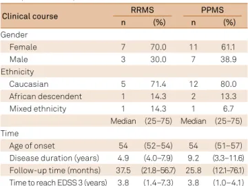
he median follow-up period was 2.2 years and 3.1 years for primary progressive multiple sclerosis (PPMS) and relaps
-ing-remitting multiple sclerosis (RRMS), respectively, while median disease time was of 9.2 for PPMS and 4.9 years for RRMS. Considering the RRMS patients, 70% had their second relapse less than a year after the irst symptom. he median EDSS scores on the irst and last evaluation for the RRMS group was 2.0 on both visits, and for the PPMS group was 2.0 at the initial visit and 7.0 on the last visit. Patients with LOMS reached the disability landmarks of EDSS 3.0, 6.0 and 7.0 in the following proportion and time: EDSS 3.0: 77.42% of patients in 3.7 years; EDSS 6.0: 58.06% in 5.1 years, and EDSS 7.0: 32.26% in 5.7 years (Tables 1 and 2). Of the 31 patients, three deaths
Table 1. Demographic and clinical data of the late onset multiple sclerosis patients with well-deined disease course.
Clinical course RRMS PPMS
n (%) n (%)
Gender
Female 7 70.0 11 61.1
Male 3 30.0 7 38.9
Ethnicity
Caucasian 5 71.4 12 80.0
African descendent 1 14.3 2 13.3
Mixed ethnicity 1 14.3 1 6.7
Median (25–75) Median (25–75)
Time
Age of onset 54 (52–54) 54 (51–57)
Disease duration (years) 4.9 (4.0–7.9) 9.2 (3.3–11.6) Follow-up time (months) 37.5 (21.8–56.7) 25.8 (12.1–76.1) Time to reach EDSS 3 (years) 3.8 (1.4–7.3) 3.8 (1.0–4.1)
occurred during the follow-up: one from gastrointestinal bleeding, one myocardial infarction and one unknown cause.
Descriptions of brain MRI scans were compatible with MS in all patients. Sixteen of them had descriptions of spinal cord MRIs, of which nine (56.3%) had one or more lesions. Only 11 patients had their CSF analyzed, of which 54.5% had
oligoclonal bands. Only six had visual evoked potential stud
-ies, with altered potentials in ive (83.3%).
LOMS versus YOMS
Twenty-two LOMS patients were compared to 44 patients with YOMS, whose initial symptom had started between 20 and 40 years of age.
We found no signiicant statistical diference between initial symptoms or functional system reported by either group on initial or inal evaluation. he predominant ini
-tial symptom in RRMS patients was brainstem in the LOMS (44%) and sensitive in the YOMS (39%), while patients with the progressive form in both groups had predominantly motor symptoms.
he progression index (calculated by dividing the EDSS score by the disease duration time) was worse in the LOMS patients, more clearly seen in the PPMS group, 0.55 versus 0.74, for younger and older onset respectively. he annual relapsing rate was higher in the younger group (median of 0.7 versus 0.51). Neither of these diferences had statistical sig
-niicance (Tables 3 and 4).
Table 4. Young onset primary progressive multiple sclerosis group comparison to the late onset primary progressive multiple sclerosis group.
PPMS (Age onset) < 50 YOPPMS ≥ 50 LOPPMS Mann-Whitney
Median (25% - 75%) Median (25% - 75%) p-value
Disease Duration (years) 13.5 (8.7–16.5) 7.2 (3.3–11.6) 0.021
Follow up (months) 86.1 (67–124.1) 25.8 (16.1–76.1) 0.005
EDSS-First Visit+ 3.5 (3.0–6.0) 3.0 (3.0–4.0) 0.219
EDSS-Last Visit++ 6.5 (6.5–8.0) 7.5 (6.5–8.0) 0.691
Time from onset to initial evaluation (months) 49.3 (30.3–73.6) 49.0 (27.7–49.6) 0.452
PI (Progression Index) 0.55 (0.43–0.81) 0.74 (0.63–1.83) 0.058
Time to reach EDSS 3 (years) 3.0 (2.1–4.1) 3.1 (0.9–4.1) 0.711
Time to reach EDSS 6 (years) 6.8 (4.6–10.5) 5.5 (2.8–8.7) 0.324
Time to reach EDSS 7 (years) 9.0 (8–14.3) 7.3 (4.3–11.6) 0.168
PPMS: primary progressive multiple sclerosis; YOPPMS: young onset primary progressive multiple sclerosis; LOPPMS: late onset primary progressive multiple sclerosis; EDSS: expanded disability status scale; n: number of patients.
Table 2. EDSS milestones of the 31 late onset multiple sclerosis patients, despite disease course.
Variable Median (25%–75%)
First visit EDSS 3 (2.5– 6.0)
Last visit EDSS 6.5 (2.5–8.0)
Time in years to EDSS 3 3.7 (0.7– 5.9)
Time in years to EDSS 6 5.1 (2.9–8.7)
Time in years to EDSS 7 5.7 (3.5–10.5)
n %
Reached EDSS 3 24 77.40
Reached EDSS 6 18 58.1
Reached EDSS 7 10 32.30
EDSS: expanded disability status scale; n: number of patients.
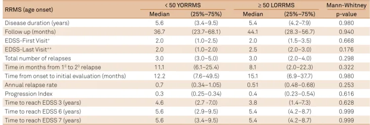
Table 3. Young onset relapsing-remitting multiple sclerosis group comparison to the late onset relapsing-remitting multiple sclerosis group.
RRMS (age onset) < 50 YORRMS ≥ 50 LORRMS Mann-Whitney
Median (25%–75%) Median (25%–75%) p-value
Disease duration (years) 5.6 (3.4–9.5) 5.4 (4.2–7.9) 0.980
Follow up (months) 36.7 (23.7–68.1) 44.1 (28.3–56.7) 0.940
EDSS-First Visit+ 2.0 (1.0–2.5) 2.0 (1.5–3.5) 0.668
EDSS-Last Visit++ 2.0 (1.0–2.0) 2.5 (2.0–3.0) 0.176
Total number of relapses 3.0 (3.0–5.0) 3.0 (2.0–4.0) 0.298
Time in months from 10 to 20 relapse 11.1 (6.1–25.4) 8.1 (2.0–22.3) 0.322
Time from onset to initial evaluation (months) 12.2 (7.6–49.5) 15.1 (6.9–37.7) 0.980
Annual relapse rate 0.7 (0.34–1.05) 0.51 (0.48–0.68) 0.253
Progression Index 0.3 (0.25–0.34) 0.4 (0.23–0.54) 0.616
Time to reach EDSS 3 (years) 4.6 (2.7 -7.0) 3.8 (1.4–7.3) 0.628
Time to reach EDSS 6 (years) 5.6 (2.9–9.5) 5.4 (4.2–8.7) 0.999
Time to reach EDSS 7 (years) 5.6 (3.4–9.5) 5.4 (4.2–8.7) 0.999
he median time to reach EDSS 3.0, 6.0 and 7.0 for the older RRMS patients was 3.8, 5.4 and 5.4 years, respectively, and in the younger group was 4.6, 5.6 and 5.6 years. In the PPMS group, the older patients reached EDSS grades 3.0, 6.0 and 7.0 in 3.1, 5.5 and 7.3 years, while the young patients reached these landmarks in 3.0, 6.8 and 9.0 years, respectively (Figure). he three EDSS milestones were reached in less time in the LOMS group compared to the YOMS, but there was no statistical signiicance (Tables 3 and 4).
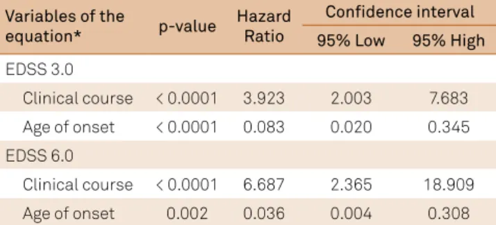
he cox regression models were applied to ind the pre
-dictive factors for reaching the EDSS grades 3.0 and 6.0 in a shorter time. We observed that patients who were 50 years and older and the ones with a primary progressive form, reached EDSS grades 3.0 and 6.0 signiicantly earlier. hese variables were co-dependent (Table 5).
All patients had brain MRI scans that fulilled dissemina
-tion in time and space, compatible with an MS diagnosis at irst visit. Spinal MRI was performed in four patients with late onset RRMS, but did not show any spinal cord abnormalities, in con
-trast to the young group, in which 90% of the scans showed MS lesions. Conversely, within the PPMS patients, spinal inlamma
-tory lesions were found in both age groups at the same propor
-tion. We observed that the positivity of oligoclonal bands in spi
-nal luid was much higher in the younger group (Table 6).
DISCUSSION
he population of Brazilian LOMS patients described in this retrospective cohort seems to be very similar to previously-published LOMS descriptions. We observed the same frequency of 4.18%, among patients with MS2,3,4,5,7; a female pre -dominance of 2.1:11,2,4,7,12,14, a Caucasian preponderance12; a similar
age of onset2,4,7,11,14 and a higher percentage of the primary progres -sive form, that reached up to 83% in some descriptions1,2,4,7,9,11,14,17,21. Interestingly, very late onset MS (deined as the irst symp
-tom at 60 years or above), represented 0.27% of our cohort. his igure is lower when compared to other series, which reported a prevalence of 0.45% to 1.33%2,3,5,9,12,17. A possible explanation for this inding is the fact that Brazilians may die of other causes before a diagnosis of MS can be made, or since MS is not a com
-mon disease in Brazil, the correct diagnosis might be delayed or never made. he few papers that report this age range, empha
-size that they have the same clinical characteristics of LOMS patients, but with a larger delay in diagnosis17,22,23. In the future with the aging world population and easier access to MRIs, very late onset MS may be more frequently described24.
Concerning disease duration, late onset PPMS had a median duration time twice as long as late onset RRMS. Conversely, when it comes to follow-up time, the oppo
-site occurs – late onset RRMS patients have a slightly lon
-ger follow-up history (3.13 versus 2.15 years). his could be explained by the longer time it usually takes to diagnose the progressive form, with a clear delay from initial symp
-tom to irst appointment at the MS clinic (4.12 years versus 1.15 years with the RRMS patients), as seen in Tables 4 and 5. he delay in diagnosis of the primary progressive form of LOMS has been observed by other authors, and may be justi
-ied by the fact that other diseases such as vascular disease of the central nervous system and cervical spondylotic myelop
-athy have a higher prevalence at this age and may present with similar symptoms7,13,14. Our short follow-up time may be due to the delay in patients being referred to a specialized MS center, and because of abandonment of the follow-up.
Similar to previously-described series, we observed a higher frequency of motor and cerebellar symptoms2,9,11,12,13,14,25,26 and
Cum Survival
1.0
0.8
0.6
0.4
0.2
0.0
Time to EDSS 3
0 20 40 60 80 100 120 140
1.0
0.8
0.6
0.4
0.2
0.0
Time to EDSS 6 10
0 20 30 40
< 50 anos > = 50 anos < 50 anos-censored > = 50 anos-censored
Highlights: 1) This is the irst Latin American study of demographic and clinical characteristics of patients with Late Onset Multiple Sclerosis (LOMS) followed at a specialized multiple sclerosis (MS) center in S̃o Paulo; 2) LOMS frequency was 4,18% of all multiple sclerosis patients; 3)Comparative analysis with a matched control group of patients with young onset MS showed that the late onset associated with a progressive course were predictors of reaching EDSS 3.0 and 6.0 in a shorter time.
a high percentage of initial multifocal involvement4,7 espe-cially compared to young adults with MS10.A possible expla
-nation isan association of the primary progressive form with
a multifocal presentation. Such an hypothesis is corrobo
-rated by a study performed exclusively with late onset RRMS patients that reported a multifocal presentation in only 11% of patients25.
In our cohort, we observed that 70% of the patients with late onset RRMS had the second relapse in less than a year, in spite of presenting with a low annual relapse rate. Diferent studies reported a similar low annual relapse rate in the late onset RRMS group, compared to the young onset RRMS group, in spite of a shorter interval between the irst and sec
-ond relapse. his low annual relapse rate in older patients could be attributed to a more inlammatory disease in young
patients1,3,12,18,23,27,28. Recovery after the irst relapse is better in younger patients and the chances of full recovery after a relapse decreases by 1% for each year older the patient is at onset9,25. his could explain why late onset RRMS patients acquire sus
-tained severe neurological disability in a short period of time. Regarding disability, 25.8% of LOMS patient were already at EDSS 6.0 at their irst appointment at the MS clinic, and on inal examination, the progressive form showed worse outcomes, concurring with previous series (Tables 4 and 5)2,20,27,29. In RRMS, the median time for LOMS patients to reach EDSS 3.0, 6.0 and 7.0 was almost identical to the younger group. On the other hand, for PPMS, the time to reach EDSS 6.0 and 7.0 was longer for the young group compared to LOMS, and the progression index diverged between LOMS and the younger group, 0.74 and 0.55,
respectively, as seen in other studies4,11,14,17,27.hese indings had no statistical signiicance, probably due to our small sample. We were able to signiicantly demonstrate that the late disease onset, as well as the primary progressive form, are predicting factors for reaching EDSS 3.0 and 6.0 in a shorter time, as two co-dependent variables, and similar to studies published1,2,11,12,18,27,29. Reaching disability milestones in a shorter period does not infer worse prognosis, consid
-ering that LOMS reached these at ages ive to 11 years later than the young adults2,18. Some authors state that disability is inluenced by the age at which the patient was irst eval
-uated, thus patients older than 50 years, tend to have the same disabilities, regardless of the age of disease onset13,15,28. In our LOMS population, oligoclonal bands proved not to assist in the MS diagnosis. Only ive out of the 10 LOMS patients were oligoclonal band positive. his lower percentage com
-pared to previous series1,7,14,17,18 is possibly due to diferences in laboratory techniques or to the disease period in which the test was done. However, our inding is in agreement with the hypothesis that older patients have lower levels of inlamma
-tory and neurodegenerative biomarkers in their CSF30. Spinal MRI seems to be an important additional diagnostic tool in LOMS patients who usually show signs of microangiopathy on brain MRI. We observed that 50% of the 12 LOMS patients submitted to spinal MRI had lesions, thus helping to diferenti
-ate between MS and vascular diseases11,14,18.Our study demon -strated that spinal MRI may be a helpful diagnostic tool only in the primary progressive LOMS group, in which we found 75% of clusters with lesions shown in spinal MRI, a high percentage, as expected in this clinical course6,11,18.
Clearly, there are limitations to our study. It is a descrip
-tion of a small popula-tion based on a retrospective review of medical records. It covers 20 years and the changes in diag
-nostic criteria may have introduced a bias in the comparison of the diferent groups of patients. Nonetheless, it is the irst Latin America description of a LOMS population and we dem
-onstrated its similarities to other cohorts previously described. he LOMS patients represent almost 5% of the MS population, and it is important to better understand its characteristics, pattern of disability progression and disease-modifying factors, such as treatment. Further studies are warranted, especially to determine how age inluences the clinical course, treatment response and permanent neurological disability.
Table 5. Cox proportion regression to ind the predictive factors for reaching the EDSS 3 and 6 in a shorter time.
Variables of the
equation* p-value
Hazard Ratio
Confidence interval 95% Low 95% High
EDSS 3.0
Clinical course < 0.0001 3.923 2.003 7.683
Age of onset < 0.0001 0.083 0.020 0.345
EDSS 6.0
Clinical course < 0.0001 6.687 2.365 18.909
Age of onset 0.002 0.036 0.004 0.308
*Variables adjusted by Gender; EDSS: expanded disability status scale.
Table 6. Spinal luid analysis for the presence of oligoclonal bands in both disease courses, and the description of altered (presence of one or more lesions) or normal spinal MRI in both disease courses.
Variable Age of onset < 50 ≥ 50 Pearson Qui-Square
n (N) % n (N) % p-value
OCB in RRMS Presence 5 (9) 56 3 (4) 75 0.506
Spinal MRI in RRMS Altered 9(10) 90 0 (4) 0 0.001
OCB in PPMS Presence 4 (4) 100 2 (6) 33 0.035
Spinal MRI in PPMS Altered 6 (8) 75 6 (8) 75 0.999
References
1. Noseworthy JH, Paty D, Wonnacott T, Feasby T, Ebers G. Multiple sclerosis after age 50. Neurology, 1983;33912):1537-44. https://doi.org/10.1212/WNL.33.12.1537
2. Tremlett H, Devonshire V. Is late-onset multiple sclerosis associated with a worse outcome? Neurology. 2006;67(6):954-9. https://doi.org/10.1212/01.wnl.0000237475.01655.9d
3. White AD, Swingler RJ, Compston DAS. Features of multiple sclerosis in older patients in South Wales. Gerontology. 1990;36(3):159-64. https://doi.org/10.1159/000213192
4. Polliack ML, Barak Y, Achiron A. Late-onset multiple sclerosis. J Am Geriatr Soc. 2001;49(2):168-71. https://doi.org/10.1046/j.1532-5415.2001.49038.x 5. Delalande S, De Seze J, Ferriby D, Stojkovic T, Vermersch P. [Late onset
multiple sclerosis]. Rev Neurol (Paris). 2002;158(11):1082-7. French. 6. Seze J, Delalande S, Michelin E, Gauvrit JY, Mackowiak MA,
Ferriby D et al. Brain MRI in late-onset multiple sclerosis. Eur J Neurol. 2005;12(4):241-4. https://doi.org/10.1111/j.1468-1331.2004.01103.x 7. Arias M, Dapena D, Arias-Rivas S, Costa E, López A, Prieto JM et al.
Late onset multiple sclerosis. Neurologia. 2011;26(5):291-6. https://doi.org/10.1016/j.nrl.2010.09.008
8. Bove R, Musallam A, Healy BC, Houtchens M, Glanz BI, Khoury S et al. No sex-speciic difference in disease trajectory in multiple sclerosis patients before and after age 50. BCM Neurology. 2013;13(73):73. https://doi.org/10.1186/1471-2377-13-73
9. Leibowitz U, Alter M, Halpern L. Clinical studies of multiple sclerosis in Israel. III. Clinical course and prognosis related to age at onset. Neurology. 1964;14(10):926-32. https://doi.org/10.1212/WNL.14.10.926 10. Confavreux C, Vukusic S, Adeleine P. Early clinical predictors and
progression of irreversible disability in multiple sclerosis: an amnesic process. Brain. 2003;126(4):770-82. https://doi.org/10.1093/brain/awg081 11. Etemadifar M, Abtahi SH, Minagar A, Akbari M, Masaeli A,
Tabrizi N. Late-onset multiple sclerosis in Isfahan, Iran. Arch Iran Med. 2012;15(10):596-8.
12. Bove RM, Healy B, Augustine A, Musallam A, Gholipour T,
Chitnis T. Effect of gender on late-onset multiple sclerosis. Mult Scler. 2012;18(10):1472-9. https://doi.org/10.1177/1352458512438236 13. Martinelli V, Rodegher M, Moiola L, Comi G. Late onset multiple
sclerosis: clinical characteristics, prognostic factors and differential diagnosis. Neurol Sci. 2004;25(Suppl 4):S350-55.3.
14. Kis B, Rumberg B, Berlit P. Clinical characteristics of patients with late-onset multiple sclerosis. J Neurol, 2008;255(5):697-702. https://doi.org/10.1007/s00415-008-0778-x
15. Liguori M, Marrosu MG, Pugliatti M, Giuliani F, De Robertis F, Cocco E et al. Age at onset in multiple sclerosis. Neurol Sci. 2000;21(4 Suppl 2):S825-9. https://doi.org/10.1007/s100720070020 16. Scalfari A, Neuhaus A, Daumer M, Ebers GC, Muraro PA. Age
and disability accumulation in multiple sclerosis. Neurology.
2011;77(13):1246-52. https://doi.org/10.1212/WNL.0b013e318230a17d
17. Hooge JP, Redekop WK. Multiple sclerosis with very late onset. Neurology. 1992;42(10):1907-10. https://doi.org/10.1212/WNL.42.10.1907 18. Harding K, Grifiths M, Wardle M, Tomassini V, Pickersgill T, Robertson N.
Late-onset multiple sclerosis in south-east wales. J Neurol Neurosurg Psychiatry. 2013;84(11). https://doi.org/10.1136/jnnp-2013-306573.26 19. Awad A, Stüve O. Multple sclerosis in the elderly patient. Drugs Aging.
2010;27(4):283-94. https://doi.org/10.2165/11532120-000000000-00000 20. Harding K, Loveless S, Wardle M, Tomassini V, Pickers T, Robertson N.
Epistatic effects on the phenotype of multiple sclerosis. J Neurol Neurosurg Psychiatry. 2013;84(11). http://dx.doi.org/10.1136/jnnp-2013-306573.30 21. Lyon-Caen O, Izquierdo G, Marteau R, Lhermitte F,
Castaigne P, Hauw JJ. Late onset multiple sclerosis: a clinical study of 16 pathologically proven cases. Acta Neurol Scand. 1985;72(1):56-60. https://doi.org/10.1111/j.1600-0404.1985.tb01547.x
22. Marra TR. Multiple sclerosis with onset after age 60. J Am Geriatr Soc. 1984;32(1):16-8. https://doi.org/10.1111/j.1532-5415.1984.tb05144.x 23. Azzimondi G, Stracciari A, Rinaldi R, D’Alessandro R,
Pazzaglia P. Multiple sclerosis with very late onset: report of six cases and review of the literature. Eur Neurol. 1994;34(6):332-6. https://doi.org/10.1159/000117073
24. Takeuchi T, Ogura M, Sato M, Kawai N, Tanihata H,
Takasaka I et al. Late-onset tumefactive multiple sclerosis. Radiat Med. 2008;26(9):549-52. https://doi.org/10.1007/s11604-008-0273-4 25. Cossburn M, Ingram G, Hirst C, Ben-Shlomo Y, Pickersgill TP,
Robertson NP. Age at onset as a determinant of presenting phenotype and initial relapse recovery in multiple sclerosis. Mult Scler. 2012;18(1):45-54. https://doi.org/10.1177/1352458511417479 26. Amador-Patarroyo MJ, Rodriguez-Rodriguez A,
Montoya-Ortiz G. How does age at onset inluence the outcome of autoimmune diseases? Autoimmune Dis. 2012;2012:251730. https://doi.org/10.1155/2012/251730
27. Poser S, Raun NE, Poser W. Age at onset, initial symptomatology and the course of multiple sclerosis. Acta Neurol Scand. 1982;66(3):355-62. https://doi.org/10.1111/j.1600-0404.1982.tb06856.x 28. Trojano M, Liguori M, Bosco Zimatore G, Bugarini R, Avolio C,
Paolicelli D et al. Age-related disability in multiple sclerosis. Ann Neurol. 2002;51(4):475-80. https://doi.org/10.1002/ana.10147 29. Cottrell DA, Kremenchutzky M, Rice GPA, et al. The natural History of multiple sclerosis: 5. The clinical features and natural history of primary progressive multiple sclerosis. Brain. 1999;122:625-39. https://doi.org/10.1093/brain/122.4.625
30. Khademi M, Dring AM, Gilthorpe JD, Wuolikainen A,