Arq Bras Cardiol 2003; 80: 217-9.
Carvalho et al Tetralogy of Fallot and hypertrophic cardiomyopathy
217
Hospital Infantil Albert Sabin - Fortaleza
Mailing address: Angela M. Férrer Carvalho - Rua Monsenhor Bruno, 777 - 60115-190 - Fortaleza, CE, Brazil - E-mail: [email protected]
English version by Stela Maris C. e Gandour
Angela Maria Férrer Carvalho, Tereza Cristina Pinheiro Diógenes, Eduardo Régis Monte Jucá, André Férrer Carvalho, Clarissa Férrer Carvalho, José Nogueira Paes Júnior
Fortaleza, CE - Brazil
Tetralogy of Fallot and Hypertrophic Cardiomyopathy: A
Rare Association
Case Report
Tetralogy of Fallot is known as the most common cyanotic congenital heart disease and has a prevalence of 10% of all congenital heart diseases. Although many other heart anomalies may coexist, the association of tetralogy of Fallot and hypertrophic cardiomyopathy is extremely rare. We report this association in a 15-month-old female, cyanotic since birth, in her first hospital admission for diagnosis and treatment of recurring cyanotic crises. In addition, a review of the literature and of the problems related to the treatment is provided.
Tetralogy of Fallot may be associated with many other congenital heart diseases, such as atrial septum defects, pulmonary valve anomalies, atrioventricular septal defect, coronary artery anomalies, some venous drainage anoma-lies, especially persistence of the left superior vena cava, and, rarely, anomalies of the left heart, such as left ventri-cular hypoplasia and aortic valve anomalies. Tetralogy of Fallot may be part of some genetic syndromes, such as the following: Down’s, Klinefelter’s, Di George, Goldenhar’s, Robinow’s, Shprintzen, conotruncal anomaly, and Cantrell pentalogy. Tetralogy of Fallot may also be associated with the use of teratogens, such as trimethadione, thalidomide, and phenylketonuria 1. When it occurs isolated, it is rarely
familial 1-3.
Hypertrophic cardiomyopathy has been described as a primary disorder of the cardiac muscle, typically isolated, although some rare reports exist of its association with septal defects, coarctation of the aorta, and pulmonary valve stenosis, which is part of the Noonan’s syndrome. One case of its association with Friedreich’s ataxia has been reported. In approximately 60% of the cases, a typical gene-tic transmission, autosomal dominant, exists 1,4,5.
The association of these 2 heart diseases is extremely rare, and, in human beings, only 2 reports (3 cases) exist in English and 2 others (3 cases) in French 6-9.
Case report
The patient is a 15-month-old mulatto female natural and coming from ltapagé, in the northeastern Brazilian state of Ceará, with a history of being born from a normal delivery in the seventh gestational month from a 22-year-old mother. Although the pregnancy was undesired, the mother denied the use of abortive drugs, medications, and the occurrence of diseases during gestation. No diabetes mellitus, cardiac anomalies, or sudden death existed in the maternal familial history, but the paternal history was completely unknown. The mother did not know the newborn’s weight at birth, nor her physical examination then, but reported that the new-born was discharged cyanotic and dyspneic on her second day of life. The infant was severely undernourished and difficult to feed. The mother denied respiratory infections. She reported worsening of the cyanosis and appearance of dyspnea after crying. These manifestations subsided spon-taneously. The mother also reported that the crises became more frequent, which made her look for medical assistance. On hospital admission, the physical examination revea-led an extremely cyanotic, dyspneic (FR 60mpm), afebrile, pale, irritated on handling, and undernourished child (weight: 6,300 g) with a significant motor deficit and absence of speech. The thorax showed an increased anteroposterior diameter, sternal bulging, preserved thoracic expandability, and physiological pulmonary auscultation. Cardiac auscultation evidenced a heart rate of 140 bpm, regular rhythm, cardiac sounds of normal intensity, and a systolic murmur audible in the entire left sternal margin. The abdomen was flabby, had no visceromegalies, and a right inguinal hernia was evidenced. The pulses were normal. The child also had clubbing of the fingers and her nails looked like the glass of a watch.
On admission, the electrocardiogram showed sinus ta-chycardia and right atrial and ventricular hypertrophy. The chest X-rays on the left posteroanterior and lateral views
218
Carvalho et al
Tetralogy of Fallot and hypertrophic cardiomyopathy
Arq Bras Cardiol 2003; 80: 217-9.
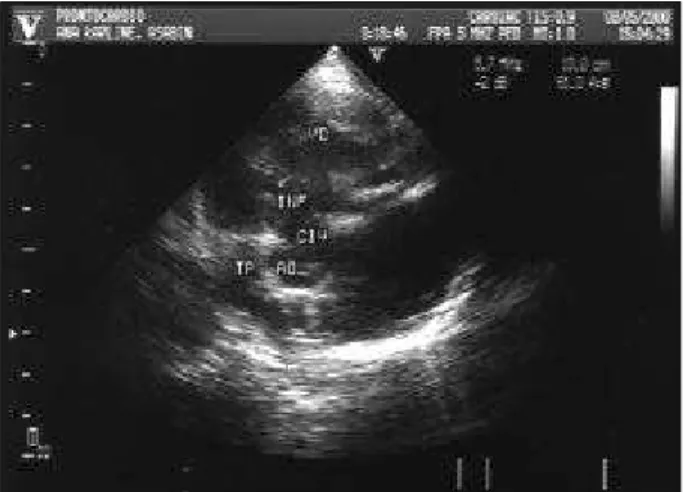
revealed cardiomegaly and a reduction in the pulmonary flow. The echocardiogram showed tetralogy of Fallot associated with severe asymmetric septal hypertrophy (figs. 1 to 4).
During hospitalization, cyanotic crises were observed and reverted with the usual treatment.
Propranolol (2 mg/kg/day) was started orally, as was the treatment for the other pediatric intercurrent events. The patient underwent palliative surgery (systemic to pulmonary artery anastomosis) and evolved with marked improvement in the cyanosis. The postoperative echocardiogram sho-wed that the systemic-pulmonary shunt was patent.
On follow-up at the age of 2 and one-half years, the child continued to be mildly cyanotic and dyspneic, but with no cyanotic crises, and was gaining weight, although a weight-height deficit persisted. The patient also showed great motor progress, was able to communicate using simple words, and was on treatment with oral propranolol (4 mg/kg/ day). The shunt was adequate.
Discussion
In English, we found the report of 3 patients with tetralogy of Fallot associated with hypertrophic
cardiomyo-pathy. One patient was a 13-year-old adolescent male, with a history of dyspnea and cyanosis, whose condition had worsened in the 4 months preceding the report, and who, after the hemodynamic study, began to have repetitive cya-notic crises. The patient underwent a successful aortopul-monary shunt on an emergency basis 6. The other 2 patients
were newborn infants with multiple congenital malforma-tions, who required a systemic-to-pulmonary artery anasto-mosis. One of the latter patients was 3 and one-half years old at the time of the report, was on chronic use of propranolol, and the other, also on chronic use of propranolol, died sud-denly at the age of 10 months 7.
In the French literature, we found 2 studies. The first study was about a newborn with multiple congenital malfor-mations diagnosed on echocardiography on the 10th day of life, who, because of mild cyanosis, was followed up until the age of 5 months, when, after aggravation of the symp-toms, underwent a hemodynamic study and treatment with propranolol. Initially, considerable improvement was obtai-ned, but later, the septal hypertrophy worseobtai-ned, and at the age of 22 months, treatment with verapamil was started 8.
The second study was about 2 adolescents aged 14 and 15
Fig. 2 – In the transverse view of the ventricles, a more marked septal hypertrophy is seen as compared with the posterior wall.
Fig. 3 – In the parasternal short-axis view, deviation of the infundibular septum to the right ventricular outflow tract is seen, causing severe infundibular stenosis with confluent pulmonary trunk and arteries.
Fig. 4 – In the apical 4-chamber view, severe asymmetric septal hypertrophy is evident. Fig. 1 – In the parasternal long-axis view, a marked septal hypertrophy with
Arq Bras Cardiol 2003; 80: 217-9.
Carvalho et al Tetralogy of Fallot and hypertrophic cardiomyopathy
219
years. The first patient had a hemodynamic diagnosis of pulmonary atresia with ventricular septal defect associated with left ventricular concentric hypertrophic cardiomyo-pathy, which evolved to heart failure, and the patient was treated with digitalis and diuretics. The second patient, who had been hemodynamically diagnosed with tetralogy of Fallot associated with left ventricular diffuse hypertrophic cardiomyopathy, underwent definitive correction of the te-tralogy of Fallot with muscle resection and died in the pos-toperative period due to cardiogenic shock 9.
The clinical treatment of tetralogy of Fallot includes an acute approach to the cyanotic crises and prophylaxis with propranolol in patients who are not considered good candi-dates for surgical treatment. In tetralogy of Fallot with a fa-vorable anatomy, total surgical correction is the treatment of choice. When surgery is necessary in patients with an unfavorable anatomy, a palliative procedure with a syste-mic-to-pulmonary artery anastomosis is performed to in-crease pulmonary blood flow 2,3.
The clinical findings in patients with hypertrophic car-diomyopathy varies. Some patients may deteriorate pro-gressively or die suddenly; others, however, may remain stable for many years. The beginning of symptoms during childhood is considered a poor prognostic signal. The treatment of choice of a symptomatic child with hypertro-phic cardiomyopathy is the adrenergic beta-blocker due to its sympatholytic effect. This medication, however, does not have any effect on hypertrophy progression and does not eliminate the risk of sudden death. Calcium channel blockers are another effective alternative. Verapamil is associated with an increase in sudden death in children under the age of 1 year, and, therefore, is not recommended in this age bracket. Its use in other children with hypertro-phic cardiomyopathy still requires deeper investigation.
Patients with arrhythmias should be treated, and amiodaro-ne has been suggested as more effective than verapamil in reducing ventricular arrhythmias. Its use has been limited to children with refractory or lethal arrhythmias due to the risk of severe secondary effects with its chronic use. In patients with bradycardia, maintenance of atrial synchrony with a pacemaker is important. Surgical treatment has been re-served for patients with severe symptoms, with gradients of 50 mmHg or greater, when medicamentous therapy does not provide adequate relief. The use of dual-chamber pacema-kers has been proposed as an alternative to surgical treat-ment with clinical and hemodynamic improvetreat-ment. The efficacy of this treatment during childhood is currently being assessed in several centers. When medical therapy fails, another alternative is heart transplantation 4,5.
Management of patients with tetralogy of Fallot asso-ciated with hypertrophic cardiomyopathy differs greatly from that of patients with tetralogy of Fallot alone, whose clinical and surgical management based on safe evidence in most cases assures good long-term results. The prognosis of patients with hypertrophic cardiomyopathy, however, is poor, mainly when the symptoms appear during childhood as already mentioned.
In our case, we chose palliative surgical treatment with systemic-to-pulmonary artery shunt and maintenance of the therapy with propranolol, adequate to the pharmacological treatment of the 2 heart diseases, because we observed a better prognosis in the preceding cases undergoing this management.
In conclusion, the recognition of tetralogy of Fallot associated with hypertrophic cardiomyopathy is of great importance, because of the therapy, which has not yet been totally established, making the surgical approach difficult, and because of the still unknown long-term prognosis.
References
1. Bum F. The aetiology of congenital heart disease. ln: Anderson RH, Macartney FJ, Shineboume EA, Tynan M, eds. Paediatric Cardiology. London: Churchill Livingstone, 1987: 15-63.
2. Neches WH, Park SC, Ettedgui JA. TetraIogy of Fallot and Tetralogy of Fallot with Pulmonary Atresia. ln: Garson A, Bricker JT, Fisher DJ, Neish SR, eds. The Science and Practice of Paediatric Cardiology. 2nd ed. Baltmore: Williams &
Wilkins, 1998: 1383-411.
3. Zuberbuhler JR. Tetralogy of Fallot. ln: Adams IH, Emmanoulides GC, Rimens-chneider IA, eds. Moss’ Heart Disease in lnfants, Chíldren and Adolescents. 5th
ed. Baltmore: Williams & Wilkins, 1995: 998-1017.
4. Denfield SW, Gajarrski RJ, Towbin JA. Cardiomyopathies. ln: Garson A, Bricker JT, Fisher DJ, Neish SR. eds. The Science and Practice of Paediatric Cardiology: 2nd ed. Baltmore: Williams & Wilkins, 1998: 1851-83.
5. Maron BJ. Cardiomyopathies. ln: Adams IH, Emmanouilides GC, Rimensch-neider TA, eds — Moss’ Heart Disease in Infants, Children and Adolescents; 5th
ed. Baltmore: Williams & Wilkins, 1995: 940-53.
6. Rao AS, Gupta SK, Reddy KN, et al. Tetralogy of Fallot with hypertrophic car-diomyopathy — an unknown combination. Indian Heart J 1989; 41: 344-7. 7. Lewin MB, Towbin JA, Thapar MK, et al. The rare association of letralogy of
Fallot with hypertrophic cardiomyopathy: report of 2 neonatal patients. Tex Heart lnst J 1997; 24: 215-7.
8. Grinnneiser O, Bourlon F, Redjimi M. Une association pathologic rare: tetralo-gie de Fallot et hypertrophie septale asymetruc. Arch Mal Coeur Vaiss 1994; 77: 577-80.