Instituto de Cardiologia do Rio Grande do Sul/Fundação Universitária de Cardiologia Mailing adress: João Ricardo M. Sant’Anna Unidade de Pesquisa do IC/FUC -Av. Princesa Isabel, 395 - 90620-001 - Porto Alegre, RS - Brazil.
Received on 9/26/98 Accepted on 2/24/99
Objective - To assess the changes in ventricular evo-ked responses (VER) produced by the decrease in left ventricular outflow tract gradient (LVOTG) in patients with hypertrophic obstructive cardiomyopathy (HOCM) treated with dual-chamber (DDD) pacing.
Methods - A pulse generator Physios CTM (Biotronik, Germany) was implanted in 9 patients with severe drug-re-fractory HOCM. After implantation, the following condi-tions were assessed: 1) Baseline evaluation: different AV delay (ranging from 150ms to 50 ms) were sequentially pro-grammed during 5 to 10 minutes, and the LVOTG (as deter-mined by Doppler echocardiography) and VER recorded; 2) standard evaluation, when the best AV delay (resulting in the lowest LVOTG) programmed at the initial evaluation was maintained so that its effect on VER and LVOTG could be assessed during each chronic pacing evaluation.
Results - LVOTG decreased after DDD pacing, with a mean value of 59 ± 24 mmHg after dual chamber pace-maker, which was significantly less than the gradient before pacing (98 + 22mmHg). An AV delay >100ms produced a significantly lower decrease in VER depola-rization duration (VERDD) when compared to an AVdelay
≤100ms. Linear regression analyses showed a significant correlation between the LVOTG values and the magnitu-de of VER (r=0.69; p<0.05) in the 9 studied patients.
Conclusion - The telemetry obtained intramyocardial electrogram is a sensitive means to assess left ventricular dynamics in patients with HOCM treated with DDD pacing.
Key words: obstructive hypertrophic cardiomyopathy,
DDD pacing intracardiac electrogram
Arq Bras Cardiol, volume 73 (nº 2), 175-179, 1999
João Ricardo M. Sant’Anna, Raquel Prati, Helmut Hutten, Günter Schreier, Peter Kastner, Renato A. K. Kalil, Paulo R. Prates, Iran Castro, Paulo C. Azambuja, Farid Faes, Ivo A. Nesralla, Max Schaldach
Porto Alegre, RS - Brazil
Ventricular Evoked Response in Patients with Hypertrophic
Obstructive Cardiomyopathy Treated with DDD Pacing
DDD pacing has been recently recognized as an alter-native therapy to reduce left ventricular outflow tract gra-dient (LVOTG) in patients with hypertrophic obstructive cardiomyopathy (HOMC) 1. Although some controversies
still remains 2, most reports show the effectiveness of this
technique to reduce (or even to eliminate) LVOTG 3,
impro-ve clinical status 4 and to increase survival 5 in patients with
HOCM. Doppler echocardiography, a non-invasive exami-nation available in most cardiac clinics, is routinely used to evaluate patients with DDD pacing. Not infrequently, however, it is difficult for the patient to come to the routine evaluation when serial or extra programming are required. An alternative means of evaluating pacing would thus be highly useful.
The summation of action potentials of a group of myocardial fibers around a lead is known as an intramyo-cardial electrogram (IMEG), a signal which is better recor-ded when fractally coated intramyocardial leads 7 are used,
and which is modified according to the status of myocardial fibers(such as under the influence of inotropic or chrono-tropic drugs) 8 or inflammatory responses (episodes of
rejection after heart transplantation) 9, and recorded with an
implantable pacemaker for posterior data processing analysis 10. The sequential analysis of an IMEG can identify
myocardial modifications, and accordingly, it has been used for monitoring rejection in patients post heart transplan-tation 11. This system, known as Cardiac Heart Acute
Rejec-tion Monitor (CHARM) 9,10 has disclosed substantial
pros-pects for a better management of heart transplant patients. The aim of this study is to present a novel system of DDD pacing suitable for obtaining intramyocardial electro-grams in patients with HOCM in an attempt to correlate possible benefits resulting from the decrease in left ventri-cular obstruction (as assessed by Doppler echocardiogra-phy) with recordings of intracardiac electrogram (recorded as ventricular evoked response - VER).
Methods
HOCM were submitted to an atrioventricular (AV) sequential DDD pacemaker implantation, model Physios (Biotronik, Germany). There were 3 males and 6 females, age ranging from 14 to 63 (mean 59) years. All patients had severe drug-refractory symptoms (syncope, effort angina, and dyspnea). Six of them were in New York Heart Association (NYHA) functional class (FC) II for dyspnea, while the others were in FC III. Current therapy consisted of calcium antagonists and
β-blocker or the association of the two. All patients had been under a regular follow-up, with routine Doppler echocar-diography, in which a progressive increase in the LVOTG could be observed. Mean LVOTG measured prior to pace-maker implantation was 92±22mmHg. Clinical characteristics of the patients are shown on table I.
DDD pacemaker lead was transvenously implanted under local anesthesia. Briefly, after venous puncture (cephalic or subclavian vein), atrial and ventricular leads were advanced up to the right atrium (RA) under fluoros-copy. The ventricular lead (TIR 60 BP or LX 60 BP/ Biotronik, Germany) was positioned far from the tricuspid valve at the right ventricular apex. The atrial lead (YP 60 BP or JP 60 BP/ Biotronik, Germany) was positioned inside the right atrial appendage. Electrophysiologic measurements were performed and proper lead insertion was undertaken after respiratory maneuvers. The leads were thus firmly fixed at the site of venous access and connected to a DDD pulse generator Physios CMT (Biotronik, Germany), the gene-rator inserted into a subcutaneous pouch at the subpec-toral region and the incision was closed. During implan-tation, the generator was programmed to VVI mode to a basic rate of 40 or 50 beats per minute, in order to assure it would remain inhibited. Patients were discharged from hos-pital on the second day after the surgery; cardiovascular medication was unchanged.
DDD mode was programmed on the 10th to 12th day of
pacemaker implantation, with an adequate pulse frequency capable of consistently capturing the atrium and main-taining a short AV delay, in order to obtain the lowest possi-ble gradient (as recorded by Doppler echocardiography).
After programming the pacemaker, the IMEG was recorded by telemetry as VER, that is, the initial intracavitary electrogram subsequent to pacemaker stimulation of the
ventricle. At the same time, LVOTG was evaluated and recorded by Doppler echocardiography. The evaluation procedures used were twofold: pacing evaluation: pace-maker was successively programmed to all AV delays avai-lable, whether long (150 to 120ms), medium (100ms) or short (<100ms, usually 75 or 50ms) during 5 to 10 minutes; chronic evaluation - the AV delay producing the lowest LVOTG was used for the final pacing programming and maintained for the following monthly evaluations. During each evaluation, the LVOTG and the VER were recorded with the previously programmed AV delay. If during that evaluation a new AV delay was considered to be more appropriate, it would be maintained for chronic pacing.
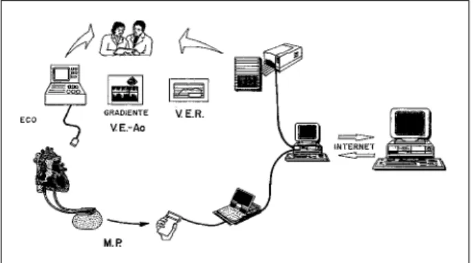
A specific software in a portable laptop base was used to program the pacemaker and record the electric signals, with each signal acquisition lasting for one minute. After adequate signals were recorded, they were sent to the cen-tral data processing site in Graz, Austria, via the Internet, along with the recordings of the corresponding LVOTG. The methodology used to analyze the VER has already been published elsewhere 12,13 (fig. 1).
Signal analyses were automatically performed, with average and representative recordings of VER, as well as the parameters related to each recording. The representative parameters were VER depolarization duration (VERDD), and the negative VER area (VERNA) (fig. 2). Analyses of the signals were sent back in 24 hours (fig. 3).
Monthly evaluations were performed up to the 6th
month of pacemaker implantation, followed by a 6-month interval evaluation. Each evaluation consisted of clinical examination, Doppler echocardiography, pacemaker programming to different AV delays and VER recordings, as previously remarked. The way to obtain the signal and the time of the examination was preserved for each patient, to maintain the same interval between the ingestion of medi-cation and the exam. In case of complimedi-cations associated with clinical deterioration, inadequate medication or pace-maker dysfunction, additional evaluations could be under-taken. The reason for the extra evaluation and any manage-ment modifications were invariably recorded.
LVOTG values obtained at the initial pacing
program-Table I - Clinical characteristics of 9 patients with hypertrophic obstructive cardiomyopathy (HOCM) during pacemaker implantation, and the maximum left ventricular outflow tract gradient, prior to and after pacemaker programming to DDD mode during the final evaluation (LVOTG)
Sex Age Main symptom Medication LVOTG (mmHg)
Initial After procedure Final
Female 41 Angina Propranolol 124 76 45
Male 47 Dyspnea Atenolol, disopyramide 117 81 46
Female 63 Dyspnea Propranolol,disopiramide 83 44 14
Female 59 Fatigue Atenolol, disopyramide 121 100 77
Female 63 Fatigue Propranolol 64 32 11
Male 14 Dizziness Verapamil 105 74
-Female 44 Fatigue Propranolol, verapamil 90 46 27
Male 44 Angina Atenolol 70 29
-ming were recorded and analyzed with a Student t test in order to identify the degree of difference significance.
To identify the influence of AV delay on the ventricular depolarization duration (VERDD), mean values of VERDD were grouped together according to AV delay classification. A paired Student t test was used to assess the difference among the distinct AV delay groups (short, medium and long). Linear regression analyses was used to test the corre-lation between the magnitude of VER and the degree of LVOT obstruction. P values <0.05 were considered to be of statistical significance.
Results
Pacemakers were programmed to DDD mode, at a rate of 70 to 75 beats per minute and an AV delay ranging from 50 to 100ms, resulting in a decrease in LVOTG from 98 ± 22 mmHg to 59±24mmHg (p<0.05). During this period, 20 evaluations were performed in 9 patients, yet 3 patients had only one evaluation performed. In the former, LVOTG measured during the final evaluation showed a value even lower than that recorded at the initial pacing evaluation (table I).
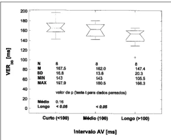
During the 20 follow-up evaluations, it was observed that long AV delays (such as 120 and 150ms) were associa-ted with higher LVOTG than medium (100ms) or short (75 and 50ms) AV delays. Accordingly, VERDD also displayed modifications in most evaluations, since it showed a decrease with long AV delays compared to medium or short AV delays (fig. 4).
Linear regression analyses showed a significant corre-lation between LVOTG and VER NA (r=0.69, p<0.05) (fig. 5).
It should be remarked that during chronic evaluations, patients with reduced LVOTG due to chronic stimulation (LVOTG <30mmHg), and those in whom AV delay modifi-cation did not decrease left ventricular obstruction, VER changes were insignificant.
Discussion
It has been reported that fractally coated leads result in better IMEG signals 7. The sensitivity of a fractally coated
lead, along with proper filtration and amplification of action potential around myocardial fibers allow a better defined electric signal which can be recorded with an implantable pacemaker 10. The intrinsic cardiac signal is called a
spon-taneous action potential, whereas the one resulting from a pacemaker is called evoked action potential. When it is originated in the ventricle it is called ventricular evoked potential (VER). These electrical signals can show modifi-cations in myocardial fibers resulting from inotropic drugs or chronotropic interventions 13,14.
In transplanted hearts with acute graft rejection confirmed by endomyocardial biopsy, a correlation has been shown between the IMEG and the degree of inflammatory process. IMEG has thus been accepted as an alternative means to grade severity of rejection episodes 14. A system
called CHARM, consisting of a DDD pacemaker, a program-mer, computers and a central analyzing site (transmitted via the Internet) is presently under clinical use. This system is likely to offer a rapid, non-invasive and accurate means of detecting acute rejection in heart transplant patients 11, thus
permitting a more effective treatment of these episodes. Considering the Physios pacemaker is capable of re-cording IMEG, and the recorded signals can be
subse-Fig. 1 - Procedures used during the study: Doppler echocardiography to record maximum left ventricular outflow tract gradient (LVOTG) and the acquisition, recording and analyzing intramyocardial electrogram, as ventricular evoked response (VER).
Fig. 2 - Variables used to assess modifications on ventricular evoked response: VERDD, representing duration of ventricular repolarization, VERNA, representing signal magnitude.
Fig. 3 - Variations on ventricular evoked response (VER ) and on left ventricular outflow tract gradient (LVOTG) recorded during acute modifications of AV delay.
AV delay : 120 ms
AV delay : 100 ms
quently analyzed, we attempted to use this pacemaker in a disease that required the use of a universal pacemaker, and further, changes in pacing programming, for instance AV delay or heart rate modifications, were likely to produce changes in myocardial conditions, such as left ventricular overload and hypertrophy. Since HOCM is a disease in which sequential pacing can reduce LVOTG 1,3-5, it was
chosen as an suitable indication to examine this system. The benefit of cardiac stimulation in HOCM can be effecti-vely shown by Doppler echocardiography 6, a non-invasive
method also used for pacemaker programming and moni-toring LVOTG changes.
In the present study, a potential correlation between VER amplitude and morphology modification and varia-tions of maximum LVOTG was assessed, not only during the initial evaluation (acute AV delay modifications) but also during chronic evaluations (an AV delay maintained for a long period).
No ethical considerations hindered the present inves-tigation, since: 1) the parameters used to program the DDD pacemaker were not distinct from those currently used for HOCM therapy; 2) implantation technique and follow-up were not unlike the usual; 3) echocardiographic evaluations were undertaken in shorter intervals than the usual, resulting in a more adequate pacing programming; 4) the acquisition of VER was accomplished by telemetry, which does not offer any risks for the patient.
The methodology used for obtaining IMEG is highly sensitive, considering that the electrical signals represent the
action potentials of m yocardial cells 8. VER can be influenced
by various acute or chronic cardiac interventions 8,10,13, and
these influences must be minimized during the study. A strict protocol was thus followed, including continuing drug therapy administered (or registering any modifications), the same time and place for echocardiographic examination, and whenever possible, the same group of investigators.
VER was the only IMEG used, from two available forms. Patients with HOCM require atrial and ventricular pacing in order to obtain optimum hemodynamic benefits; using spontaneous action potential would result in loss of ventricular stimulation and consequently, LVOTG changes. Using VER permits a predetermined frequency to be used when obtaining the signal, thus eliminating one the variables capable of changing IMEG morphology 13,16,17.
During initial evaluation, AV delay variation resulted in a decrease in LVOTG in most patients. The signal (and the LVOTG) was obtained 5 minutes after pacemaker program-ming. At first it was decided to prolong the interval during signal programming and acquisition to 10 minutes, assu-ming that in some patients the lead might be situated further from the right ventricular apex, left or interventricular sep-tum, and consequently the hemodynamic benefit deter-mined by AV delay modification ( that is, the decrease in LVOTG) would take longer to influence right ventricular dynamics. Against that hypothesis is the idea that VER would translate global modifications on left ventricular geometry thus reflecting instantaneous left ventricular dynamics, even when the lead is distant from that chamber. Prolonging adjustment periods has not disclosed any variations among the comparable signals, and they have further increased evaluation time, so that a 5 minute time after programming the pacemaker is considered satisfactory for obtaining VER.
The selection of the adequate AV delay during follow-up evaluations was modified along the study, but the criteria used to define long (150 or 120ms) medium (100) or short value (75 or 50ms) was always maintained. On the other hand, it was noted that a lengthy echocardiographic exami-nation (resulting from a complete comprehensive study, followed by repeated generator programming and LVOTG evaluations and VER recordings) caused discomfort for the patient and was not practical, since they could induce result variations. Alternatively, minor variations of AV delay, mainly ranging from 100ms to 75ms and from 75ms to 50ms resulted in insignificant changes in LVOTG, and
conse-Fig. 4 - Mean value of VERDD for distinct AV delays, observed in 8 initial evalua-tions (or acute evaluaevalua-tions). Please note that ventricular depolarization period (VERDD) was significantly increased for a shorter AV delay (<100ms), when compared with a medium AV delay (100ms) or a longer one (>100ms).
Fig. 5 - Graph showing the significant correlation between VER magnitude (VER
NA)
1. Futterman LG, Lemberg L. New indications for dual chamber pacing: hyper-trophic and dilated cardiomyopathy. Amer J Critical Care 1995; 4: 82-7 2. Nishnamura RA, Tursty JM, Hayes DL, et al. Dual-chamber pacing for
hyper-trophic obstructive cardiomyopathy: a randomized, doble-blind, crossover study. J Am Coll Cardiol 1997; 29: 345-41.
3. Sadoul N, Simon JP, de Chillou C, et al. Long-term dual-chamber pacing improves hemodynamic function in patients with obstructive hypertrophic cardiomyo-pathy. PACE 1993; 16(5-parte 2): 1120.
4. Fazanapazir L, Cannon RO, Tripodi D, Panza JA. Impact of dual-chamber permanent pacing in patients with obstructive hypertrophic cardiomyopathy with symptoms refractory to verapamil and beta-adrenergic blocker therapy. Circulation 1992; 85: 2149-61.
5. Fananapazir L, Epstein ND, Curiel RV, Panza JA, Tripodi D, McAreavey D. Long-term results of dual-chamber (DDD) pacing in obstructive hypertrophic cardiomyopathy. Circulation 1994; 90: 2731-42 .
6. Shah PM, Gramiak R, Adelman AG, Wigle ED. Echocardiographic assesses-ment of the effects of surgery and propranolol on the dynamics of outlow obstruction in hupertrophic subaortic stenosis. Circulation 1972;45:516- 21. 7. Wetzig, T, Fröhlich R, Bolz A, et al. Messung und Analyse monophasischer Aktionspotentiale mittels fracktal beschichteter Elektroden - Teil II. Biomed Technik 1995; 40: 160-7.
8. Yan S, Blomström-Lundqvist C, Olsson SB. Monophasic action potentials: Concepts to practical applications. J Cardiovasc Electrophysiol 1994;3:287-308. 9. Hutten H, Schreier G, Auer T, Iberer F, Tscheliessnigg KH, Schaldach M. Analysis of epimyocardial ECGs from transplanted hearts. Biotronik Review 1994; 5: 1-3.
References
10. Auer T, Schreier G, Hutten H, Tscheliessnigg KH, Iberer F, Schaldach M. Analysis of the ventricular evoked response for use in cardiac transplanting monitoring. Cardiac Pacing. Anais do EUROPACE 97 - 7th Sympsium on
Cardiac Pacing. (Atenas, Grécia). Bolonha. Itália: Monduzzi Editore SPA, 1995: 495-9.
11. Brofman PRS, Lucchese FA, Leães PE, et al. Monitoração não-invasiva do paciente após tranplante cardíaco: Novos aspectos metodológicos. Progress in Biomedical Research 1997; 2(supl. B): 69-75.
12. Hutten H, Schaldach M. Automatic pacing becames a technical reality. Biomedizinische Technik 1996; 41: 1-11.
13. Wetzig T, Bolz A, Hardt R, et al. Validation of monophasic action potentials for detecting neurohomral influences using special DDD-pacemakers. Biotronik Review 1995; 6: 1-3.
14. Eick RET, Whalley, DW, Rasmussen HH. Connections: Heart disease, cellular electrophysiology, and ion channels. FASEB-Journal 1992; 5: 2568-80. 15. Billing ME (chaiman da International Society for Heart Transplantation). A
working formulation for the standardization of nomenclature in the diagnosis of heart and lung rejection: Heart Rejection Group Study. J Heart Transplantation 1990; 9: 587-93.
16. Hardman SMC, Young GE, Walker T, Biggs WA, Seed MIM, Noble A. A multi-electrode catheter for simulaneous pacing and registration of endocardial monophasic action potential. Erop JCPE 1991; 2: 75-82.
17. Schereier G, Hutten H. Remote intramyocardial electrogram analysis for non-invasive monitoring after heart transplantation. Med Biol Eng Comput 1996; 34: 205-6.
quently on VER. Thus, higher ranges of AV delay changes (such as from 150ms, 100ms and 50ms) resulted in more marked changes of LVOTG (as predictable from the accu-racy of the evaluation method) and VER.
VER modifications due to AV delay modifications showed 2 distinct patterns: 1) they were particularly striking in patients in whom a decrease in AV delay resulted in a significant reduction of LVOTG. The corresponding VER showed a higher duration of depolarization, as seen in figure 4; 2) small variations in LVOTG, as shown in patients with a very favorable response to DDD pacing and who previously presented a maximum systolic gradient of 30 mmHg, resulted in minor VER modifications.
Theses findings support the idea that the VERDD can be used to help select optimum AV delay (AV delay produ-cing the lowest LVOTG).
Serial evaluations along the study have shown that a progressive decrease in LVOTG occurred in all patients. Similarly, AV delay variation, patients who showed marked modification in LVOTG also disclosed important modifi-cation in VER. In one of the patients, it was observed that an inotropic-negative drug (calcium antagonist) resulted
in a higher decrease in LVOTG, together with a modifica-tion in VER.
The correlation observed between the LVOTG and the magnitude of electrical activity of the heart (determined by the VERNA) can have several explanations, yet to be proven. A modification in left ventricular shape or a decrease in wall stress due to the decrease in interventricular pressure might be a possible explanation, however these hypothesis were not assessed in the present study.
Although the present study is limited due to the small number of patients and evaluations, our findings suggest that VER is sensitive enough means to detect hemodynamic changes resulting from AV delay modifications in patients with HOCM. In case these findings are confirmed by larger studies, non-invasive recordings of VER through pace-maker telemetry could become a useful method of program-ming optimum AV delay and monitoring DDD pacing as a therapy in patients with HOCM.