222
Ebaid et al
Ebstein's anomaly with coarctation of the aorta
Arq Bras Cardiol volume 73, (nº 2), 1999
Instituto do Coração do Hospital das Clínicas - FMUSP.
Mailing address: Munir Ebaid - Incor - Av. Enéas C. Aguiar, 44 - 05403-000 - São Paulo, SP, Brazil
Received on 2/22/99 Accepted on 4/24/99
Munir Ebaid, Estela Azeka, Nana Miura Ikari, Eduardo A. Sosa, Miguel Barbero Marcial, Edmar Atik
São Paulo, SP - Brazil
Ebstein’s Anomaly with Coarctation of the Aorta.
An Unusual Association
Brief report
Ebstein’s anomaly with coarctation of the aorta is an extremely unusual condition. In this report, the clinical and surgical features of 3 male patients, aged 7 months, 4 years and 14 years, are discussed. All patients were in situs solitus. The first 2 patients had atrioventricular and ventriculoarterial discordanceand progressed to heart failure in the neonatal period. The third had atrioven-tricular and ventriculoarterial concordance, as well as Wolf-Parkinson-White syndrome, with frequent episodes of paroxysmal tachycardia.
The 3 patients underwent surgery for correction of the coarctation of the aorta. The patient with atrioventri-cular and ventriculoarterial concordance underwent tricuspid valvuloplasty using a DeVega-like technique. In addition, ablation of 2 anomalous pathways (Kent bundle), which were detected by the electrophysiologic study, was also subsequently performed.
The 3 patients showed a good postoperative outcome for 2 years, although, in those with discordance, the sur-gical procedure did not influence the dysplasia of the tricuspid valve, because this valve showed light to mode-rate dysfunction.
Congenital anomalies characterized by tricuspid valve stenotic lesions or regurgitation, alone or combined with other abnormalities, are unusual. The most frequent defect involving this valve is the so-called Ebstein’s anomaly, first described by Wilhelm Ebstein in 1866 1.
This anomaly has a prevalence of 0.5% among the different congenital heart diseases 1. Studies show that lesions such as pervious foramen ovale and atrial septal defect (ASD) are almost always present. Other types of associations, such as stenosis or atresia of the pulmonary valve, ventricular septal defect and transposition of the great arteries, are uncommon 2,3. However, the association of Ebstein’s anomaly with coarctation of the aorta is described as extremely rare, mainly when ventriculoarterial concordance is present 4.
This report describes 3 individuals with Ebstein’s anomaly and coarctation of the aorta, 2 of them in discor-dance and the remaining 1 in atrioventricular and ventri-culoarterial concordance (AV).
Report of the Cases
Patients in atrioventricular and ventriculoarterial discordance - Case 1 – A 7-month-old infant with a past medical history of dyspnea on strenuous effort and a heart murmur detected on the 10th day of life. The physical examination showed an infant in good health, with slight tachydyspnea and without cyanosis. His heart rate (HR) was 160bpm; his systemic blood pressure in the upper limbs was 110/70mmHg. His lungs showed no rales.
On physical examination, visible pulsations at the precordium were noted and his heart auscultation showed a split 2nd heart sound of increased intensity, as well as a moderate systolic murmur in the mitral area, irradiating to the axilla. His liver was felt 4cm below the right costal margin. He had a strong pulse in the upper limbs and no pulse in the lower limbs.
His chest X-ray showed a moderately enlarged cardiac silhouette, as well as an increased pulmonary vasculature. The electrocardiogram (ECG) revealed sinus rhythm and a pattern of significant left ventricular hypertrophy. The vectorcardiogram, however, was consistent with biventricular hypertrophy with predominance of the left-sided chamber.
The Doppler echocardiography showed AV and ven-triculoarterial discordance combined with Ebstein’s anomaly of the tricuspid valve, ASD and coarctation of the aorta, with a pressure gradient of 40 mmHg between the ascending and the descending aorta.
The angiocardiographic and hemodynamic evalua-tions confirmed the clinical diagnosis.
At that time, due to the lack of clinically significant consequences of the regurgitation of the left AV valve, only the correction of the coarctation of the aorta with termino-terminal anastomosis was performed.
Arq Bras Cardiol volume 73, (nº 2), 1999
Ebaid et al Ebstein's anomaly with coarctation of the aorta
223 Case 2 – A 4-year-old male patient with a past medical
history of congestive heart failure that had started in the 1st week of life. At that time, he was hospitalized for 12 days and was discharged with a prescription for digitalis. He remai-ned asymptomatic until his current age, when a heart mur-mur was detected, and he was then referred for diagnostic tests. He was in good health, his breathing was normal, and he did not have cyanosis. His pulse was strong in the upper limbs but absent in the lower limbs. His blood pressure was 150/90mmHg in the upper limbs. His lungs showed no rales. The auscultation of the precordium revealed a 1st heart sound more audible in the mitral area, a split 2nd heart sound, with increased intensity in the pulmonary area, and a moderately intense systolic heart murmur with diastolic vibrations in the mitral area.
The chest X-ray revealed an enlarged cardiac silhoue-tte and an increased pulmonary vasculature.
The ECG performed when the patient was 10 days old showed a QRS complex of the qR type and a positive T wave in lead V1 (fig. 1). At the age of 4 years, his ECG showed a pattern of left ventricular hypertrophy with QS morphology from lead V1 to lead V3 and R morphology in leads V5 and V6 (fig. 1).
Doppler echocardiography, combined with the hemo-dynamic study, revealed AV and ventriculoarterial discor-dance, coarctation of the aorta and tricuspid regurgitation of moderate repercussion. The pressure gradient between the ascending and the descending aorta was 55mmHg. The patient underwent termino-terminal anastomosis to correct just the aortic coarctation. Currently, he remains in good health.
Patient with atrioventricular and ventriculoarterial concordance - Case 3 - A 14-year-old male teenager with a heart murmur detected at the age of 2 months. At that time, he was diagnosed with coarctation of the aorta. The hemodynamic study revealed an increased end-diastolic
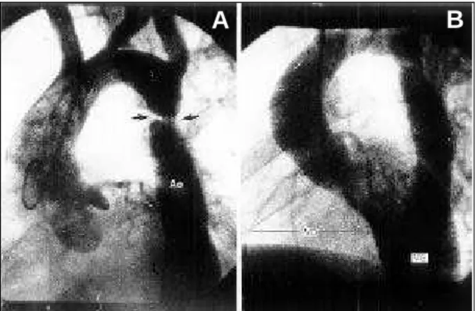
pressure of the left ventricle (LV) and normal pressures in the right chambers as follows: mean right atrial pressure 7, right ventricular pressure (26/6mmHg), pulmonary artery pressure (28/14/20mmHg), mean pulmonary capillary pressure (13 mmHg), left ventricular pressure (105/20mmHg) and pressure of the ascending aorta (105/70/85mmHg). The angiographic study showed a moderate constriction of the initial portion of the descending aorta and an enlarged right ventricular chamber (fig. 2). The patient underwent surgery at the age of 6 years for correction of the coarctation of the aorta; the technique of termino-terminal anastomosis was used. He remained asymptomatic until the age of 14, when he was admitted to the Emergency Department of the Clinic due to a syncopal episode.
On physical examination at admission, he was in good health, his respiration was normal, he was not pale and did not have cyanosis; his HR was 70bpm; his systemic blood pressure was 120/70mmHg. His lungs showed no rales. His precordium did not show any deformities and his heart auscultation revealed a regular heart rhythm, with a slight regurgitation murmur in the tricuspid area. His pulse was symmetric and of normal amplitude in the 4 limbs.
The chest X-ray revealed a moderately enlarged cardiac silhouette and a normal pulmonary vasculature.
The ECG showed a pre-excitation pattern (Wolf-Parkinson-White) of the B type and the vectorcardiogram revealed a QRS shifted to the left, backwards and upwards, with counterclockwise rotation at the horizontal and frontal planes, as well as a clearly delayed conduction time in the initial and end portions. An electrophysiological study was performed in this patient and revealed 2 anomalous bundles (one anteroseptal and the other posterolateral).
The echocardiography revealed Ebstein’s anomaly with moderate tricuspid regurgitation and effective aortic isthmoplasty, without pressure gradient at the site of the surgical correction of the coarctation. The hemodynamic and angiographic studies confirmed the diagnosis of Ebstein’s anomaly.
Fig. 1 – A) Case 2 - electrocardiogram showing a qR pattern in lead V1 due to atrioven-tricular and ventriculoarterial discordanceand hypertrophy of the ventricle placed on the right (left ventricle) on the 10th day of life; B) electrocardiogram of the same patient
when he was 4 years old, showing hypertrophy of the ventricle placed on the left as a result of the coarctation of the aorta combined with the malformation of the left atrio-ventricular valve (right ventricle), in addition to anterosuperior divisional heart block.
A
B
224
Ebaid et al
Ebstein's anomaly with coarctation of the aorta
Arq Bras Cardiol volume 73, (nº 2), 1999
Ablation of the anomalous bundles and tricuspid valvuloplasty according to the De Vega technique were performed. The immediate electrocardiographic and vectorcardiographic studies showed morphology of right bundle-branch block (RBBB) with the features observed in Ebstein’s anomaly (fig. 3). Two years later, the patient remains in good health and is not receiving any medication. Fig. 3 – A) Electrocardiogram of the patient with atrioventricular and ventri-culoarterial concordance, revealing pre-excitation syndrome; B) electrocardiogram performed after the ablation of the anomalous bundles, showing a morphology of right bundle-branch block, a common finding in Ebstein’s anomaly.
A
B
Discussion
Coarctation of the aorta is one of the most unusual abnormalities associated with Ebstein’s anomaly 5.
In patients with Ebstein’s anomaly that show AV and ventriculoarterial discordance, the coarctation of the aorta is less unusual than in those with concordance 6; this may occur due to the lower anterograde flow in the ascending aorta during the intrauterine life.
The coarctation of the aorta aggravates the clinical findings, especially when it plays a direct role in the malfor-mation of the tricuspid valve, as occurs with ventriculo-arterial discordance. In fact, the 2 patients with discordance had heart failure during the neonatal period.
Arrhythmias are common in Ebstein’s anomaly and occur in more than 50% of these patients. The most frequent are supraventricular paroxysmal tachyarrhythmias and atrial flutter or fibrillation, as a result of right atrial dysfunc-tion or anomalous muscular bundles, as observed in the 3rd patient. In that case, the electrophysiologic study revealed 2 anomalous pathways that were responsible for the episo-des of paroxysmal tachycardia. Of note in this case was the ECG with a morphology of RBBB after the ablation of the bundles; this is a common finding in Ebstein’s anomaly.
The initial decision of correcting only the coarctation of the aorta in the 3 cases and of adopting a conservative approach in relation to the tricuspid valve regurgitation in the 2 patients in whom the transposition had been corrected took into account the grade of dysfunction. This dysfunc-tion was mild compared with the moderate hemodynamic repercussion, and was expected to decrease with the elimi-nation of the aortic obstruction; this actually occurred. Only the 3rd patient required surgical therapy for the AV valve because this valve would not be effectively improved with the surgical therapy of the coarctation alone.
In conclusion, Ebstein’s anomaly and coarctation of the aorta are rarely associated, mainly when a patient presents with AV and ventriculoarterial concordance.
1. Ebstein W. Uber einen sehr seltenen fall von insufficienz der valvula tricus-pidalis, belingt durch eine angeborene hoch-gradige Missildung derselben. Arch Anat Physiol 1866; 33: 238-54.
2. Ebaid M, Vila JH, Pedroso CO, Stolf NAG, Verginelli G, Macruz R. Anomalia tipo Ebstein associada a comunicação interventricular. Relato de 4 casos. Arq Bras Cardiol 1990; 34: 129-33.
3. Ferreira SMF, Ebaid M, Aiello VD. Ebstein’s malformation of the tricuspide and mitral valvr associated with hypoplasia of ascending aorta. Int J Cardiol 1991; 33: 170-2.
References
4. Salkar HR, Salkar RG, Sengupta PP. Ebstein’s Anomaly with coarctation of the aorta – an unusual association. Indian Heart J 1996; 48: 283-4.
5. Radford DJ, Graff RF, Nielson GH. Diagnosis and natural history of Ebstein’s anomaly. Br Heart J 1985; 54: 517-22.