w w w . r b o . o r g . b r
Original
Article
Distal
femoral
cut
in
total
knee
arthroplasty
in
a
Brazilian
population
夽
Marcos
Areias
Vieira
Costa,
Alan
de
Paula
Mozella
∗,
Hugo
Alexandre
de
Araujo
Barros
Cobra
InstitutoNacionaldeTraumatologiaeOrtopedia(INTO),RiodeJaneiro,RJ,Brazil
a
r
t
i
c
l
e
i
n
f
o
Articlehistory: Received6June2014 Accepted10July2014 Availableonline12June2015
Keywords: Kneearthroplasty Alignmentinarthroplasty Femoralcut
a
b
s
t
r
a
c
t
Objective:Todeterminetheidealangleformakingthedistalfemoralcutintotalknee arthro-plastyinaBrazilianpopulation.
Methods:Panoramic radiographs of the lower limbs bearing weight from 79 patients (57womenand22men)werestudied,totaling107kneeswithanindicationfortotalknee arthroplasty.Thefemoralanatomicalaxis,femoralmechanicalaxisandcervical-diaphyseal angleweretracedout.Theangleofthefemoralcutwasdeterminedfromthemeetingpoint betweenthefemoralanatomicalandmechanicalaxes.Theidealdegreeoffemoralvalgus wascomparedbetweenmenandwomenandbetweenkneespresentingvarusand val-gusalignmentofthelowerlimb.Theidealdistalfemoralcutwasalsocorrelatedwiththe cervical-diaphysealangle.
Results:The idealfemoralvalgusanglerangedfrom4.2to8.6degrees,witha meanof 6.3degrees.Therewasnostatisticallysignificantdifferenceinthedistalfemoralcutbetween patientswithcoronalvarusandvalgusalignment(p=0.180).Comparingmenandwomen, therewasnostatisticallysignificantdifferenceregardingtheidealfemoralvalgusbetween thegroups(p=0.057).Thecervical-diaphysealanglepresentedaninverserelationshipwith thedistalfemoralcut.
Conclusions: Themeananglebetweenthefemoralmechanicalandanatomicalaxeswas 6.3degree.Neitherpreoperativecoronalalignmentnorsexhadanyinfluenceonthedistal femoralcut.Thecervical-diaphysealanglepresentedaninverserelationshipwiththedistal femoralcut.
©2014SociedadeBrasileiradeOrtopediaeTraumatologia.PublishedbyElsevierEditora Ltda.Allrightsreserved.
夽
WorkdevelopedattheKneeSurgeryCenter,InstitutoNacionaldeTraumatologiaeOrtopedia(INTO),RiodeJaneiro,RJ,Brazil. ∗ Correspondingauthor.
E-mail:[email protected](A.d.P.Mozella). http://dx.doi.org/10.1016/j.rboe.2015.05.007
Corte
femoral
distal
na
artroplastia
total
de
joelho
na
populac¸ão
brasileira
Palavras-chave: Artroplastiadojoelho Alinhamentoemartroplastia Cortefemoral
r
e
s
u
m
o
Objetivo: Determinaroânguloidealparafeituradocortefemoraldistalnaartroplastiatotal dojoelhoempopulac¸ãobrasileira.
Métodos: Foramestudadasradiografiaspanorâmicas comcargadosmembrosinferiores em79pacientes(57mulherese22homens),numtotalde107joelhoscomindicac¸ãode artroplastiatotal.Foramtrac¸adosoeixoanatômicofemoral(EAF),oeixomecânicofemoral (EMF)eoângulocervicodiafisário(âCD).Oângulodocortefemoraldistalfoideterminado pelo encontroentreoEMFeoEAF.Ovalordovalgofemoralidealfoicomparadoentre homensemulhereseentrejoelhoscomalinhamentoemvaroevalgodomembroinferior. Ocortefemoraldistalidealfoicorrelacionadoaindacomoângulocervicodiafisário. Resultados: Oângulodovalgofemoralidealvarioude4,2até8,6graus, commédia de 6,3. Ocorte femoraldistal nãomostrou diferenc¸a quandocomparados pacientescom alinhamentocoronalemvaroevalgo,semsignificânciaestatística(p=0,180).Quando com-paradoshomensemulheres,ovalgofemoralidealnãomostroudiferenc¸aentreosgrupos estatisticamentesignificante(p=0,057).Oângulocervicodiafisáriomostrourelac¸ãoinversa comocortefemoraldistal.
Conclusões: Amédiadoânguloentreoseixosmecânicofemoraleanatômicofemoralfoide 6,3graus.Alinhamentocoronalpré-operatório,assimcomoosexo,nãoexerceuinfluência nocortefemoraldistal.Oângulocervicodiafisáriomostrourelac¸ãoinversacomocorte femoraldistal.
©2014SociedadeBrasileiradeOrtopediaeTraumatologia.PublicadoporElsevier EditoraLtda.Todososdireitosreservados.
Introduction
Numerous studies have shown a correlation between the durabilityoftotalkneearthroplasty(TKA)andrestorationof thenormallimbalignment.1–3Itisbelievedthatrestorationof themechanicalaxiswithamaximumvariationof3◦ toward varusorvalgusisassociatedwiththebestresultsfromTKA.1–6 However,someauthorshavedemonstratedthatpostoperative alignmentsofthelimboutsideoftheinterval of±3◦ inthe coronalplaneareobservedinupto30%ofthecases.7–9
Innormalknees,thetibialjointsurfaceisatavarusangleof approximately3◦inrelationtothemechanicalaxis,whilethe femoralsurfaceisatavalgusangleof9◦.Historically,attempts havebeen madetoreproducethisanatomicalalignmentof thekneeintotalarthroplastybycuttingthetibiaatavarus angle.However,severalstudieshavedemonstratedthattibial componentsplacedatvarusanglesgreaterthan5◦tendtofail duetomedialcollapse.3,10
IncorrectalignmentofTKAhasbeenidentifiedasacause oflong-termcomplications,including acceleratedwear,11,12 premature mechanical loosening of the implant1,13,14 and patellofemoralproblems15–17suchaspatellofemoral instabil-ityandpatellarfracture.
Thus,itisrecommendedthatthetibialcomponentshould beimplantedperpendicularlytothemechanicalaxisofthe tibiainthecoronalplane.Thefemoralcomponentisusually implantedatavalgusangleof5◦to6◦,whichisthesizeofangle supposedlynecessaryforreestablishinganeutralmechanical axisinthelimb.
Theaimofthepresentstudywastomeasuretheidealangle formaking thedistalfemoralcutinBrazilianpatientswho
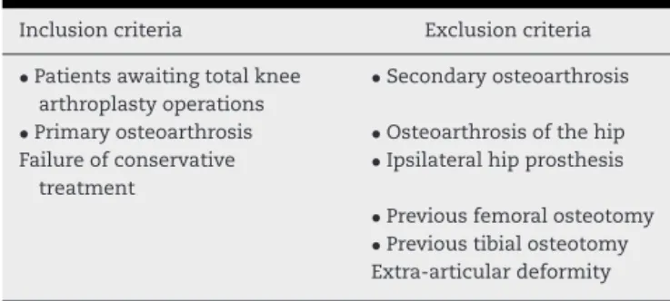
Table1–Inclusionandexclusioncriteria.
Inclusioncriteria Exclusioncriteria
•Patientsawaitingtotalknee arthroplastyoperations
•Secondaryosteoarthrosis
•Primaryosteoarthrosis •Osteoarthrosisofthehip Failureofconservative
treatment
•Ipsilateralhipprosthesis
•Previousfemoralosteotomy
•Previoustibialosteotomy Extra-articulardeformity
underwent TKAattheKneeSurgeryCenterofthe National InstituteofTraumatologyandOrthopedics(INTO).
Material
and
methods
Between August 2011and February 2012,panoramic radio-graphson79patients(22menand57women)wereanalyzed, thustotaling107limbs,inaccordancewiththeinclusionand exclusioncriterialistedinTable1.
Thisstudywassubmittedforevaluationandapprovalby ourinstitution’sresearchethicscommittee.
Radiographic
evaluation
Fig.1–Panoramicradiographofthelowerlimbs.(1) Femoralanatomicalaxis.(2)Femoralmechanicalaxis.(3) Cervicodiaphysealangle.(4)Idealfemoralvalgus.
performedwiththepatients positionedwiththeirlimbsat neutralrotationandmaximumextension.
Inalltheradiographicexaminations,wedefined:(1)the anatomicalaxisofthefemoraldiaphysis;(2)themechanical femoralaxis;and(3)thecervicodiaphysealangle.
Themechanicalaxisofthefemoraldiaphysiswasdefined byastraightlinethatjoinedthecentersoftwocirclesthat weretangentialtothemedialandlateralcorticalboneofthe femur.Thefirstcirclewaslocated2cmdistallytothelesser trochanter.Thesecondwasatthejunctionbetweenthedistal metaphysisandthefemoraldiaphysis,asdeterminedusing Heim’ssquare.18
Themechanicalaxiswasdefinedinaccordancewiththe currentconceptsintheliterature,asastraightlinepassing throughthecenterofthefemoralheadtothemidpointofthe widthofthedistalfemur.
Theideal distal femoral cut angle corresponded to the intersection between the anatomical axis and the femoral mechanicalaxis(Fig.1).
Measurementof theangle formedbetween the femoral anatomicalaxisandthelineofthefemoralneckdefinedthe cervicodiaphysealangle.Two circlesthatweretangentialto thelowercortexanduppercortexofthefemoralneckwere usedtotraceoutthelineoftheneck.
Themeasurementswerealwaysmadebytwoevaluatorsat differenttimesusingthesameinstrumentswithprecisionof theorderofmillimeters.
Statistical
analysis
Statisticalanalysiswasperformedwiththeaimofevaluating thedegreeofsignificanceoftheparametersmeasured.The Shapiro-WilksWtestwasusedtoevaluatewhetherthe vari-ancehadnormaldistributionandtheLevenetestwasused toassessitshomoscedasticity.Thedistalfemoralcutangles andthecervicodiaphysealangleswereconsideredtohave nor-maldistributionandStudent’sttestwasusedtocomparethe means.TheStatistica8.0softwarewasusedforthestatistical calculations.
Results
Seventy-ninepatients(22menand57women)werestudied, withatotalof107limbs.Thepatients’meanagewas67years, witharangefrom58to86.Surgerywasperformedontheright sidein53casesandontheleftsidein54cases.
Seventy knees presented alignment with varus angles between 3◦ and 20◦ (mean 8.4◦; standard deviation 3.5◦). Twenty-sixkneespresentedpreoperativealignmentwith val-gusanglesbetween2.7◦and16◦(mean6.6◦;standarddeviation 3.1).Neutralpreoperativealignmentwasobservedin11cases. Theidealfemoralvalgusanglerangedfrom 4.2◦ to8.6◦, withameanof6.3◦.Fig.2showsthedistributionoftheideal distalfemoralcutforthepatientsstudied.
Themalepatientspresentedanidealdistalfemoralcutof 6.6◦(rangefrom4.9◦to8◦),whileforthewomen,6.2◦wasthe idealangleforthedistalfemoralcut(rangefrom4.2◦to8.6◦). Amongthe patients with preoperativevarus alignment, theidealdistalfemoralcutwas6.2◦(rangefrom4.2◦to8.4◦). Amongthepatientswithpreoperativevalgusalignment,the idealfemoralvalgusanglewas6.5◦(rangefrom4.2◦to8.6◦).
Thecervicodiaphysealanglerangedfrom114.3◦to138.3◦, withameanof127.2◦.Fig.3correlatesthedistalfemoralcut andcervicodiaphysealanglevaluesforeachpatient.
Among the male patients, the mean cervicodiaphyseal anglewas127.5◦ (rangefrom118.1◦ to138.3◦),whileamong thewomenitwas127◦(rangefrom114.3◦to136.5◦).Themean cervicodiaphyseal angleidentifiedamongthepatientswith preoperativevarusalignmentwas127.2◦ (rangefrom114.3◦ to136.5◦),whileamongthosewithpreoperativevalgus align-ment,itwas127.1◦(rangefrom115.1◦to138.3◦).
0 5 10 15 20 25 30 35 40
No. of patients
4 5 6
Distal femoral cut
7 8 9
Cervicodiaphyseal angle General
110 4 4.5 5 5.5 6 6.5 7 7.5 8 8.5 9
115
120
125 y=–0.1061x +19.87
Femoral valgus
130
135
140
Fig.3–Correlationbetweenthedistalfemoralcutandthecervicodiaphysealangle.
StatisticalanalyseswereperformedusingStudent’sttest witha95%confidenceinterval.Nostatisticaldifferenceinthe idealdistalfemoral valgusvaluesor the cervicodiaphyseal anglewasobservedbetweenthemenandwomen(p=0.57). Thestatisticalanalysisontheseanglesalsodidnotshowany statisticaldifferenceincomparingthepreoperativevarusand valgusdeformities(p=0.18).
Discussion
Theanalysisontheidealfemoralvalgusangleshowedsmall absoluteanglevalues.Thus,preciseradiographicstandards needtobeused inpanoramic radiographs,especially with regardtocontrollingtheexternalrotationofthelowerlimbs whiletheexaminationisbeingperformed.Radiographswith rotationaldeviationofthelowerlimbs,whichmostfrequently occursduringexternalrotation,producelargerfemoral val-gusangles becauseoftheanatomicalbowingofthe femur alongthesagittalaxis.Thisbowingalsoimpedesproper mea-surementoftheanatomicalaxisofthefemoralcanal.Forthis reason,onlyradiographswithperfectrotationalcontrol,such thatthelessertrochanterdidnotappearandthepatellawas centralizedontheknee,wereincludedinthisstudy.19
Anothercomplicatingfactorindeterminingtheangleswas extra-articulardeformity,whichalteredtheaxesandangles analyzed unpredictably. Radiographs with extra-articular deformitywereexcludedfromthestudy.
OneoftheobjectivesofTKAistorestoreneutralalignment ofthelowerlimbthroughmakingbonecutsperpendicularly tothemechanicalaxesofthefemurandtibia.20Itisacommon practiceamongmanysurgeonstousethesamedistalfemoral cutangleforallpatientsandtoassumethatthereisminimal variationintheanglebetweenthemechanicaland anatom-icalaxesofdifferentpatients’knees.However,somestudies haveadvocatedpreoperativeplanningusingpanoramic radio-graphsasameansofobtainingalignmentthatismoreprecise andindividualized.21,22
Themeanvalueofthedistalfemoralcutthatwasfound forthepresentstudypopulationdidnotshowanystatistical differenceinrelationtowhatwasfoundbyResendeetal.23 inanotherBrazilianpopulation.If wehadempiricallyused the mean anglefound forthe patientsin thisstudy, allof themwouldhavehadacceptablealignment,whiletakinginto accountapermissibleerrorofupto3◦,asputforwardinthe literature.24,25 Thisdivergesfrom the data ofoneBrazilian author,whofoundthat19.7%ofthepopulationoperatedhad insufficientalignment,basedonanacceptableerrorof3◦in thecoronalplan.23
Despiteatendencyforthedistalfemoralcuttobegreaterin menthaninwomen(6.6◦versus6.2◦),therewasnostatistical differencebetweenthegroups,whichisconcordantwiththe currentliterature.23,26,27
Weidentifiedaninversetrendbetweenthe cervicodiaphy-sealanglevaluesandtheidealdistalfemoralcutvalues.This wasduetothegreaterdistanceofthediaphysisfromthe cen-tralaxisofthebodyinthefemoralneckswithgreatervarus angleandthesmallerdistanceofthediaphysisfromthe cen-tralaxisofthebodyinthefemoralneckswithgreatervalgus angle.
Thepreoperativecoronalalignmentdidnotsignificantly correlatewiththedistalfemoralcutinthisstudy.Thedistal femoralcutvaluewasrelatedtotheanatomicalfactorsofthe femur,withoutusinganytibialparametertodetermineit.This makesusthinkthattheoverallalignmentofthelimbdoes notinfluencethedistalcut.Ontheotherhand,Deakinetal.20 demonstratedarelationshipbetweenthedistalfemoralcut and thealignment ofthe lowerlimb,whichshould beless than 6◦ invalguscasesandgreater than6◦ inseverevarus cases.
Conclusion
Thepreoperativecoronalalignmentandsexdidnothave anyinfluenceonthedistalfemoralcut.
Thecervicodiaphyseal anglehadaninverserelationship withthedistalfemoralcut.
Conflicts
of
interest
Theauthorsdeclarenoconflictsofinterest.
r
e
f
e
r
e
n
c
e
s
1. BargrenJH,BlahaJD,FreemanMA.Alignmentintotalknee arthroplasty.Correlatedbiomechanicalandclinical observations.ClinOrthopRelatRes.1983;(173):178–83. 2. BäthisH,PerlickL,TingartM,LüringC,ZurakowskiD,GrifkaJ.
Alignmentintotalkneearthroplasty.Acomparisonof computer-assistedsurgerywiththeconventionaltechnique.J BoneJointSurgBr.2004;86(5):682–7.
3. JefferyRS,MorrisRW,DenhamRA.Coronalalignmentafter totalkneereplacement.JBoneJointSurgBr.1991;73(5): 709–14.
4. LotkePA,EckerML.Influenceofpositioningofprosthesisin totalkneereplacement.JBoneJointSurgAm.1977;59(1): 77–9.
5. RandJA,CoventryMB.Ten-yearevaluationofgeometrictotal kneearthroplasty.ClinOrthopRelatRes.1988;232:168–73. 6. RitterMA,FarisPM,KeatingEM,MedingJB.Postoperative
alignmentoftotalkneereplacement.Itseffectonsurvival. ClinOrthopRelatRes.1994;(299):153–6.
7. PetersenTL,EnghGA.Radiographicassessmentofknee alignmentaftertotalkneearthroplasty.JArthroplasty. 1988;3(1):67–72.
8. MahaluxmivalaJ,BankesMJ,NicolaiP,AldamCH,AllenPW. Theeffectofsurgeonexperienceoncomponentpositioning in673PressFitCondylarposteriorcruciate-sacrificingtotal kneearthroplasties.JArthroplasty.2001;16(5):635–40. 9. MielkeRK,ClemensU,JensJH,KershallyS.Navigationin
kneeendoprosthesisimplantation–preliminaryexperiences andprospectivecomparativestudywithconventional implantationtechnique.ZOrthopIhreGrenzgeb.2001;139(2): 109–16.
10.TewM,WaughW.Tibiofemoralalignmentandtheresultsof kneereplacement.JBoneJointSurgBr.1985;67(4):551–6. 11.EckhoffDG,PiattBE,GnadingerCA,BlaschkeRC.Assesing
rotationalalignmentintotalkneearthroplasty.ClinOrthop RelatRes.1995;1995(318):176–81.
12.WasielewskiRC,GalanteJO,LeightyRM,NatarajanRN, RosenbergAG.Wearpatternsonretrievedpolyethylenetibial insertsandtheirrelationshiptotechnicalconsiderations duringtotalkneearthroplasty.ClinOrthopRelatRes.1994; 299:31–43.
13.HoodRW,VanniM,InsallJN.Thecorrectionofknee alignmentin225consecutivetotalcondylarknee replacements.ClinOrthopRelatRes.1981;(160):94–105. 14.MorelandJR.Mechaismsoffailureintotalkneearthroplasty.
ClinOrthopRelatRes.1988;1988(226):49–64.
15.BergerRA,RubashHE,SeelMJ,ThompsonWH,CrossettLS. Deteriningtherotationalalignmentofthefemoral
componentintotalkneearthroplastyusingtheepicondylar axis.ClinOrthopRelatRes.1993;1993(286):40–7.
16.ArimaJ,WhitesideLA,McCarthyDS,WhiteSE.Femoral rotationalalignment,basedontheanteroposterioraxis,in totalkneearthroplastyinavalgusknee.Atechnicalnote.J BoneJointSurgAm.1995;77(9):1331–4.
17.FiggieHE3rd,GoldbergVM,FiggieMP,InglisAE,KellyM, SobelM.Theeffectofalignmentoftheimplantonfractures ofthepatellaaftercondylartotalkneearthroplasty.JBone JointSurgAm.1989;71(7):1031–9.
18.HeimUF.Definingtheboundarybetweendiaphysisand metaphysisusingquadrantmeasurement.Acontributionto theclassificationanddocumentationoffracturesoflong tubularbonesexemplifiedbythedistaltibia.Unfallchirurg. 1987;90(6):274–80.
19.SkyttäET,HaapamäkiV,KoivikkoM,HuhtalaH,RemesV. Reliabilityofthehip-to-ankleradiographindeterminingthe kneeandimplantalignmentaftertotalkneearthroplasty. ActaOrthopBelg.2011;77(3):329–35.
20.DeakinAH,BasanagoudarPL,NunagP,JohnstonAT,Sarungi M.Naturaldistributionofthefemoralmechanical-anatomical angleinanosteoarthriticpopulationanditsrelevancetototal kneearthroplasty.Knee.2012;19(2):120–3.
21.RauhMA,BoyleJ,MihalkoWM,PhillipsMJ,Bayers-TheringM, KrackowKA.Reliabilityofmeasuringlong-standinglower extremityradiographs.Orthopedics.2007;30(4):299–303. 22.PatelDV,FerrisBD,AichrothPM.Radiologicalstudyof
alignmentaftertotalkneereplacement.Shortradiographsor longradiographs?IntOrthop.1991;15(3):209–10.
23.RezendeFC,FerreiraMC,DebieuxP,FrancioziCE,LuzoMV, CarneiroM.Éseguroocortefemoraldistalemartroplastia totaldojoelhocom5◦a6◦devalgoempiricamentena
populac¸ãogeriátricabrasileira?RevBrasOrtop.2013;48(5): 421–6.
24.AkagiM,OhM,NonakaT,TsujimotoH,AsanoT,Hamanishi C.Ananteroposterioraxisofthetibiafortotalknee arthroplasty.ClinOrthopRelatRes.2004;420:213–9. 25.McGroryJE,TrousdaleRT,PagnanoMW,NigburM.
Preoperativehiptoankleradiographsintotalknee arthroplasty.ClinOrthopRelatRes.2002;2002(404):196–202. 26.HsuRW,HimenoS,CoventryMB,ChaoEY.Normalaxial
alignmentofthelowerextremityandload-bearing distributionattheknee.ClinOrthopRelatRes.1990;(255): 215–27.