SOCIEDADE BRASILEIRA DE ORTOPEDIA E TRAUMATOLOGIA
w w w . r b o . o r g . b r
Original
article
Correlation
between
Ahlbäck
radiographic
classification
and
anterior
cruciate
ligament
status
in
primary
knee
arthrosis
夽
Glaucus
Cajaty
Martins
a,∗,
Gilberto
Luis
Camanho
b,
Leonardo
Marcolino
Ayres
a,
Eduardo
Soares
de
Oliveiras
aaHospitalFederaldeIpanema,Servic¸odeOrtopediaeTraumatologia,RiodeJaneiro,RJ,Brazil
bUniversidadedeSãoPaulo,FaculdadedeMedicina,DepartamentodeOrtopediaeTraumatologia,SãoPaulo,SP,Brazil
a
r
t
i
c
l
e
i
n
f
o
Articlehistory:
Received24January2016 Accepted18February2016 Availableonline30December2016
Keywords:
Anteriorcruciateligament Kneearthrosis
Arthroplasty,knee
a
b
s
t
r
a
c
t
Objective:Tocorrelatethe Ahlbäckradiographic classificationwiththeanterior cruciate
ligament(ACL)statusinkneearthritispatients.
Methods:Thestudyevaluated89kneesofpatientswhounderwenttotalkneearthroplasty
duetoprimaryosteoarthritis:16maleand69females,withmeanage69.79years(53–87 years).OsteoarthritiswasclassifiedradiographicallybytheAhlbäckradiographic classifi-cationintofivegrades.TheACLwasclassifiedinthesurgeryaspresentorabsent.The correlationofACLstatusandAhlbäckclassificationwasassessed,aswellasthoseofACL statusandtheparametersage,gender,andtibiofemoralangulation(varus–valgus).
Results:Incasesofvarusknees,therewasacorrelationbetweengradesItoIIIandACL
pres-encein41/47(86.7%)casesandbetweengradesIVandVandACLabsencein15/17(88.2%) cases (p<0.0001). In valgus knees, no statistically significant correlation wasobserved betweentheACLstatusandtheAhlbäckclassification.Inthepresentstudy,absenceof theACLwasmorecommoninmen(9/17;52%)thaninwomen(19/72;26%).
Conclusion: Incasesofmedialosteoarthritis,theAhlbäckradiographicclassificationisa
usefulparametertopredictACLstatus(presenceorabsence).Ingonarthritisingenuvalgum, ACLstatuswasnotpredictedbyAhlbäck’sclassification.
©2016SociedadeBrasileiradeOrtopediaeTraumatologia.PublishedbyElsevierEditora Ltda.ThisisanopenaccessarticleundertheCCBY-NC-NDlicense(http:// creativecommons.org/licenses/by-nc-nd/4.0/).
夽
StudyconductedattheHospitalFederaldeIpanema,RiodeJaneiro,RJ,Brazil,andattheDepartmentofOrthopedicsandTraumatology, FacultyofMedicine,UniversityofSãoPaulo,SãoPaulo,SP,Brazil.
∗ Correspondingauthor.
E-mail:glaucusc@terra.com.br(G.C.Martins).
http://dx.doi.org/10.1016/j.rboe.2016.02.012
Correlac¸ão
entre
a
classificac¸ão
radiográfica
de
Ahlbäck
e
o
estado
de
conservac¸ão
do
ligamento
cruzado
anterior
em
gonartrose
primária
Palavras-chave:
Ligamentocruzadoanterior Artrosedojoelho
Artroplastiadojoelho
r
e
s
u
m
o
Objetivo:Correlacionaraclassificac¸ãoradiográficadeAlhbäckcomoestadodeconservac¸ão
doligamentocruzadoanterior(LCA).
Métodos: Avaliados85pacientes(89joelhos)submetidosàartroplastiatotaldejoelhopor
osteoartroseprimária.Foram16homense69mulherescommédiade69,79anos(53a87). Aosteoartrosefoisubdivididaemcincograusdeacordocomaclassificac¸ãoradiográficade Alhbäck.OLCAfoiavaliadonacirurgiacomopresenteouausente.Foifeitaacorrelac¸ão entreoestadodoLCAeaclassificac¸ãodeAlhbäck.Foitambémanalisadaacorrelac¸ãoentre oestadodoLCAeosparâmetrosidade,sexo,angulac¸ãotibiofemoral(varo-valgo).
Resultados: Noscasosdejoelhovaro,foiobservadaumacorrelac¸ãoentreosgrausIatéIIIe
apresenc¸adoLCAem41/47(86,7%)casos,bemcomoentreaausênciadoLCAeosgrausIV eVem15/17(88,2%)casos(p<0,0001).Poroutrolado,noscasosdejoelhovalgonãohouve relac¸ãoestatisticamentesignificanteentreapresenc¸aouausênciadoLCAeaclassificac¸ão deAlhbäck.Nestasérie,foiobservadoqueaausênciadoLCAfoimaiscomumentreos homens9/17(52%)doqueemmulheres19/72(26%).
Conclusões: Noscasosdegonartrosedocompartimentomedial,aclassificac¸ãodeAlhbäck
éparâmetroconfiávelparapreveracondic¸ãodoLCA(presenteouausente).Noscasosde gonartroseemgenuvalgonãoseobservoucorrelac¸ãoentreaclassificac¸ãodeAlhbäckea condic¸ãodoLCA.
©2016SociedadeBrasileiradeOrtopediaeTraumatologia.PublicadoporElsevier EditoraLtda.Este ´eumartigoOpenAccesssobumalicenc¸aCCBY-NC-ND(http:// creativecommons.org/licenses/by-nc-nd/4.0/).
Introduction
TheAhlbäckradiographicclassificationforkneeosteoarthritis wasoriginallydescribedbytheauthor1in1968andmodified in1992byKeyeset al.,2 who subdividedit into fivegrades (I–V).Thisclassificationisthemostcommonlyusedby ortho-pedicsurgeons,notonlytoassessthedegreeofradiographic involvement, but also to monitor disease progression and assistinsurgicalplanning.
Despite the widespread use of this classification, some studiescriticizetheinadequatelevelsofinterobserver agree-ment with different degrees of experience. However, this classificationismorereproduciblewhenusedbyexperienced observers.3,4
Oneofthefactorstobetakenintoaccountbeforeasurgical procedureinthe treatmentofkneearthrosis isthe preser-vationstatusofthe anteriorcruciateligament(ACL). Keyes etal.,2intheirwork,recommendedosteotomyor unicompart-mentalprosthesisforcaseswithintactACL.WhentheACLis compromised,totalkneearthroplastywouldbeindicated.
Inthisclassicstudy,2 althoughtheauthorsanalyzed200 casesofmedialarthrosisoftheknee,inonly25%ofthecases (50knees)wasthepresenceorabsenceoftheACLevaluated anddescribed.Nootherstudiesevaluatingthesame correla-tionwereretrievedintheliterature.Thisraisesthequestion ofwhattherelationshipbetweenACLpreservationstatusand Ahlbäckclassificationwouldeffectivelybe.
Thepossibility ofdeterminingtheACLpreservation sta-tuspreoperativelythrougharadiographicexaminationofthe knee based on Alhbäck classificationwould be relevant in
choosingthemostappropriatesurgicaltechnique2forcases ofkneearthrosis.Thisinformationbecomesmoresignificant whenconsideringtheincreasingtrendtouseimplantsthat aim topreserve the stillintact structuresincases ofknee arthrosis.Examplesofthistrendincludetheuseof unicom-partmentalprosthesis5and prosthesesthatallow preserva-tionofbothcruciateligaments,currentlyunderevaluation.6,7 Thepresentstudyaimedtocorrelatethe Ahlbäck radio-graphic arthrosis classification with the ACL preservation status(absent/present).
ThesecondarygoalwastocorrelatetheACLpreservation statuswiththeparametersage,gender,andtibiofemoralangle (varus/valgus).
Material
and
methods
ThisstudywasapprovedbytheResearchEthicsCommitteeof thishospitalandfollowstheHelsinkiConventionnorms.
Eightyfive(89knees)consecutivepatientswhounderwent totalkneearthroplastywithsubstitutionoftheposterior cru-ciateligamentbetweenNovember2010andNovember2014 werestudied.Ofthe85patients,16weremaleand69female; theiragerangedfrom53to87years(mean69.79).
Onlycasesofprimaryarthrosisinindividualsover50years were included. Patients who had undergone previous knee osteosynthesissurgery,osteotomies,andarthrotomieswere notincluded,aswellascasesofosteonecrosis,rheumatologic disease,orpost-traumaticsequelae.
Table1–Ahlbäckradiologicalclassification.
Grade Anteroposteriorradiography Profileradiography
I Jointspacenarrowing –
II Jointspaceobliteration –
III Bonecontactsmallerthan5mm Normalposteriorregion
IV Bonecontactbetween5and10mm Posteriorosteophytes
V Severesubluxation Anteriorsubluxationofthetibiagreaterthan10mm
proximal tibia. A goniometer was used to measure the tibiofemoralangle,whichisformedbytheintersectionofthe femoraland tibial anatomical axes, asdescribed byKraus etal.8 and Hinmanet al.9 Whenthe tibiofemoralaxiswas divertedtothemedialcompartment,theknee was consid-eredtobeinvarus.Whenitwasonthelateralcompartment, thekneewasconsideredtobeinvalgus.In25(28%)cases,the kneewasdescribedasvalgus,andin64(72%),asvarus.
Patientswithvaruskneeweredividedintotwosubgroups: varusequaltoorabove10◦,andvarusbelow10◦.Patientswith
valguskneeweredividedintotwosubgroups:valgusequalto orabove15◦,andvalgusbelow15◦.
Theabovementionedradiographswereclassifiedintofive gradesofarthrosis using the Ahlbäck1 classification(1968) modifiedbyKeyesetal.2(Table1).
Twospecialistsurgeonswithover15yearsexperiencein arthroplastyanalyzedtheradiographicparameters(Ahlbäck classificationandtibiofemoralangle).Thetwoevaluators ana-lyzedtheX-rayssimultaneously,workingtogether.Incaseof disagreement,athirdcolleaguewithequalexperiencehelped determining the final result. During the process of radio-graphicanalysis,the evaluatorsdid nothaveaccesstothe identificationordataofpatients.TheAhlbäckclassification wasavailableforconsultationbytheevaluatorsthroughout theprocessofradiographsanalysis.
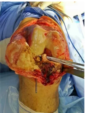
Fig.1–PresentACL,shownonthesurgicalclamp.
Table2–CorrelationbetweentheACLpreservation statusandclinical-radiographicparameters.
Variable ACLpreservationstatus
Absent Present Statisticalanalysis
Gender
Male 9(52%) 8(48%) p=0.044
Female 19(26%) 53(74%)
Age 70.1+6.7 69.6=−7.9 p=0.38
TFAng
Varus 21(35.5%) 38(64.5%) p=0.274
Valgus 7(28%) 18(72%)
Varus
<=10 16(29.6%) 38(69.4%) p=0.275
>10◦ 5(50%) 5(50%)
Valgus
<15◦ 2(15.4%) 11(84.6%) p=0.202
≥15 5(41.6%) 7(58.4%)
TFAng,tibiofemoralangle;ACL,anteriorcruciateligament.
Duringsurgery,thestateofACLpreservationwasassessed bytheleadauthor.Onlythepresenceorabsenceofthe liga-mentwasrecorded(Fig.1).TheACLwasclassifiedasabsent whentherewascompletelossofcontinuityofitsfibers.When thefibersofthisligamentstillpresentedcontinuityfromits femoralorigintoits tibialinsertion,theligament was con-sidered present. In the case of ACL presence, no attempt was made to classify the case according to the degree of macroscopicdegeneration(preservedvs.degenerated),asthis classificationisextremelysubjective.10
TheACL condition(presenceor absence)wascorrelated withthefivegradesAhlbäckclassification;theassessmentin varusandvalguskneeswasmadeseparately.
ThepresenceorabsenceoftheACLwascorrelatedwiththe parametersage,gender,andtibiofemoralangle.
Statisticalanalysis
Thechi-squaredandFisher’sexacttestswereusedfor anal-ysisofparametricdata.TheKruskal–WallisandtheG2-Wilks testswereusednonparametricdata.p-Values<0.05were con-sideredassignificant.
Results
In27/89patients(30.4%),theACLwasnotdetectableduring surgery;itwaspresentintheremaining62cases.
Table3–CorrelationbetweentheACLpreservationstatusandtheAhlbäckclassification(varus).
Ahlbäckclassification ACLpreservationstatus Statisticalanalysis
Absent Present Total
I 0 7 7 p<0.0001
II 3 8 11
III 3 26 29
IV 10 2 12
V 5 0 5
ACL,anteriorcruciateligament.
Table4–CorrelationbetweentheACLpreservationstatusandtheAhlbäckclassification(valgus).
Ahlbäckclassification ACLpreservationstatus Statisticalanalysis
Absent Present Total
I 1 8 9 p=0.307
II 1 3 4
III 1 3 4
IV 4 3 7
V 0 1 1
ACL,anteriorcruciateligament.
TheACLwasabsentin7/25(28%)ofcasesofvalgusknee andin16/64(35.5%)ofvarusknee,withnostatistical signifi-canceattheG2-Wilkstest(p=0.24).
No statistically significant correlation was observed between varus greater than or less than 10◦ and the ACL
preservationstatusaccordingtoFisher’stest(p=0.202). Val-guskneegreaterthanorlessthan15◦wasalsonotcorrelated
withthestateoftheACL(Fisher’sexacttest,p=0.275). IndividualswithpreservedACL had amean age of69.9 years(SD±7.9);inturn,themeanageofpatientswithabsent ACLwas70.1(SD±6.7),withnostatisticaldifferenceby Stu-dent’st-test(p=0.385).
Incasesofkneearthrosiswithvarusdeformity,theanalysis ofthecorrelationbetweentheAhlbäckradiographic classifi-cationandthe ACLstatusindicatedarelationshipbetween gradesIthroughIIIandthepresenceofACLin41/47(86.7%) ofcases;andbetweengradesIVandVandtheabsenceofthe ACLin15/17(88.2%)cases(G2-Wilkstest;p<0.0001;Table3).
In cases of valgus deformity, no statistically significant correlationwasobservedbetweentheAhlbäckradiographic classificationandtheconditionoftheACL.Inkneesclassified asgradesIthroughIII,theACLwaspresentin14/17(82.4%); inthoseclassifiedasgradeIVandV,theACLwaspresentin 4/8(50%)patients(Table4).
Discussion
Ideally,aclassificationinthemedicalfieldshouldbeableto identifytheseverityoftheinjuryassessedandhavepredictive value,aswellasassistintherapeuticindication.Furthermore, itshouldbesimple,easytoremember,andpresenthighlevels ofinter-andintra-observeragreement.3,11Inpractice,sucha classificationisrarelyavailable.
Studiesassessingradiographicclassificationsthat evalu-atearthriticdegenerationinkneeshavedemonstratedthat
narrowingofthetibiofemoraljointspacewasthemost sen-sitiveparameterindetectingarticularinvolvement.12–15This parameterpresentshighintra-andinter-observercorrelation, andismorereliabletoradiographicallygradearthrosisthan subchondralsclerosis.13
TheKelgren–Lawrence(KL),14,15thejointspacenarrowing (JSN),14and theAmerican CollegeofRheumatology(ACR)14 classificationsarereliableforearlydiagnosisofknee arthro-sisandformonitoringtheclinical-radiographicevolution.For thesereasons,theclassificationsarewidelyusedby rheuma-tologistsintheclinicalmanagementofkneedisorders.
When arthrosisprogresses,nolongerbeingamenableto conservativetreatmentandrequiringsurgicaltreatment,the radiographicchangesalsoworsen.TheKL,JSN,andACR rat-ingswouldnolongerbesousefultotheorthopedicsurgeon,as thehighergradesdescribedbythemarenotdetailedenough toaidinthechoiceofthemostappropriatesurgicaloption. Forexample,grade4,thehighestintheKLclassification,is definedbyanevidentreductioninthetibiofemoralarea,with evidentsubchondralsclerosisandosteophytes.Thisdefinition correspondstogradeIIintheAhlbäckclassification.15
Althoughwidely used,somestudies3,4 indicate that the Ahlbäckclassificationpresentslowreproducibilityandpoor differentiationbetweengradesIthroughIII.
Weidow et al.4 reported that, forAhlbäck classification purposes, signs of bone contact in radiographs are more important than the tibiofemoral narrowing measurement. These authorsdescribed thatpatientswhostilldisclosean evidentradiographic tibiofemoralspace and who wouldbe initiallyclassifiedasgradeIcouldpresentsignificantbone fric-tionandjointwearduringsurgery.Infact,suchacasewould functionallybeagradeIII.Inotherwords,theseresearchers haveshownthatitwouldbedifficulttodifferentiateAhlbäck gradesIthroughIII.
generallybepresent,whileingradeIV,theACLinjurywould determineagreaterdestructionofthemedialplateauinits centralandposteriorportions,andwouldeventuallyevolve toanteriorsubluxationofthetibia(gradeV).GradesIVand Vwouldpresentsimilaritiesandwouldbe,bydefinitionand radiographically,significantlydifferentfromgradesItoIII.
OrthopedicsurgeonswidelyusetheAhlbäckclassification, adoptingitasaguidelineforthechoiceofsurgicaltreatment. AccordingtoKeyesetal.,2gradesIthroughIIIareamenable totreatmentbyosteotomyorunicompartmentalprosthesis, although a total prosthesis can be safely used when fac-torssuchasageandlevelofphysicalactivityaretakeninto account.Inturn,duetotheassociationofACLinsufficiency andmoreseverejointdestruction,gradesIVandVshouldbe treatedwithtotalkneeprosthesis.
In their seminal article, Keyes et al.,2 despite having assessed200casesofmedialkneearthrosis,describeinthe text only50 knees inwhich the presenceof the ACL was actuallyassessed;thereisnoreferenceinthisregardforthe remaining150cases. Thisfactraises the questionofwhat therealcorrelationbetweentheACLpreservationstatusand Ahlbäckclassificationeffectivelywouldbe.Theoriginal arti-clegivestheinitialimpressionthattheACLwouldbepresent andfunctionalinalmost100%ofcasesclassifiedasAhlbäck IthroughIII.Sinceanextensivesearchinliteraturecouldnot findanyotherstudiesthatassessedthecorrelationbetween thisclassificationandthepresenceoftheACL,whichwould confirmordisprovethefindingsofKeyesetal.,2theauthors decidedtoconductthepresentstudy.
Theabsence ofACL leads to greater destruction ofthe medialkneeplateau.2,16Moschellaetal.,16inastudyofthe jointwearpatternin70varuskneesundergoingTKA, demon-stratedthat incaseswheretheACL waspresent, thejoint wearwouldbecentralinthemedialplateau.However,when theACLwasdeficient,thewearwouldbeinthe anteroposte-riorplaneofthisplateau,andthereforewider.Similarfindings werereportedbyGarridoetal.17
Leeet al.18 reported thatACL absence wouldbe associ-atedwithasignificantinvolvementofthearticularcartilageof thecontralateralcompartmentinpatientswithmedialknee arthrosis.Thiswouldindicateahigherseverityduetothejoint involvementofbothkneeplateaus.
Inthepresentstudy,itwasshownthattheACLwaspresent in86.7% ofcasesdescribed asAhlbäckgradesI, II, and III whilein gradesIV and V the ACL was absent in 88.2% of casesinvarusknees;thisdifferencewasstatistically signif-icant(p<0.0001).TheseresultsareinagreementwithKeyes etal.2 and demonstratethatthe Ahlbäckclassificationcan providearelativelyreliableideaoftheACLconditionandis thereforeusefulinsurgicalplanningincasesofmedial com-partmentkneearthrosis.
Inthis study,the Ahlbäckclassificationwas notable to predict the presence or absence ofthe ACL inknees with valgusdeformityand arthrosis.In gradesIthroughIII, the ACLwaspresent in82.4%ofthe cases,similartotherates foundinkneeswithvarusdeformity,whileingradeIVand V,it wasabsentinonly50%.These lessreliableresultsare partiallyduetothefactthatthe radiographicfemoralbone contactcannotbeadequatelyshowninosteoarthritisofthe lateralcompartment.4ThisfactshowsthattheuseofAhlbäck
classification would not be suitable for assessing cases of gonarthrosisinvalgus,4despiteitswidespreaduseineveryday clinicalpractice.3,4,11
Inthepresentstudy,theACLwasabsentin30.3%ofthe sample, similarto that observedbyAllain et al.10 and Lee etal.,18whodescribedabsenceofACLin40%and39%, respec-tively,intheirseriesofkneearthroplasties.
NostudiesthatassessedtheACLpreservationstatusand itscorrelationwithclinicalorradiographicparameterswere retrievedintheliterature.Inthepresentstudy,itwas statisti-callyshownthatmaleshadahigherprevalenceofabsentACL observedduringkneearthroplastysurgerythanwomen(52% vs.26%).
There is a growing movement in orthopedicsfor surgi-calprocedurestobeaslittleaggressiveaspossible;thegoal is to intervene effectively in the affected and pathology-generatingstructures. Thistrendisevidentinthecasesof medialgonarthrosisinwhichtheACLiscompetent;inthese cases,osteotomyorpartialarthroplastyproceduresare pre-ferredtototalkneearthroplasty.2,5,18Anothercurrentareaof studyistheuseoftotalkneeprosthesiswithmaintenanceof bothcruciateligaments,whichmustobviouslybefunctional fortheproperfunctioningofthearthroplasty.6,7Thesetrends highlighttheneedforassessingACLintegrityduringsurgical planning.
It is important to note that ACL integrity can also be evaluatedbymagneticresonanceimaging.12,14Nevertheless, despitebeinganexcellentimagingmethod,itisanexpensive methodwithalongwaitingqueueandunfortunatelyitis sel-domaccessibletopatientsfrom theBrazilianPublicHealth System(SistemaÚnicodeSaúde[SUS]),exceptforsome uni-versityhospitalsand reference centers.Thisreinforcesthe importanceofinformationthatcanbeobtainedthrough sim-plekneeradiographs.
Alimitationofthepresentstudyisthefactthatitisbased ontheuseofAhlbäckclassification,whichsomeauthors3,4,11 describeashavinglowlevelsofintra-andinter-observer corre-lation.Inordertoincreasethereliabilityofthisstudy,thecases wereclassifiedinagreementbytwospecialistsinkneesurgery whohaveextensiveexperienceinusingthisclassification.3,4 Furthermore,theresultswereassessedintwomore homoge-neousgroups(AhlbäckgradesItoIIIandanotherformedby gradesIVandV),whichwouldincreasethereproducibilityand reliability.3,4Apositivefindingofthepresentstudyisthefact thattheresultsconfirmedtheusefulnessoftheAhlbäck clas-sificationtopredictACLpreservationstatusinvarusknees, whichhasnotbeendemonstratedincasesofvalgusknees.
Conclusions
1. In the case of medial compartment gonarthrosis, the AhlbäckclassificationisareliableparametertopredictACL status(presentorabsent).
2. Invalguskneearthrosis,theACLstatuswasnotpredicted bytheAhlbäckclassification.
Conflicts
of
interest
r
e
f
e
r
e
n
c
e
s
1. AhlbäckS.Osteoarthrosisoftheknee.Aradiographic investigation.ActaRadiolDiagn(Stockh).1968;277 (Suppl.):7–72.
2. KeyesGW,CarrAJ,MillerRK,GoodfellowJW.Theradiographic classificationofmedialgonarthrosis.Correlationwith operationmethodsin200knees.ActaOrthopScand. 1992;63(5):497–501.
3. GalliM,DeSantisV,TafuroL.ReliabilityoftheAhlbäck classificationofkneeosteoarthritis.OsteoarthritisCartilage. 2003;11(8):580–4.
4. WeidowJ,CederlundCG,RanstamJ,KärrholmJ.Ahlbäck gradingofosteoarthritisoftheknee:poorreproducibilityand validitybasedonvisualinspectionofthejoint.ActaOrthop. 2006;77(2):262–6.
5. GoodfellowJ,O’ConnorJ.Theanteriorcruciateligamentin kneearthroplasty.Arisk-factorwithunconstrainedmeniscal prostheses.ClinOrthopRelatRes.1992;276:245–52.
6. RiesMD.EffectofACLsacrifice,retention,orsubstitutionon kinematicsafterTKA.Orthopedics.2007;30(8Suppl.):74–6.
7. ChristenM,AghayevE,ChristenB.Short-termfunctional versuspatient-reportedoutcomeofthebicruciatestabilized totalkneearthroplasty:prospectiveconsecutivecaseseries. BMCMusculoskeletDisord.2014;6(15):435.
8. KrausVB,VailTP,WorrellT,McDanielG.Acomparative assessmentofalignmentangleofthekneebyradiographic andphysicalexaminationmethods.ArthritisRheum. 2005;52(6):1730–5.
9. HinmanRS,MayRL,CrossleyKM.Isthereanalternativeto thefull-legradiographfordeterminingkneejointalignment inosteoarthritis?ArthritisRheum.2006;55(2):306–13.
10.AllainJ,GoutallierD,VoisinMC.Macroscopichistological assessmentsofthecruciateligamentsinarthrosisofthe knee.ActaOrthopScand.2001;72(3):266–9.
11.VilardiAM,MandarinoM,VeigaLT.Evaluationof reproducibilityfrommodifiedAhlback’sclassificationfor kneeosteoarthrosis.RevBrasOrtop.2006;41(5):157–61.
12.WadaM,BabaH,ImuraS,MoritaA,KusakaY.Relationship betweenradiographicclassificationandarthroscopicfindings ofarticularcartilagelesionsinosteoarthritisoftheknee.Clin ExpRheumatol.1998;16(1):15–20.
13.GüntherKP,ScharfHP,PuhlW,WillauschusW,KalkeY, GlückertK,etal.Reproducibilityofradiologicdiagnosisin gonarthrosis.ZOrthopIhreGrenzgeb.1997;135(3):197–202.
14.WuCW,MorrellMR,HeinzeE,ConcoffAL,WollastonSJ, ArnoldEL,etal.ValidationofAmericanCollegeof Rheumatologyclassificationcriteriaforkneeosteoarthritis usingarthroscopicallydefinedcartilagedamagescores. SeminArthritisRheum.2005;35(3):197–201.
15.PeterssonIF,BoegårdT,SaxneT,SilmanAJ,SvenssonB. Radiographicosteoarthritisofthekneeclassifiedbythe AhlbäckandKellgren&Lawrencesystemsforthe tibiofemoraljointinpeopleaged35–54yearswithchronic kneepain.AnnRheumDis.1997;56(8):493–6.
16.MoschellaD,BlasiA,LeardiniA,EnsiniA,CataniF.Wear patternsontibialplateaufromvarusosteoarthriticknees. ClinBiomech(Bristol,Avon).2006;21(2):152–8.
17.GarridoCA,SampaioTCF,FerreiraFS.Estudocomparativo entreaclassificac¸ãoradiológicaeanálisemacroe
microscópicadaslesõesnaosteoartrosedojoelho.RevBras Ortop.2011;46(2):155–9.

