Pediatric minor head trauma: do cranial CT scans change
the therapeutic approach?
Felipe P. Andrade,I,* Roberto Montoro Neto,IRenan Oliveira,IGabriela Loures,IILuana Flessak,IRoberta Gross,I Camille Donnabella,IAndrea Puchnick,IIILisa Suzuki,IIRodrigo RegaciniI,II
IUniversidade Anhembi Morumbi, Laborato´rio de Simulac¸a˜o, Sa˜o Paulo/SP, Brazil.IIHospital Infantil Sabara´, Departamento de Diagno´stico por Imagem, Sa˜o Paulo/SP, Brazil.IIIUniversidade Federal de Sa˜o Paulo (UNIFESP), Departamento de Diagno´stico por Imagem, Sa˜o Paulo/SP, Brazil.
OBJECTIVES: 1) To verify clinical signs correlated with appropriate cranial computed tomography scan indications and changes in the therapeutic approach in pediatric minor head trauma scenarios. 2) To estimate the radiation exposure of computed tomography scans with low dose protocols in the context of trauma and the additional associated risk.
METHODS:Investigators reviewed the medical records of all children with minor head trauma, which was defined as a Glasgow coma scale X13 at the time of admission to the emergency room, who underwent
computed tomography scans during the years of 2013 and 2014. A change in the therapeutic approach was defined as a neurosurgical intervention performed within 30 days, hospitalization,412 hours of observation, or neuro-specialist evaluation.
RESULTS:Of the 1006 children evaluated, 101 showed some abnormality on head computed tomography scans, including 49 who were hospitalized, 16 who remained under observation and 36 who were dismissed. No patient underwent neurosurgery. No statistically significant relationship was observed between patient age, time between trauma and admission, or signs/symptoms related to trauma and abnormal imaging results. A statistically significant relationship between abnormal image results and a fall higher than 1.0 meter was observed (p=0.044). The mean effective dose was 2.0 mSv (0.1 to 6.8 mSv), corresponding to an estimated additional cancer risk of 0.05%.
CONCLUSION:A computed tomography scan after minor head injury in pediatric patients did not show clinically relevant abnormalities that could lead to neurosurgical indications. Patients who fell more than 1.0 m were more likely to have changes in imaging tests, although these changes did not require neurosurgical inter-vention; therefore, the use of computed tomography scans may be questioned in this group. The results support the trend of more careful indications for cranial computed tomography scans for children with minor head trauma.
KEYWORDS: Computed Tomography; Minor head trauma; Pediatric; CT Scan; Radiology.
Andrade FP, Neto RM, Oliveira R, Loures G, Flessak L, Gross R, et al. Pediatric minor head trauma: do cranial CT scans change the therapeutic approach? Clinics. 2016;71(10):606-610
Received for publication onDecember 19, 2015;First review completed onMarch 24, 2016;Accepted for publication onAugust 4, 2016 *Corresponding author. E-mail: [email protected]
’ INTRODUCTION
Head Trauma is responsible for more than 1.0 million emergency room (ER) visits, 95,000 hospitalizations, 7,000 deaths and 29,000 permanent disabilities, and in the United States (US) alone, costs surpass US $1.0 billion in hospital care annually (1-3). It is the leading trauma related morbidity/mortality in children worldwide and accounts for most indications of computed tomography (CT) scans in this group (4).
The "golden hour" concept in the trauma scenario is com-mon knowledge in the medical community (2). Clinical signs of intracranial brain injury (IBI) in children are less reliable, which increases the use of CT scans, which are a highly sensitive imaging tests that can detect early IBI or help to safely discharge patients with head traumas (1-4).
However, up to 97% of CT findings are negative and less than 1% require neurosurgical intervention in this scenario (1,5,6). The short-term disadvantages of indiscriminate CT use include higher health costs, more sedation procedures, increased length of stay in the ER and additional dissatisfac-tion of parents. The major long-term disadvantage of CT use is premature exposure to ionizing radiation, which is associated with an increased risk of cancer and mortality (4). Despite these disadvantages, the use of CT scans after minor head trauma in children more than doubled from 1995 to 2005 (7,8).
DOI:10.6061/clinics/2016(10)09
Copyright&2016CLINICS–This is an Open Access article distributed under the terms of the Creative Commons License (http://creativecommons.org/licenses/by/ 4.0/) which permits unrestricted use, distribution, and reproduction in any medium or format, provided the original work is properly cited.
To establish clinical criteria for neuroimaging after minor head trauma in pediatric patients, Kuppermann et al. pro-posed ‘‘The Pediatric Emergency Care Research Network (PECARN)’’, the largest cohort study on the topic, which has been used by other authors a reference standard that objectively defines clinically important brain injuries. How-ever, significant variations in neuroimaging indications after mild pediatric head trauma still persist, with rates ranging from 5 to 70% in the US (3,5). Recent studies support the idea of observation in the ER in favor or avoiding CT scans, and the PECARN requires more clinical evidence to better define important brain injuries (9,10).
Although CT scans represent only approximately 11% of all radiological images in the US and 4% in Europe, dose levels administered in CT scans may have an influence on the stimulation of genetic mutations and carcinogenesis (11), and this information should be known and available to patients and their physicians (12).
The effective dose of ionizing radiation is primarily used to compare the cumulative risk (stochastic effect) associated with exposure to ionizing radiation and this risk requires special attention, particularly when repeated examinations are performed (13).
This study aims to include not only the clinical variables that could affect decision-making about cranial CT scans in pediatric minor head traumas but also to address one of the most important issues that involves performing an imag-ing test: does the result change the therapeutic approach and justify the risks associated with exposure to ionizing radiation?
’ MATERIALS AND METHODS
A cross-sectional study was performed at Hospital Infantil Sabará, a tertiary care pediatric hospital in São Paulo, Brazil. The researchers reviewed the medical records of patients admitted to the ER with head trauma who underwent CT scans in the years 2013 and 2014 and included only those with a score on the Glasgow Coma Scale (GCS)X13, corresponding to a minor head trauma. We did not exclude patients with trauma in other areas of the body associated with head trauma or those who could be identified as potential victims of abuse.
Information on demographics, physical examination find-ings, symptoms, mechanism and time of trauma, medical management and discharge data were obtained from hos-pital records. The CT scan interpretations were obtained from the final reports of attending radiologists. Subgaleal hematoma or extracranial changes were not considered as relevant findings. The main relevant changes considered were extra-axial hemorrhage (epidural, subdural and ara-chnoid hemorrhage), intra-axial hemorrhage (intracerebral or intraventricular hemorrhage) and fractures. Any other possible intracranial changes were also considered includ-ing pneumocephalus, brain edema and cerebral herniation, among others.
The variables collected for analysis of their relationship with changes in the therapeutic approach after imaging were age, gender, length of loss of consciousness, mechanism of trauma, vomiting, seizure, nausea, headache, drowsiness, dizziness, cranial hematomas, cranial laceration and visual alterations.
Changes in the therapeutic approach after CT scans included the following: neurosurgical intervention within
30 days, hospitalization, 412 hours of observation, and
neuro-specialist evaluation.
The dose-length product (DLP) was used to determine the effective dose according to the new International Com-mission on Radiological Protection (ICRP) recommenda-tions (12,14). All CT scans was performed using a low-dose protocol (80 kV).
Statistical analyses were performed with the Statistical Package for Social Sciences (SPSS), version 16.0, IBM®, USA. All variables were descriptively analyzed. All data were summarized as numbers and percentages (%) or means and standard deviations according to the variable type.
The chi-square test or Fisher’s exact test were used to determine the association between the physical examination findings, symptoms, mechanism of trauma, medical manage-ment and discharge data and the image findings. Student’s t test for independent variables was used to determine the association between the timing of trauma and hospitaliza-tion and the image findings. The results were considered significant when thep-value was less than 0.05 (po0.05).
Ethics
This study was conducted according to the Declaration of Helsinki and was approved by the Hospital Infantil Sabará Institutional Review Board.
’ RESULTS
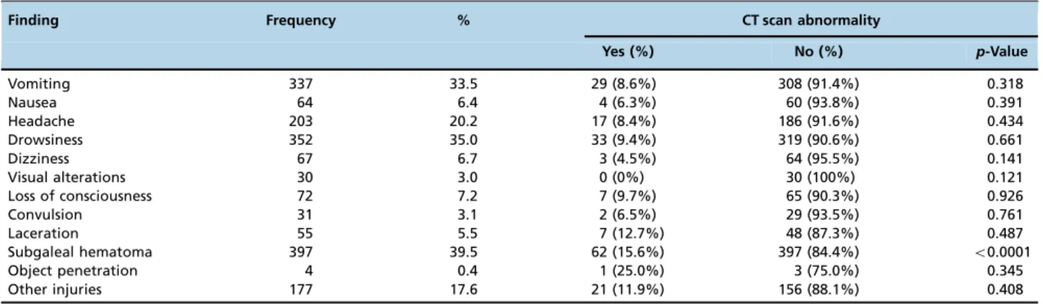
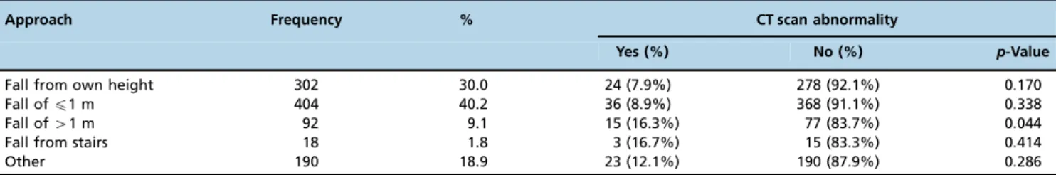
A total of 1,006 children comprised our study population, which included patients aged 0 to 17.83 years, with a mean age of 3.9±3.7 years, including 545 (54.2%) boys and 461 (45.8%) girls. The trauma-admission time ranged from 0.1 to 336.0 hours (mean of 15.0±35.0 hours). The mean length of stay in the hospital was 9.0±21.4 hours, ranging from 0.30 to 192.0 hours. Table 1 shows the frequency of signs and symptoms and the probability (%) of any abnormal imaging results related to these variables. Regarding the trauma mechanism, 81.1% of the injuries were falls, of which 40.2% corresponded to falls ofp1 m.
The following signs and symptoms were surveyed: vomit-ing, nausea, headache, drowsiness, dizziness, visual alterations, loss of consciousness, convulsion, laceration, and penetration of objects. No type of injury described in the tables as other was significantly associated with abnormal imaging results. In the imaging studies, 887 (88.2%) patients underwent only a CT scan. The other 119 patients completed imaging exami-nations consisting of x-ray examiexami-nations (9.4%), ultrasound examinations (0.6%), Magnetic Resonance Imaging (MRI) (0.9%), or combined x-ray and ultrasound examinations (0.9%). Of the 1006 patients undergoing imaging examinations, only 101 (10.0%) showed abnormal imaging findings. Cranial fractures were the most common finding (Table 2). Most patients were discharged after imaging examinations (84.7%) and no patient underwent neurosurgery (Table 3).
(po0.0001) and the need for 412 hours of observation
(p=0.001).
Subjects were divided in two groups according to trauma-admission time (p2 hours and42 hours) and then analyzed
according to imaging findings. Of the 101 patients with abnormal imaging findings, 33.7% presented a trauma-admission time p2 hours. A significant difference was not observed between patients with and without imaging abnorm-alities with regard to trauma-admission time (p=0.979).
A statistically significant association was found between abnormal imaging results and fall height (Tables 2 and 4). Among the 101 patients with abnormal imaging results, 35.6% suffered falls ofp1 m in height, and 14.9% suffered falls of41 m in height. Of all children who suffered falls of
p1 m, 8.9% presented abnormal imaging results; however, of all children who suffered falls of41 m, 16.3% were revealed
to have abnormal imaging findings.
Among the 101 children with abnormal imaging results, 48.5% were hospitalized, 15.8% underwent observation and 35.6% were dismissed (Table 3). In total, 112 patients were referred to a specialist; of these, 41.1% showed abnormalities on imaging and 58.9% had a normal CT scan.
Information about the DLP was recovered for 357 (35.5%) CT scans. The DLP ranged from 13.2 to 797.8 mGycm
(mean of 401.0±163.1 mGycm), which is equivalent to an effective dose of 2.0 mSv (0.1-6.8 mSv) and corresponds to an estimated additional cancer risk of 0.05%.
’ DISCUSSION
This two-year cross-sectional study showed that the time between trauma and admission and clinical data, such as vomiting, nausea, headache, drowsiness, dizziness, visual disturbances, loss of consciousness, seizures, laceration and penetration of objects had no significant effect in predicting possible changes in cranial CT findings after minor head trauma in pediatric patients. No significant association was found between abnormal imaging results and patient age; therefore, unlike other studies (5,9,12,15,16), we did not categorize patients as infants and children. Furthermore, the only variable that showed a statistically significant associa-tion with abnormal imaging results and a change in the medical approach was a fall from a height of41.0 m.
According to our findings, standard clinical references used in previous studies (PECARN) (3), such as altered mental status, loss of consciousness, severe headache and vomiting, did not contribute to the decision to perform a CT scan in the context of head trauma. Some difficulties in the interpretation of those variables may be present. For instance, variations in mental status could be due to several reasons, such as an undiagnosed pathology, the mechanism of injury was frequently not witnessed, or the report of patient loss of consciousness was not clear. In addition, multiple studies (17) that exclusively examined vomiting and headache as criteria for performing a CT scan missed an important IBI. Approximately one quarter of preverbal children hospitalized for head trauma are victims of child abuse; therefore, information about the injury is even less reliable (10,15).
In addition, a review of the literature reveals no con-sensual definition of what constitutes a‘‘minor head trauma’’. Usually, different studies refer to minor trauma as cases in which children have GCS scores ranging from 13 to 15,
Table 2-Correlation between imaging findings and mechanism of trauma.
Mechanism of trauma
Imaging findings
Extra-axial hemorrhage
Intra-axial hemorrhage
Intracranial hemorrhage and fracture
Fracture Other
Fall from own height
7 2 10 3 2
Fall ofp1 m 6 2 4 24 0 Fall of41 m 0 1 5 8 1 Fall from stairs 1 0 0 2 0
Other 9 4 6 4
Total 23 9 25 41 3
Pearson Chi-Squarep=0.050
Table 3-Frequency of therapeutic approach adopted for victims of minor head trauma.
Approach Frequency % CT scan abnormality Yes (%) No (%)
Dismissed 849 84.7 36 (4.2%) 813 (95.8%) Observation 59 5.9 16 (27.1%) 43 (72.9%)
Admitted 98 9.7 49 (50.0%) 49 (50.0%)
Surgery 0 0 — —
Return to ER 46 4.6 9 (19.6%) 37 (80.4%) Specialist referral 112 11.1 46 (41.1%) 66 (58.9%)
Table 1-Frequency of clinical history and physical exam findings in victims of minor head trauma and their correlation with abnormal imaging findings.
Finding Frequency % CT scan abnormality
Yes (%) No (%) p-Value
Vomiting 337 33.5 29 (8.6%) 308 (91.4%) 0.318
Nausea 64 6.4 4 (6.3%) 60 (93.8%) 0.391
Headache 203 20.2 17 (8.4%) 186 (91.6%) 0.434
Drowsiness 352 35.0 33 (9.4%) 319 (90.6%) 0.661
Dizziness 67 6.7 3 (4.5%) 64 (95.5%) 0.141
Visual alterations 30 3.0 0 (0%) 30 (100%) 0.121
Loss of consciousness 72 7.2 7 (9.7%) 65 (90.3%) 0.926
Convulsion 31 3.1 2 (6.5%) 29 (93.5%) 0.761
Laceration 55 5.5 7 (12.7%) 48 (87.3%) 0.487
Subgaleal hematoma 397 39.5 62 (15.6%) 397 (84.4%) o0.0001
Object penetration 4 0.4 1 (25.0%) 3 (75.0%) 0.345
although this slight variation increases the risk of IBI from approximately 2-3% in children with a GCS score of 15 to 7-8% in patients with a GCS score of 14 and to nearly 25% in children with a GCS score of 13 (3,16).
Skull fracture was the most prevalent imaging abnormality found in our study. The results corroborate the well-estab-lished fact that no correlation exists between skull fracture and IBI (18) because, in most cases, fractures due to mild trauma are small and do not produce significant misalign-ment. Thus, we discourage the use of cranial x-rays in pediatric minor head trauma patients, as it will not change the subsequent treatment.
Most children who were hospitalized, under observa-tion 412 hours, or referred to a neuro-specialist showed
some abnormality on imaging tests, and the differences were significant for the last two approaches. When CT scans present any changes, even without clinical significance, the attending physician tends to place the child under observa-tion or request a neuro-specialist evaluaobserva-tion. Thus, concerns about exposure to ionizing radiation must be weighed against the benefits of these interventions because surgery is not typically required.
Some patients who were referred to the neuro-specialist or were hospitalized returned later to undergo a control CT scan. This follow-up image was not included in our data and can be considered a limitation of this study.
The increase in the baseline risk of long-term malignancy is the main argument in favor of more careful use of cranial CT scans in the context of minor head trauma, particularly in the pediatric population, in which the tissues are more sensitive to ionizing radiation (19,20,21). These patients are subjected to repeated CT scans and have a longer lifetime to manifest a radiation-induced malignancy (2,6). However, low ionizing radiation protocols expose children to very low doses of radiation, and although not part of the scope of this paper, a CT scan in the context of trauma can help reassure parents of pediatric trauma victims, reduce observation time or the probability of hospitalization, and allow the attending physician to safely discharge the child.
CT scans performed after pediatric minor head injury did not show clinically significant abnormalities that could lead to neurosurgical indications.
The mechanism of trauma plays a more important role than isolated clinical signs and symptoms, patient age, or trauma-admission time in changing the medical approach in pediatric minor head trauma cases. According to the findings of this article, victims who fell more than 1.0 m were more likely to have changes in imaging tests, although this did not necessarily require neurosurgical intervention; therefore, the use of CT scans may be questioned in this group.
The results of this study support the trend of more cautious indications for cranial CT scans for children with
minor head trauma. Imaging protocols based on clinical data in the context of pediatric minor head trauma require greater external validation to ensure reliability and applicability because the variables involved may be subjective.
’ AUTHOR CONTRIBUTIONS
Andrade FP designed the project, performed data collection, and wrote the manuscript. Neto RM designed the project and performed the data collection. Oliveira R performed the data collection and statistical analysis. Loures G revised the manuscript and performed data collection. Flessak L, Donnabella C performed data collection and revised the manuscript. Gross R performed data collection and statistical analysis. Puchnick A assisted in the writing of the manuscript and performed statistical analysis. Suzuki L designed the project, performed data collection and analyzed the CT images. Regacini R designed the project, assisted in the discussion and conclusion and analyzed the CT images.
’ REFERENCES
1. Atabaki SM, Stiell IG, Bazarian JJ, Sadow KE, Vu TT, Camarca MA, et al. A clinical decision rule for cranial computed tomography in minor pediatric head trauma. Arch Pediatr Adolesc Med. 2008;162(5):439-45, http://dx.doi.org/10.1001/archpedi.162.5.439.
2. Shutzman SA, Greenes DS. Pediatric minor head trauma. Ann Emerg Med. 2001;37(1):65-74, http://dx.doi.org/10.1067/mem.2001.109440. 3. Kuppermann N, Holmes JF, Dayan PS, Hoyle JD Jr, Atabaki SM,
Holubkov R, et al. Pediatric Emergency Care Applied Research Net-work (PECARN). Identification of children at very low risk of clinically-important brain injuries after head trauma: a prospective cohort study. Lancet. 2009;374(9696):1160-70, http://dx.doi.org/10.1016/S0140-6736 (09)61558-0.
4. Mathews JD, Forsythe AV, Brady Z, Butler MW, Goergen SK, Byrnes GB, et al. Cancer risk in 680,000 people exposed to computed tomography scans in childhood or adolescence: data linkage study of 11 million Australians. BMJ. 2013;346:f2360, http://dx.doi.org/10.1136/bmj.f2360. 5. Mannix R, Bourgeois FT, Schutzman SA, Bernstein A, Lee LK.
Neuro-imaging for pediatric head trauma: do patient and hospital character-istics influence who gets imaged? Acad Emerg Med. 2010;17(7):694-700, http://dx.doi.org/10.1111/j.1553-2712.2010.00797.x.
6. Hennelly KE, Mannix R, Nigrovic LE, Lee LK, Thompson KM, Mon-uteaux MC, et al. Pediatric traumatic brain injury and radiation risks: a clinical decision analysis. J Pediatr. 2013;162(2):392-7, http://dx.doi.org/ 10.1016/j.jpeds.2012.07.018.
7. National Center for Health Statistics Centers for Disease Control and Prevention. Public use data file, emergency department file, 2005. National Hospital Ambulatory Medical Care Survey. Hyattville, MD http://ftp.cdc.gov/pub/Healt_Statistcs/NCHS/Datasets/NHAMCS/ readme05.text.
8. National Center fo Healt Statistcs Centers for Disease Control and Pre-vention. Public use data file, emergency department file, 1995. National Hospital Ambulatory Medical Care Survey. Hyattville, MD http:// ftp.cdc.gov/pub/Healt_Statistcs/NCHS/Datasets/NHAMCS/readme05. text.
9. Schonfeld D, Fitz BM, Nigrovic LE. Effect of the duration of emergency department observation on computed tomography use in children with minor blunt head trauma. Ann Emerg Med. 2013;62(6):597-603, http://dx.doi.org/10.1016/j.annemergmed.2013.06.020.
10. Medwid K, Couri GG. How accurate are clinical decision rules for pediatric minor head injury? Ann Emerg Med. 2012;60(3):278-9, http://dx.doi.org/10.1016/j.annemergmed.2012.01.029.
11. 1990 Recommendations of the International Commission on Radiological Protection. Ann ICRP. 1991;21(1-3):1-201.
Table 4-Frequency of therapeutic approach adopted for victims of minor head trauma and correlation with abnormal imaging findings.
Approach Frequency % CT scan abnormality
Yes (%) No (%) p-Value
Fall from own height 302 30.0 24 (7.9%) 278 (92.1%) 0.170
Fall ofp1 m 404 40.2 36 (8.9%) 368 (91.1%) 0.338
Fall of41 m 92 9.1 15 (16.3%) 77 (83.7%) 0.044
Fall from stairs 18 1.8 3 (16.7%) 15 (83.3%) 0.414
12. Deak PD, Smal Y, Kalender WA. Multisection CT protocols: sex- and age-specific conversion factors used to determine effective dose from dose-length product. Radiology. 2010;257(1):158-66, http://dx.doi.org/10.1148/ radiol.10100047.
13. Struelens L, Vanhavere F, Bosmans H, Van Loon R, Mol H. Skin dose measurements on patients for diagnostic and interventional neuror-adiology: a multicentre study. Radiat Prot Dosimetry. 2005;114(1-3):143-6, http://dx.doi.org/10.1093/rpd/nch537.
14. The 2007 recommendations of the International Commission on Radio-logical Protection. ICRP publication 103. Ann ICRP 2007;37(2-4):1-332. 15. Kuppermann N. Pediatric head trauma: the evidence regarding
indica-tions for emergent neuroimaging. Pediatr Radiol. 2008;38 Suppl 4:S670-4. 16. Quayle KS, Jaffe DM, Kuppermann N, Kaufman BA, Lee BC, Park TS, McAlister WH. Diagnostic testing for acute head injury in children: when are head computed tomography and skull radiographs indicated? Pediatrics. 1997;99(5):E11, http://dx.doi.org/10.1542/peds.99.5.e11.
17. Dayan PS, Holmes JF, Atabaki S, Hoyle J Jr, Tunik MG, Lichenstein R, Alpern E, Miskin M, Kuppermann N; Traumatic Brain Injury Study Group of the Pediatric Emergency Care Applied Research Network (PECARN). Association of traumatic brain injuries with vomiting in children with blunt head trauma. Ann Emerg Med. 2014;63(6):657-65. 18. Wing R, James C. Pediatric head injury and concussion. Emerg Med Clin
North Am. 2013;31(3):653-75, http://dx.doi.org/10.1016/j.emc.2013.05.007. 19. Frush DP, Donnelly LF, Rosen NS. Computed tomography and
radia-tion risks: what pediatric health care providers should know. Pediatrics. 2003;112(4):951-7.
20. Brenner DJ, Hall EJ. Computed tomography--an increasing source of radiation exposure. N Engl J Med. 2007;357(22):2277-84, http://dx.doi. org/10.1056/NEJMra072149.