QUEIROZ RM ETAL.
488 REV ASSOC MED BRAS 2017; 63(6):488-491
IMAGE IN MEDICINE
Thrombosed aneurysm of saphenous vein coronary artery
bypass grafting
RODOLFO MENDES QUEIROZ1*, ROGÉRIO NASTRI FILHO1, MARCUS ANTÔNIO FEREZ1,2, MAURO JOSÉ BRANDÃODA COSTA1,
CLAUDIO BENEDINI LAGUNA1, MARCUS VINICIUS NASCIMENTO VALENTIN1
1Radiology and Imaging Diagnosis Department, Documenta – Hospital São Francisco, Ribeirão Preto, SP, Brazil 2Intensive Care Center Department, Hospital São Francisco, Ribeirão Preto, SP, Brazil
SUMMARY
Study conducted at Documenta – Centro Avançado de Diagnóstico por Imagem, Hospital São Francisco,
Ribeirão Preto, SP, Brazil Article received: 11/25/2016 Accepted for publication: 12/1/2016 *Correspondence: Address: Rua Bernardino de Campos, 980 Ribeirão Preto, SP – Brazil Postal code: 14015-130 [email protected]
http://dx.doi.org/10.1590/1806-9282.63.06.488
We describe the case of a male patient, aged 76 years, referred for cardiac inves-tigation due to retrosternal chest pain and dyspnea. He had a history of acute myocardial infarction and angioplasties in the last 30 years, including a saphenous vein coronary artery bypass grafting (SVCABG). Echocardiogram showed hy-poechoic oval formation near the right ventricle, suggesting a pericardial cyst. Computed angiotomography revealed a predominantly fusiform and thrombosed aneurysmal dilation of the SVCABG to the right coronary artery. SVCABG an-eurysms are very rare and potentially fatal. They usually appear in the late post-operative period, and patients are often asymptomatic. On radiography, it is frequently presented as enlargement of the mediastinum, with echocardiography, computed tomography and magnetic resonance imaging being very useful for diagnosis. Coronary angiography is the gold standard to detect these cases. Our report illustrates a rare situation arising late from a relatively common surgery. Due to its severity, proper recognition in the routine assessment of patients with a similar history is essential.
Keywords: aneurysm, bypass, coronary, saphenous, myocardium.
C
ASE REPORTMale patient, 76 years old, underwent complementary in-vestigation through imaging examinations due to complaints of retrosternal chest pain and mild dyspnea several months ago. The patient reported hospitalization and clinical treat-ment of acute myocardial infarction one month before. The patient had hypertension, dyslipidemia, was a former smok-er and had a history of two othsmok-er acute myocardial infarctions prior to 2010. The patient’s history included saphenous vein coronary artery bypass grafting (SVCABG) for about 30 years, two percutaneous transluminal coronary angioplasty procedures, one with stenting to clear the SVCABG, and surgical repair of abdominal aortic aneurysm with endo-prosthesis for approximately 4 years. Reports of catheteriza-tions prior to 2015 described new SVCABG occlusion.
A chest radiograph showed only a metallic sternal suture and a small stent near the cardiac silhouette (Figure 1A and B).
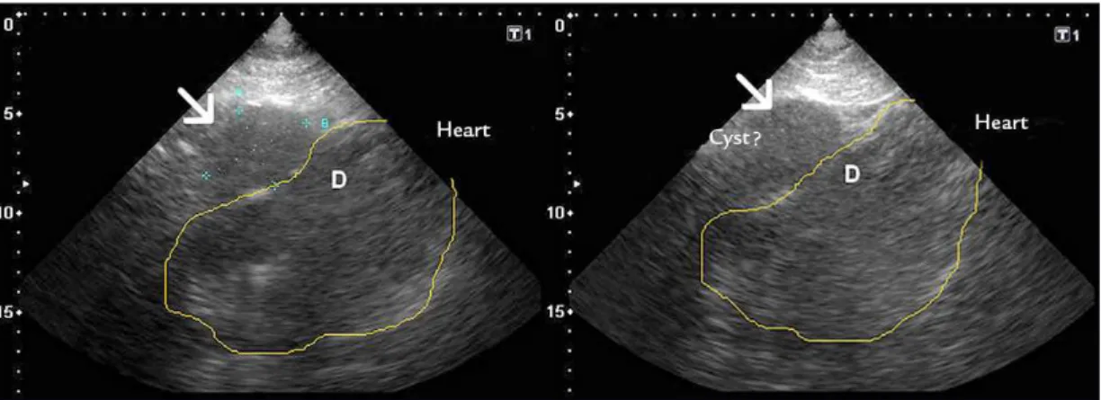
The echocardiogram showed a hypoechoic, elon-gated oval image, adjacent to the right cardiac chambers,
causing a slight extrinsic compression on diastole (Figure 2A and B), apparently without flow according to color Doppler investigation. The diagnostic possibility of peri-cardial cyst was raised.
THROMBOSEDANEURYSMOFSAPHENOUSVEINCORONARYARTERYBYPASSGRAFTING
REV ASSOC MED BRAS 2017; 63(6):488-491 489
FIGURE 1 Chest X-ray in posteroanterior (A) and proile (B) views, showing mainly the metallic sternotomy (arrow heads) and the small stent
along the cardiac silhouette (small arrow).
FIGURE 2 A and B. Echocardiogram showing a hypoechoic oval image (thin arrow) adjacent to the right cardiac chambers that causes slight
compression in the latter.
FIGURE 3 Computed tomography of the chest, images acquired before (A, B, C) and after (D, E, F) the administration of intravenous contrast medium, showing aneurysms of aortocoronary saphenous vein bypass grafts to the right coronary artery (*) and the small stent in its implanta-tion surgical ostium (thin double arrowheads). The aneurysm is characterized by a large oval and elongated formaimplanta-tion in the middle mediastinum, hypoattenuating, with lobulated contours and in close contact with the right heart chambers. It presents peripheral parietal calciications, without enhancement after intravenous iodine contrast.
A
A B
A
D
B C
F E
B
QUEIROZ RM ETAL.
490 REV ASSOC MED BRAS 2017; 63(6):488-491
D
ISCUSSIONAneurysmal formations, also known as “saphenous bridges,” are rare in SVCABG. These cases should be differentiated from ectasia, which does not exceed 1.5 times the normal vessel size and is observed in about 14% of patients 5-7 years after surgery.1-4
They can be divided into pseudoaneurysms and true aneurysms. Pseudoaneurysms do not involve all the al layers of the vascular wall. They occur earlier and usu-ally close to anastomoses, often related to trauma, surgi-cal technique problems, infections and the preparation of the vein.1 True aneurysms usually occur later,
consist-ing of true distended walls, often associated with athero-sclerosis, hyperlipidemia, endothelium weakness, and smoking.1 Aneurysms are commonly diagnosed 10 years
after revascularization (69%).5
The incidence of aneurysmal formations in SVCABG varies between 0.07 and 2%,6,7 with the most common
being true aneurysms (61-86%).3,5,7,8 The two types of
aneurysm predominate in men (76-86%) between 50-75 years.3,5,7,8 Approximately 45-66% of the patients have
symptoms, especially chest pain, angina and dyspnea.5
The discovery of aneurysmal formations often occurs incidentally due to mediastinal enlargement identified on chest radiography performed for other reasons.4,6,9 The
golden standard of diagnosis is coronary angiography,1
but echocardiography, computed tomography and mag-netic resonance imaging can also be used.1 The latter two
evaluate size, permeability and the relationship with neighboring structures.9
The most-affected SVCABGs are those directed to the right coronary arteries (38%) and the left anterior descend-ing artery (25%).5 They can become quite large,4,7 with a
mean diameter between 6.0 and 7.0 cm, often with inter-nal thrombi or thrombosed (61%).5
Approximately 36% of cases develop with complica-tions such as rupture with consequent hemothorax and hemopericardium,1,6,9 thromboembolism, fistulas, acute
myocardial infarction, heart failure and compression of adjacent structures,1,4-7,9
Surgical treatment with new revascularization is rec-ommended even in asymptomatic patients, due to the high mortality rate in case of rupture. Conservative ther-apy such as the use of polytetrafluoroethylene-coated stents, hemodynamic embolization with coils or amplatzer vascular plug is advised in individuals with a very high surgical risk, followed by imaging tests.1-9
Our report illustrates a rare situation arising late from a relatively common surgery. Due to its severity, proper recognition in the routine assessment of patients with a similar history is essential.
R
ESUMOAneurisma trombosado de bypass aortocoronariano de veia safena
Descrevemos o caso de paciente do sexo masculino, 76 anos, em avaliação cardiológica em razão de dor torácica retroesternal e dispneia. Antecedente de infartos agudos do miocárdio e angioplastias nos últimos 30 anos, in-cluindo um bypass aortocoronário de veia safena (BACVS) ou “ponte de safena”. Em ecocardiograma, observou-se formação ovalada alongada hipoecoica junto ao ventrí-culo direito, podendo sugerir um cisto pericárdico. An-giotomografia computadorizada do tórax evidenciou uma dilatação aneurismática predominantemente fusiforme e trombosada de “ponte de safena” para artéria coronária direita. Aneurismas de BACVS são raríssimos e potencial-mente fatais. Geralpotencial-mente, surgem em um período tardio pós-cirúrgico, sendo seus portadores muitas vezes assin-tomáticos. Na radiografia, frequentemente se apresentam como alargamento do mediastino, sendo a ecocardiogra-fia, a tomografia computadorizada e a ressonância mag-nética muito úteis no diagnóstico. A angiografia corona-riana é o padrão-ouro na detecção. Este relato ilustra uma situação rara decorrente tardiamente de uma cirurgia relativamente comum, e por causa de sua gravidade torna--se essencial o seu reconhecimento na rotina de avaliação
de pacientes com antecedentes semelhantes.
Palavras-chave: aneurisma, bypass, coronária, safena,
miocárdio.
R
EFERENCES1. Albuquerque MG, Farran JA, Pereira CAP, Romano ER, Brotto INM, Romano MLP. Giant aneurysm of saphenous vein bypass for right coronary after angioplasty. Arq Bras Cardiol. 2012; 99(2):125-7.
2. Frazier AA, Qureshi F, Read KM, Gilkeson RC, Poston RS, White CS. Coronary artery bypass grafts: assessment with multidetector CT in the early and late postoperative settings. Radiographics. 2005; 25(4):881-96.
3. Kalimi R, Palazzo RS, Graver LM. Giant aneurysm of saphenous vein graft to coronary artery compressing the right atrium. Ann Thorac Surg. 1999; 68(4):1433-7.
THROMBOSEDANEURYSMOFSAPHENOUSVEINCORONARYARTERYBYPASSGRAFTING
REV ASSOC MED BRAS 2017; 63(6):488-491 491
5. Ramirez FD, Hibbert B, Simard T, Pourdjabbar A, Wilson KR, Hibbert R, et al. Na-tural history and management of aortocoronary saphenous vein graft aneurysms: a systematic review of published cases. Circulation. 2012; 126(18):2248-56. 6. Fukui T, Suehiro S, Shibata T, Sasaki Y, Minamimura H, Kinoshita H.
Aortocoronary saphenous vein graft aneurysm in redo coronary artery bypass grafting: report of a case. Surg Today. 1998; 28(3):321-4.
7. Dieter RS, Patel AK, Yandow D, Pacanowski-Jr JP, Bhattacharya A, Gimelli G, et al. Conservative vs. invasive treatment of aortocoronary saphenous
vein graft aneurysms: Treatment algorithm based upon a large series. Cardiovasc Surg. 2003; 11(6):507-13.
8. Almanaseer Y, Rosman HS, Kazmouz G, Giraldo AA, Martin J. Severe di-latation of saphenous vein grafts: A late complication of coronary surgery in which the diagnosis is suggested by chest X-ray. Cardiology. 2005; 104(3):150-5.