1. Fisioterapeuta; Mestranda em Fisioterapia pela UFSCar.
2. Fisioterapeuta; Mestre em Engenharia Biomédica pela UFPB; Douto-rando em Fisioterapia pela UFSCar; Professor do Departamento de Fi-sioterapia da UFPB.
3. Acadêmica de Fisioterapia da UFPB, João Pessoa, PB. 4. Fisioterapeuta.
5. Fisioterapeuta; Mestranda em Engenharia de Produção pela UFPB. 6. Fisioterapeuta; Mestre em Ergonomia pela UFPB; Professor do
Depar-tamento de Fisioterapia do UNIPÊ, João Pessoa, PB. Approved in 14/6/07.
Correspondence to: Catarina de Oliveira Sousa, Rua João Batista de Arru-da, 138, Vila Brasília – 13566-120 – São Carlos. SP. Tels. de contato: (16) 3361-4618 / 3351-8768. E-mail: [email protected]
Electromyograhic activity in squatting at
40°, 60° and 90° knee flexion positions
Catarina de Oliveira Sousa1, José Jamacy de Almeida Ferreira2, Ana Catarina L. Veras Medeiros3,
Antônia Hermínia de Carvalho4, Rosana Cavalcante Pereira5, Dimitri Taurino Guedes6 and Jerônimo F. de Alencar2
O
RIGINALA
RTICLEKeywords: Biomechanics. Rehabilitation. Muscular activity. Closed kinetic chain. ENGLISH VERSION
ABSTRACT
The aim of this study was to compare the electromyographic (EMG) activity of the femoris rectus, femoris biceps, tibialis anteri-or and soleus muscles in squatting, associating the trunk in erect position with two angles of knee flexion (40° and 60°) and the trunk at 45° flexion with three angles of knee flexion (40°, 60° and 90°). All associations were performed with and without additional load (10 kg). The sample was composed of 12 healthy individuals with mean age of 21.1 ± 2.5 years and weight of 62.8 ± 7.4 kg. The EMG of the cited muscles was isometrically registered in 10 squat-ting positions. For statistical analysis, Friedman Two-Way ANOVA and the Newman-Keuls Post-Hoc test were used. The results showed co-activation between the femoris rectus and femoris bi-ceps muscles with the trunk in flexion and at 40° of knee flexion and between the femoris rectus and soleus muscles in the other positions considered (p < 0.05). It was also possible to observe co-activation between tibialis anterior and femoris biceps muscles with knee at 40° and 60° of flexion, with the trunk erect and in flexion and between the tibialis anterior and soleus muscles in the other positions (p < 0.05). Concerning isolated muscular activation, high-er knee flexion in squatting was an important factor to greathigh-er mus-cles activation, except for the soleus. Trunk position and the addi-tional load of 10 kg have influenced in the muscular activation of the femoris rectus at 60° of knee flexion, in which the erect trunk provided more activation. The femoris biceps presented greater activation when the knee was in 40° of flexion and the trunk flex-ioned. The co-activation between the femoris rectus and biceps with the trunk in flexion, and between the femoris rectus and sole-us in the other positions, lead to new possibilities of exercises in rehabilitation.
INTRODUCTION
During many years, physiotherapists dedicated to rehabilitation of athletes have used open kinetic chain exercises (OKC), in which the movements are developed with the free distal segment(1), in
order to strengthen the lower extremity(2). However, a better
un-derstanding of kinesiology and biomechanics provided the current application of the closed kinetics chain exercises (CKC), which have
been incorporated to the rehabilitation protocols, especially the knee joint(2). This kind of exercise involves multi-articular movements
performed with steady distal extremity(1).
Some suppositions are presented for the use of CKC exercises: from the biomechanical view point, it is suggested that these exer-cises are safer and produce stress and forces which offer less risk to the recovering structures when compared with the exercises in OKC. The co-activation or co-contraction of the agonist and antag-onist muscles occur during the movements in CKC in order to pro-vide articular stabilization(3-4). Moreover, the exercises in CKC are
equally efficient in the force production in the quadriceps femoris, when compared with the exercises in OKC(5).
Co-contraction has been positively seen as something neces-sary for gains in dynamic stability. Due to its applicability, the co-contraction measurement becomes relevant in several fields which have as aim the interest in the human movement(4,6-7).
Among the exercises in CKC, squatting, triple flexion of the low-er extremity has been considlow-ered effective in the development of the hip, knee and ankle muscles through the increase of quadri-ceps, hamstrings and triceps surae activity(8). In spite of that, it
should be carefully used in individuals with patelo-femoral (PF) and posterior crossed ligament alterations (PCL), especially in wider knee flexion angles, an occasion in which translation and compres-sion forces increase in this joint(9).
During squatting, the gravity line is dislocated behind the knee axis, increasing the flexor torque(10-11). The hamstrings promote
sta-bilization in the knee with a posterior traction in the tibia in order to counterpart the anterior force imposed by the quadriceps(12). The
degree of activation in which the hamstring muscles act in the pelvis, probably depends on the knee and hip angles as well as individual muscular lengths(13). Therefore, the hamstrings tension
may be increased with a slight anterior chest flexion, which also moves the gravity center forward, decreasing the knee flexion torque and decreasing hence the tibia translation force and the patelo-femoral compression in the joint(14).
Still in squatting, a flexion torque is also created in the ankle when the gravity line is dislocated forward to the talocrural joint. The soleus muscle slows down the ankle dorsiflexion and creates a knee extension torque, forcing the tibia backwards, which mini-mizes the anterior shear force in this joint(15). Therefore, all the
ki-netic chain of the lower extremity is recruited by the application of an axial force in the distal segment.
Escamilla(8) recommends a breadth of 0° to 50° of knee flexion
for the squatting exercises used in rehabilitation, since it is the breadth in which anterior lower shear forces occur in the tibio-femoral joint. However, experiments performed with athletes dem-onstrated that squatting exercises with thigh parallel to the ground (0°-100°) do not cause injuries and instability in healthy knees(16).
On the other hand, the use of external loads tends to increase the shear force(17) and, probably, demand greater activity of the
It is important to understand and compare the muscular activi-ties which occur in the knee joint during squat exercise in order to determine positions of better muscular balance, ligament tension and articular compression(3). Electromyographic activity (EMG) has
been widely used in analysis studies of the human movement with the purpose to investigate muscular function through acquisition and analysis of electric signals produced by the muscles. The EMG breadth estimated by the mean of the corrected signal or by the RMS (root mean square) reflects the recruiting pattern or activa-tion of the motor units which control a given muscle. Moreover, there is a relationship very close to the linear between the EMG and the force generated by the muscle, especially, in isometric contractions(18-20).
As the EMG reflects the degree of muscular activation in a giv-en momgiv-ent of isometric contraction, in this investigation an exper-imental protocol was used with the aim to compare the EMG ac-tivity of the rectus femoris, biceps femoris, anterior tibialis and soleus muscles in squatting associated with straight trunk posi-tion with 2 angles of knee flexion (40° and 60°), and the flexed trunk position at 45° with 3 angles of knee flexion (40°, 60° and 90°). These knee flexion angles were adopted as they present the inferior, medium and superior thresholds of the breadth stages considered safe for rehabilitation and sportive training(8). All
combi-nations were performed with and without load (10 kg) in order to verify its influence in muscular co-activation. These combinations of knee and trunk positions were adopted since they present sim-ilarity with exercises used in rehabilitation. Additionally, the degree of stabilizer synergy of the muscles examined has not been com-pletely clarified yet.
METHODS
The sample consisted of 12 healthy individuals from both gen-ders (six males and six females), mean age of 21.1 ± 2.5 years and weight of 62.8 ± 7.4 kg, who did not participate in any regular physical activity program. The individuals did not present osteomyo-articular deformities or diseases of the lower extremities which would interfere in the performance of the experimental protocol. The subjects were informed about the aim and purpose of the study and signed a free and clarified consent form for participation in the study. This form was according to the norms established by the resolution 196/96 from the National Health Board (NHB) and ap-proved by the Ethics and Research Committee of the Institution where the research was conducted.
The used hardware involves two biological amplifiers with two independent channels, built in differential configuration and high common mode rejection ratio (CMRR > 90 dB), high entrance im-pedance (> 10 MΩ), low noise (< 5 µV RMS), with selected gain of 1000 times, passing band with 10 to 490 Hz frequency. The elec-tromyographic signals acquisition and processing were performed through the software BioMed, using a sample frequency of 1000 Hz. Moreover, for data pick up, storing and processing, an A/D con-verser plaque with 16 channels and resolution of 12 bits per chan-nel were used. All hardware and the software were developed in the Laboratory of Biological Signals Processing and Acquisition of the Nucleus of Studies and Technology in Biomedical Engineering (NETEB/UFPB).
One cm of diameter, Ag/Ag-Cl auto-adhesive disposable elec-trodes (SkinTact F-521), were attached to the skin, which had been previously trichotomized and cleaned with alcohol for removal of residues and grease excess, aiming to diminish the skin impen-dence and consequently improving the register conditions of the EMG(14,21).
For the biceps femoris muscle, the electrodes were attached on the muscular body, 12 cm above the popliteal line; for the rec-tus femoris, they were attached 12 cm above the superior edge of the patella(22). For the soleus muscle, the electrodes were attached
4 cm below the inferior edge of the gastrocnemius muscle, on the median line of the muscular body; for the anterior tibialis, the first electrode was attached 4 cm below and 1 cm lateral to the tibial tuberosity. For all muscles, the distance between the electrodes of register was of 3 cm(23).
The reference electrodes positions were: for the biceps femo-ris(22) and rectus femoris, at the patellar tendon of the same side;
for the soleus on the third medium of the anterior face of the leg and for the anterior tibialis, on the medial malleolus.
For normalization of the electromyographic signals during squat-ting, the electromyographic activity in maximal voluntary isometric contraction (MVIC) of each muscle was previously picked up.
The test position for MVIC of the rectus femoris and biceps fem-oris muscles was with the subject seated with knee on 60° flexion using a Bonnet chair adapted and locked in that angle. For the so-leus and anterior tibialis, the individual was seated with knee and ankle positioned at 90°. In order to avoid the calcaneum rising dur-ing the plantar flexion, and the forefoot’s durdur-ing dorsiflexion, a con-straint band which passed over the knee and was attached on a wooden platform over the ground was used.
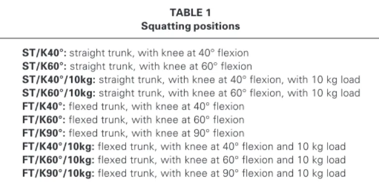
The subjects were then instructed and familiarized concerning the test positions during squatting, in which they should remain during 10 s for simultaneous pick up of the EMG for each muscle. For the breadth of the EMG, the 2 initial and final seconds of each signal were discarded and the mean of the 6 s piece of the ratified sign was calculated. The sequence of the evaluations ac-cording to each test position was obtained through a drawing, al-lowing a 2 min rest between each position. The EMG was regis-tered only on the right inferior limb, since it was the dominant limb of all subjects. The test positions are described in table 1.
TABLE 1 Squatting positions
ST/K40°: straight trunk, with knee at 40° flexion
ST/K60°: straight trunk, with knee at 60° flexion
ST/K40°/10kg: straight trunk, with knee at 40° flexion, with 10 kg load
ST/K60°/10kg: straight trunk, with knee at 60° flexion, with 10 kg load
FT/K40°: flexed trunk, with knee at 40° flexion
FT/K60°: flexed trunk, with knee at 60° flexion
FT/K90°: flexed trunk, with knee at 90° flexion
FT/K40°/10kg: flexed trunk, with knee at 40° flexion and 10 kg load
FT/K60°/10kg: flexed trunk, with knee at 60° flexion and 10 kg load
FT/K90°/10kg: flexed trunk, with knee at 90° flexion and 10 kg load
Squatting with straight trunk and knee at 90° of flexion was not performed due to the poor balance of the subjects in this position, caused by the dislocation of the gravity center of the body back-wards.
The use of an external load had the aim to simulate the handled load carrying, frequent in some routine and sportive activities. The 10 kg load was adopted according to McAtammey and Corlett(24),
who report that the use of a load of this magnitude does not offer additional risks of musculoskeletal injuries, neither for men or wom-en.
For stabilization of the knee angles, a 2-armed 30 cm long pro-tractor with steady angles of 40°, 60° e 90° was placed on the sagittal level, parallel to the medium lateral line of the thigh and leg with the axis on the head of the fibula. For measurement of the hip flexion angle, a universal goniometer at 45° angle was used, with its axis laterally positioned at the major trochanter level, and arms parallel to the lateral medium lines of the trunk and the thigh, on the sagittal level.
According to Hammond et al.(6), the muscular co-activation
statistically significant difference in the muscular activation per-centage, as calculated for the data normalized by the MVIC in each test position.
The data statistical analysis was performed with the aid of the GB-STAT School Pack software, version 1997. The median and in-terquartilic breadth were calculated for the descriptive statistics. Since the normality requisites (Shapiro-Wilk test) and variances homogeneity (Levene’s F test) have not been confirmed, a Two-Way ANOVA by Friedman for non-parametric data was applied. The two-way ANOVA was performed (muscles x situations) comparing the different muscles in the different situations (trunk, knee and load angles) and one-way ANOVA (situations) comparing the differ-ent situations for each muscle. Since ANOVA showed significant difference, the Newman-Keuls post hoc test was used for identifi-cation of the differences. For all tests the adopted significance lev-el was 5% (p < 0.05). In the results description, the normalized EMG data will be always used and therefore, the values will be expressed as EMG breadth percentage in MVIC.
RESULTS
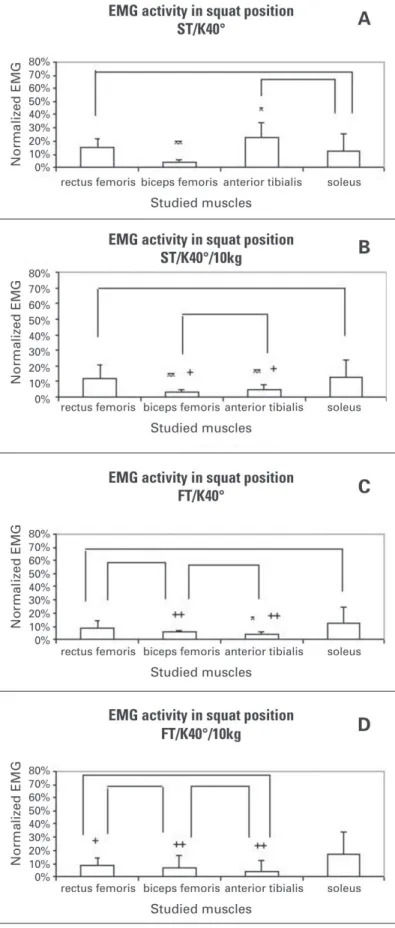
In the ST/K40° position (figure 1A), the rectus femoris present-ed higher activation (15.3%) than the biceps femoris muscle (3.4%), and lower than the anterior tibialis muscle (22.7%). In this posi-tion, there was co-activation between the rectus femoris (15.3%) and soleus (11.4%) muscles, and between the anterior tibialis (22.7%) and soleus muscles (11.4%). In the ST/K40°/10kg position (figure 1B), the rectus femoris muscle also presented higher acti-vation (11.67%) than the biceps femoris (3.0%) and anterior tibia-lis muscles (4.5%). The soleus muscle presented higher activation (12.7%) comparing with the biceps femoris and the anterior tibia-lis. There was co-activation between the rectus femoris and sole-us msole-uscles and between the biceps femoris (3.0%) and anterior tibialis muscles (4.5%).
In the ST/K40° position (figure 1C), the rectus femoris muscle presented higher activation (8.4%) comparing with the anterior tib-ialis one (3.9%). The soleus muscle presented higher activation (12.2%) comparing with the biceps femoris (5.2%) and the anteri-or tibialis muscle (3.9%). There was co-activation between the rec-tus femoris and biceps femoris, recrec-tus femoris and soleus mus-cles, and biceps femoris ans anterior tibialis muscles. In the ST/ K40°/10kg position (figure 1D), the soleus muscle (17.5%) present-ed higher activation comparing with the rectus femoris (8.1%), the biceps femoris (6.7%) and anterior tibialis muscles (4.4%). There was co-activation between the rectus femoris and biceps femoris, rectus femoris and anterior tibialis muscles, and biceps femoris and anterior tibialis muscles.
In the ST/K60° position (figure 2A), the biceps femoris muscle presented lower EMG activity (4.1%) than the rectus femoris (15.6%), anterior tibialis (19.4%) and soleus muscles (11.4%). There was co-activation between the rectus femoris and anterior tibialis muscles, rectus femoris and soleus muscles and anterior tibialis and soleus muscles. In the ST/K60°/10kg position, the biceps fem-oris muscle also presented lower activation (5.2%) than the rectus femoris (18.0%), anterior tibialis (27.9%) and soleus muscles (17.6%). Likewise, there was co-activation between the rectus femoris and anterior tibialis, rectus femoris and soleus and soleus and anterior tibialis muscles (figure 2B).
In the ST/K60° position (figure 2C), the rectus femoris muscle presented higher EMG activity (11.4%) comparing with the biceps femoris (4.2%) and the anterior tibialis muscles (5.0%). The soleus muscle presented higher activation (17.4%) when compared with the biceps and anterior tibialis muscles. There was co-activation between the rectus femoris and soleus and biceps femoris and anterior tibialis muscles. In the ST/K60°/10kg position (figure 2D), equal to the previous position, the rectus femoris presented high-er activity (10.2%) comparing with the biceps femoris (5.9%), and
Figure 1 – A, B, C and D – EMG activity of the studied muscles in the squatting positions ST/K40°, ST/K40°/10kg, FT/K40° and FT/K40°/10kg, re-spectively.
* p < 0.05, ** p < 0.01, concerning the rectus femoris; + p < 0.05; ++ p < 0.01, concerning the soleus; ___ co-activation
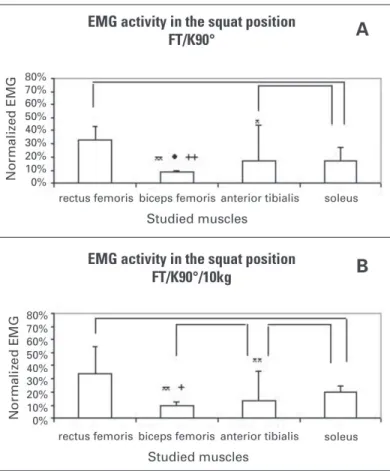
In the ST/K90° position (figure 3A), the rectus femoris present-ed higher activation (32.1%) than the biceps femoris (8.0%), and
anterior tibialis muscles (17.0%). The anterior tibialis presented higher activation than the biceps femoris, and the soleus, higher activation (17.2%) only concerning the biceps femoris. There was co-activation between the rectus femoris and soleus and anterior tibialis and soleus muscles. In the ST/K90°/10kg position (figure 3B), the rectus femoris presented higher EMG activity (34.1%) compared with the biceps femoris (9.2%) and the anterior tibialis muscles (13.3%). The soleus muscle (19.2%) presented higher activity only concerning the biceps femoris. There was co-activa-tion between the rectus femoris and soleus, biceps femoris and anterior tibialis and soleus and anterior tibialis muscles.
Figure 2 – A, B, C and D – EMG activity of the studied muscles in the squatting positions ST/K60°, ST/K60°/10kg, FT/K60° and FT/K60°/10kg, re-spectively.
* p < 0.05, ** p < 0.01, concerning the rectus femoris; + p < 0.05; ++ p < 0.01, concerning the soleus; • p < 0.05; •• p < 0.01, concerning the anterior tibialis ___ co-activation
Figure 3 – A and B – EMG activity of the studied muscles in the squatting positions FT/K90° and FT/K90°/10kg, respectively.
* p < 0.05, ** p < 0.01, concerning the rectus femoris; + p < 0.05; ++ p < 0.01, concerning the soleus; • p < 0.05, concerning the anterior tibialis ___ co-activation
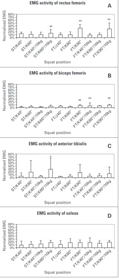
When the activation of each muscle was analyzed in each test position, it was observed that the rectus femoris presented higher activations in the St/K60°/10kg (15.2%), FT/K90° (33.2%) and FT/ K90°/10kg positions (29.7%), concerning the remaining positions, as seen in figure 4A.
According to figure 4B, the biceps femoris muscle presented the highest electromyographic activities in the FT/K40°/10kg (6.7%), FT/K90° (8.0%) and FT/K90°/10kg positions (9.2%) compared with the positions with straight trunk.
In figure 4C, it is observed that the anterior tibialis muscle pre-sented higher electromyographic activity in the ST/K60°/10kg po-sition (27.9%) only concerning the FT/K40° (3.9%) and FT/K40°/ 10kg positions (4.4%). The FT/K90° (17.0%), FT/K90°/10kg (13.3%) and ST/K60° positions (19.4%) also presented considerable activa-tion breadths, despite not being statistically significant.
flexed with knee at 40° of flexion. This result was expected, once with trunk flexion the hamstring muscles are more tensed and with its length closer to the optimum state for its activation(14). When
the trunk is flexed and the knee is at 60° and 90°, the extensor strength demand is very high and requires a higher activation of the rectus femoris comparing with the biceps, and it has its activa-tion decreased due to the approximaactiva-tion of its origin and inseractiva-tion in this position. On the other hand, the biceps femoris muscle pre-sented the lowest activation between the studied muscles in the majority of the test positions, especially with straight trunk, which corroborates the results by Kvist and Gillkist(25), who have observed
low activation of hamstrings concerning the quadriceps, both in closed and open kinetics chain exercises. In this same way of think-ing, Shields et al.(26), when studying the co-contraction between
the quadriceps femoris and the hamstrings in the squat exercise in three load levels, have verified that the activity of the quadriceps muscle was higher than the hamstring’s for all levels, having also a quadriceps/hamstrings ratio ranging from 2.3 to 3.0.
Concerning the soleus muscle, it is observed that it presented a co-activation concerning the rectus femoris in all squat positions, except for the flexed trunk and knee at 40° of flexion with load. When the ankle position during the tests is analyzed, it is observed that the higher the squat depth, the higher the demand of dorsi-flexor torque which needs to be balanced with a growing activa-tion of the plantar flexors, especially the soleus. The acactiva-tion of this muscle in CKC provides a posterior traction force in the tibia, close to its origin site and can represent a balance factor to the tibia anteriorization trend in the cases of injury of ACL, and an important alternative for the knee stabilization to be emphasized in ACL post-reconstruction rehabilitation programs(15).
Escamilla et al.(3), quantified the muscular actions in normal
trained subjects, in CKC (squatting and leg press) and in OKC activ-ities, and observed the rectus femoris co-activation with the gas-trocnemius in CKC, which eccentrically contracts in order to con-trol the ankle dorsiflexion during the knee flexion and perform plantar flexion during the extension. The authors justified the gas-trocnemius activation during the extension as a synergetic action with the biceps femoris for knee posterior stabilization. Likewise, Kvist and Gillquist(25) performed an investigation in which they
ana-lyzed the muscular activation and tibial translation in subjects with and without injury of the ACL, during squatting in three load levels and variable positioning of the gravity center in relation to the an-kle. It was observed that the quadriceps and the gastrocnemius simultaneously acted and this mechanism was considered impor-tant to increase knee stability.
Nevertheless, the gastrocnemius has origin in the femoral con-doles and if its origin was not balanced by the soleus action in CKC, a femur posteriorization may occur, leading to a tendency to knee hyperflexion, and consequently greater tension in the ACL(15).
The anterior tibialis presented a co-activation with the biceps femoris in the following positions: straight trunk with knee flexed at 40° with load, flexed trunk with knee at 40° of flexion with and without load, and flexed trunk with knee at 60° of flexion with and without load positions, which could have happened in order to sta-bilize the ankle and keep the body balance, once an action of this muscle directly over the knee is not observed. In the squatting positions with straight trunk and 40° of knee flexion without load, and in the positions of straight trunk and 60° of knee flexion with and without load, co-activation of the anterior tibial with the soleus has occurred, probably in the trial to stabilize the ankle articulation. Concerning the muscles activation independently, it can be ob-served that the knee flexion angle is an important factor for deter-mination of a higher activation of the studied muscles, where 90° > 60° > 40°. Probably, the higher activation is the result of the need of a stronger eccentric contraction in the trial to surpass the flexor torque in the three joints of the lower limb during squatting. Figure 4 – A, B, C and D – EMG activity of the rectus femoris, biceps
femoris, anterior tibialis and soleus muscles, respectively, in the studied squatting positions.
** p < 0.01 * p < 0.05
DISCUSSION
Concerning the rectus femoris muscle, its activation was im-proved with the increase of the knee flexion angle to 90°. Evidence on this fact has been found by Escamilla et al.(9), when observing
that in CKC exercises, higher activity of the rectus femoris is pro-duced between 83-95° of knee flexion. This angulation increases the compressive forces in the tibiofemoral and patelofemoral joints(10). Concerning the ligamentar tension, Beynnon and
Flem-ing(27) analyzed several activities in OKC and CKC, in subjects with
normal ACL, in which the ACL tension was measured in vivo. The authors stated that the CKC activities performed in the 0° to 40° breadth, presented the lowest levels of tension over the ACL, while the OKC activities in the breadths close to total knee extension, presented the highest tensions. Still about this aspect, Beynnon et al.(28), compared rehabilitation protocols in which the exercises
which caused higher tension in the ACL should be earlier (acceler-ated protocol) or intermediately introduced (non-acceler(acceler-ated pro-tocol). However, contrary to what the authors have reported con-cerning the ACL tension in the previous study(27), the exercises in
OKC were introduced (week 1) earlier than the CKC exercises (week 6-8), in the two protocols. Moreover, the squatting breadth used (0 to 90°) was not that considered safer concerning the ACL tension (0 to 40°). Nonetheless, after two years of follow-up in the last study, the authors reported results similar concerning antero-pos-terior relaxation, when the two protocols were compared.
Concerning the biceps femoris, the highest activities occurred in the positions with flexed trunk and knee at 90° of flexion, with and without load, which could have occurred due to the increase of the hip torque, demanding a greater extensor action of the ham-strings, maximal gluteus and magnus adductor (ischial fibers)(8).
This greater activity of the biceps femoris may mean increase in posterior tibial traction forces, reducing the potential tension in the anterior cruciate ligament (ACL) and increasing the tension in the PCL(3,10).
The anterior tibialis presented its highest activation with straight trunk, which probably occurred due to the need for a higher mus-cular contraction in this position of higher instability of body bal-ance.
The soleus muscle presented a similar activation pattern in all squatting positions, which implies in the need of constant activa-tion of this muscle in order to control the dorsiflexor torque in the test positions. Additionally, due t its tibial origin location, it is as-sumed that the soleus may act slowing the knee flexion and limit-ing a higher anterior tibial translation. This findlimit-ing is accordlimit-ing to the study developed by Elias et al.(15) who considered the soleus
muscle a functional antagonist of the ACL. This soleus result, as-sociated with what has been verified for the biceps femoris, repre-sents an indication of posterior tibial contention forces which can be activated in squatting.
Concerning the trunk position, its influence over the activation of the studied muscles was evaluated considering the same knee flexion angle and the same load situation. In this aspect, the rectus femoris had the highest activation when the 60° angle of knee flexion is associated with 10 kg load and straight trunk position, compared with the flexed trunk position. This higher activation prob-ably occurred due to a better length-tension ratio of the rectus femoris and by the need of controlling the higher flexor torque in this position, considering the posterior dislocation of the gravity center(8). Concerning the biceps femoris, in the 40° angle with 10
kg load and flexed trunk, higher activation comparing with the straight trunk position has occurred. Thus result may have occurred due to better length-tension ratio as well as by the dislocation of the gravity center forward, increasing the muscular strength de-mand for the control of flexor torque in the hip joint(3). Although the
subjects have shown greater easiness in performing squatting with flexed trunk, this fact did not determine differences in the muscu-lar activation in all positions.
The additional load of 10 kg influenced in the activation of the rectus femoris and biceps femoris muscles for the knee flexion angles of 60 and 40°, respectively, with higher muscular activation with load increase. For the other muscles, the additional load did not cause any influence, suggesting caution concerning the load increase, since it may represent a significant increase in the shear antero-posterior forces of the tibiofemoral joint(17).
Despite the relevance of these results, this study presented some limitations concerning the selected sample as well as the adopted experimental protocol. Concerning the sample, the fact the subjects are healthy, with heterogeneous anthropometric char-acteristics, as well as the lack of objective evaluation of the strength of the subjects, leads us to recommend caution in the generaliza-tion of these results. Concerning the experimental protocol, it was performed in static positioning conditions, and therefore, the re-sults may be different if applied to a dynamic protocol.
CONCLUSION
Squatting is a multiarticular exercise in CKC, where a simulta-neous hip, knee and ankle flexion occurs, causing a co-contraction of several muscles, which represents an important factor for the dynamic stability. In this investigation, in concordance with some studies presented in the literature, significant muscular co-activa-tion between the rectus and biceps femoris muscles occurred only in the positions with flexed trunk, which reinforces the need of this trunk positioning during performance of squatting exercises providing lower tensions over the ACL, especially in angles higher than 60° of knee flexion. Additionally, the co-activation between the rectus femoris and soleus muscles open perspectives for the inclusion of specific strengthening of the latter in rehabilitation pro-grams, as a way of balancing the anterior tibial translation forces, and consequently reducing the tension on the ACL.
The increase of the squatting depth has also remarkably increased the activation difference of the rectus femoris, especially, concern-ing the biceps femoris, which can indicate an imbalance of forces, where a disproportional increase of the extensor force compared with the flexor one occurs. Since the knee extensor action tends to cause anterior tibial translation, the use of deep squats in the early post-surgical period of the ACL reconstruction would not be recommended due to greater torque demand played over the ex-tensor group during squatting exercises with knee flexed beyond 40°.
This study had an innovative aspect, since it has analyzed the interaction of many muscles of the anterior and posterior muscular chains which act in the dynamic stabilization of the knee joint in CKC, associating it to the trunk positioning with or without load and to many knee flexion degrees. However, the study was per-formed in healthy subjects using static positioning. Thus, further studies are needed with the purpose to establish a relationship between muscular activation and the magnitude of tibial transla-tion forces at dynamic positransla-tioning conditransla-tions which include sub-jects with deficient ACL, in order to validate the use of these find-ings in rehabilitation programs.
All the authors declared there is not any potential conflict of inter-ests regarding this article.
REFERENCES
1. Steindler A. Kinesiology of the human body under normal and pathological con-ditions. Springfield, IL: Charles C Thomas; 1977. p. 63.
2. Hopkins JT, Ingersoll CD, Sandrey MA, Bleggi SD. An electromyographic com-parison of 4 closed chain exercises. J Athl Train. 1999;34:353-7.
4. Fonseca ST, Silva PLP, Ocarino JD, Ursine PGS. Análise de um método eletro-miográfico para quantificação de co-contração muscular. Rev Bras Ciên e Mov. Brasília 2001;9:23-30.
5. Fitzgerald GK. Open versus closed kinetic chain exercise: issues in rehabilitation after anterior cruciate ligament reconstrutive surgery. Phys Ther. 1997;77:1747-54.
6. Hammond MC, Fitts SS, Kraft GH, Nutter PB, Trotter MJ, Robinson LM. Co-contraction in the hemiparetic forearm: quantitative EMG evaluation. Arch Phys Med Rehabil. 1988;69:348-51.
7. Frost G, Dowling J, Dyson K, Bar-Or O. Cocontraction in three age groups of children during treadmill locomotion. J Electromyogr Kinesiol. 1997;7:179-86. 8. Escamilla RF. Knee biomechanics of the dynamic squat exercise. Med Sci Sports
Exerc. 2001;33:127-41.
9. Escamilla RF, et al. Effects of technique variations on knee biomechanics during the squat and leg press. Med Sci Sports Exerc. 2001;33:1552-66.
10. Wilk KE, Escamilla RF, Fleisig GS, Barrentine SW, Andrews JR, Boyd ML. A comparison of tibiofemoral joint forces and electromyographic activity during open and closed kinetic chain exercises. Am J Sports Med. 1996;24:518-27. 11. Van Eijden TM, deBoer W, Weijs WA. The orientation of the distal part of the
quadriceps femoris muscle as a function of the knee flexion-extension angle. J Biomech. 1985;18:803-9.
12. Isear JA Jr, Erickson JC, Worrell TW. EMG analysis of lower extremity muscle recruitment patterns during an unloaded squat. Med Sci Sports Exerc. 1997;29: 532-9.
13. Li Y, McClure PW, Pratt N. The effect of hamstring muscle stretching on stand-ing posture and on lumbar and hip motions durstand-ing forward bendstand-ing. Phys Ther. 1996; 76:836-45.
14. McCaw ST, Melrose DR. Stance width and bar load effects on leg muscle activ-ity during the parallel squat. Med Sci Sports Exerc. 1999;31:428-36.
15. Elias JJ, Faust AF, Chu Y, Chao EY, Cosgarea AJ. The soleus muscle acts as an agonist for the anterior cruciate ligament: an in vitro experimental study. Am J Sports Med. 2003;31:241-6.
16. Chandler TJ, Wilson GD, Stone MH. The effect of the squat exercise on knee stability. Med Sci Sports Exerc. 1989;21:299-303.
17. Hattin HC, Pierrynowski MR, Ball KA. Effect of load, cadence, and fatigue on tibio-femoral joint force during a half squat. Med Sci Sports Exerc. 1989;21:613-8.
18. Seki K, Narusawa M. Relation between the size of motor units and the spectral characteristics of their action potentials. Electroencephalogr Clin Neurophysiol. 1998;109:436-43.
19. Bigland B, Lippold DCJ. The relationship between force, velocity and integrated electrical activity in human muscles. J Physiol. 1954;260:267-77.
20. Farina D, Fosci M, Merletti, R. Motor unit recruitment strategies investigated by surface EMG variables. J Appl Physiol. 2002;92:235-47.
21. Earl JE, Schmitz RJ, Arnold BL. Activation of the VMO and VL during dynamic mini-squat exercises with and without isometric hip adduction. J Electromyogr Kinesiol. 2001;11:381-6.
22. Wang RY, Chan RC, Tsai MW. Effects of thoraco-lumbar electric sensory stimu-lation on knee extensor spasticity of persons who survived cerebrovascular ac-cident (CVA). J Rehabil Res Dev. 2000;37:73-9.
23. Araujo RB, Duarte M, Amadio AC. Estudo sobre a variabilidade do sinal eletro-miográfico intra e interindivíduos durante a contração isométrica. Apresentado no 7o Congresso Brasileiro de Biomecânica; 1997 Maio 28-30; Campinas – São
Paulo; 1997. p. 128-30.
24. McAtamney L, Corlett EN. RULA: a survey method for the investigation of work-related upper limb disorders. Appl Ergon. 1993;24(2):91-9.
25. Kvist J, Gillquist J. Sagittal plane knee translation and electromyographic activity during closed and open kinetic chain exercises in anterior cruciate ligament-deficient patients and control subjects. Am J Sports Med. 2001;29:72-82.
26. Shields RK, Madhavan S, Gregg E, Leitch J, Petersen B, Salata S, et al. Neuro-muscular control of the knee during a resisted single-limb squat exercise. Am J Sports Med. 2005;33:1520-6.
27. Beynnon BD, Fleming BC. Anterior cruciate ligament strain in-vivo: a review of previous work. J Biomech. 1998;31:519-25.