1Residence Program in Ophthalmology, Hospital Federal de Bonsucesso, Bonsucesso, RJ, Brazil. 2Department of Ophthalmology, Universidade do Sul de Santa Catarina, Florianópolis, SC, Brazil. 3Department of Pediatrics, Universidade do Sul de Santa Catarina, Florianópolis, SC, Brazil.
Received for publication 21/12/2015 - Accepted for publication 18/01/2016 The authors declare no conflicts of interests.
The study was conducted at Hospital Regional de São José Dr. Homero de Miranda Gomes (HRSJ), located in the municipality of São José (Santa Catarina), Brazil.
RESUMO
Objetivo: Avaliar a prevalência da retinopatia da prematuridade (ROP) em recém-nascidos (RN) prematuros (Idade Gestacional (IG) < 37 semanas) e/ou peso ao nascimento (PN) £ 1500g e os que possuem fatores de risco, nascidos no HRSJ entre janeiro de 2007
e janeiro de 2011. Método: Estudo transversal, retrospectivo, analítico e observacional. Os dados foram obtidos a partir de
prontu-ários no Hospital Regional de São José Dr. Homero de Miranda Gomes. Resultados: Observou-se a presença de retinopatia em
37,81% dos RNs, sendo o estágio 1 o mais prevalente. Verificou-se que não houve diferença estatística entre os sexos (p=0,993). A presença da ROP foi maior no grupo com PN < 1000 gramas (83,33%), avaliados com mais de 6 semanas de vida e com IG menor que 32 semanas (49,48%). Os fatores de risco com significado estatístico foram: oxigenioterapia, ventilação mecânica, persistência do canal arterial, asfixia perinatal, síndrome do desconforto respiratório, transfusão sanguínea, hemorragia intraventricular, sepsis,
infecção neonatal e doença da membrana hialina. Conclusão: Conclui-se que o fator sexo e gestação múltipla não tiveram significância
estatística. Os RNs com menor PN e IG tem um maior risco de desenvolver ROP. Em relação à oxigenioterapia, a prevalência nos expostos é maior e proporcional ao tempo de utilização de oxigênio.
Descritores: Retinopatia da prematuridade/epidemiologia; Fatores de risco; Prevalência; Oxigenioterapia
Epidemiologic profile of preterm infants with
retinopathy of prematurity in the Dr. Homero de
Miranda Gomes Regional Hospital in São José
Perfil epidemiológico dos recém-nascidos
prematuros com retinopatia da prematuridade no Hospital
Regional de São José Dr. Homero de Miranda Gomes
Mara Barreto Theiss
1; Astor Grumann Júnior
2, Marise Regina Wiethorn Rodrigues
3ABSTRACT
Objectives: To evaluate the prevalence of retinopathy of prematurity (ROP) in premature newborns (gestational age < 37 weeks) and / or birth weight £ 1,500g and those with risk factors, born at the Dr. Homero de Miranda Gomes Regional Hospital in São José (HRSJ) between January 2007 and January 2011. Methods: Cross-sectional, retrospective, observational and analytical study. Data were obtainedfrom medical records at the HRSJ. Results: The presence of 37.81% of retinopathy in newborns was observed, with stage 1 being the mostprevalent. No statistical difference was found between the sexes (p = 0.993). The presence of ROP was higher in the group with PN < 1,000grams (83.33%), evaluated over 6sixweeks of age and with gestational ages less than 32 weeks (49.48%). Risk factors with statisticalsignificance were: oxygen therapy, mechanical ventilation, patent ductus arteriosus, perinatal asphyxia, respiratory distress syndrome,blood transfusions, intraventricular hemorrhage, sepsis, neonatal infection and hyaline membrane disease. Conclusion: It is concludedthat: the gender factor and multiple pregnancy were not statistically significant. The newborns with lower birth weight and gestational agehave an increased risk for developing ROP. Regarding oxygentherapy, the prevalence is higher in the exposed and proportional to theperiod of oxygen.
INTRODUCTION
R
etinopathy of Prematurity (ROP) is one of the leadingcauses of preventable childhood blindness, being
responsible for 50 thousand blind children worldwide(1).
It affects preterm newborns (gestational age < 37 weeks), and its severity offers an inversely proportional relation to the
gestational age (GA) and birth weight(2,4).
It is defined as a vasoproliferative disease, and is developed
from immature retinal vasculature(5). Tracing newborns (NBs) in
risk allows the identification of severe forms of the disease, and early treatment reduces the risk of vision loss.
As described earlier, the immature retina favors the formation of neovascular tissue, which can develop into fibrovascular proliferation toward the vitreous, forming membranes and retinal traction. Said traction can result in retinal detachment and development of low visual acuity of varying
degree(6,7). Thus, it is extremely important in the implementation
of a screening of ROP in NBs and early treatment to reduce the
long-term consequences of the disease(8).
The main risk factors for the development of retinopathy are prematurity and low birth weight. However, there are other risk factors such as Apgar score lower than 7, fluctuation in oxygen levels in the first weeks of life, use of oxygen therapy, the need for mechanical ventilation, blood transfusion, patent ductus arteriosus, respiratory distress syndrome, child being small for gestational age (SGA), intraventricular hemorrhage, perinatal asphyxia, multiple pregnancy, sepsis and meningitis(2,9,11).
The ICROP defined the disease according to its severity (staging 1 to 5), location (zones I to III), length in hours (from 1 to 12 hours), with or without Plus disease (arteriolar dilatation and venous tortuosity) whose presence would be an indicator of disease activity(12).
This study was proposed to assess the prevalence and staging of ROP and the associated factors in preterm NBs (IG < 37 weeks) and/or birth weight £ 1500g and those who have risk factors, assesses in the HRSJ from January 2007 to January 2011.
METHODS
Cross-sectional and retrospective study. Approved by
the Research Ethics Committee (REC) of Hospital Regional de São José Dr. Homero de Miranda Gomes (HRSJ) under record No. 47/10. Due to the specificity of the research (data from medical records) it was not possible to obtain the Informed Consent Form from each patient, but an Agreement on Use of Data was signed.
The study was conducted at HRSJ, which is a State reference in the treatment of ROP. A census was carried out in the period from January 2007 to January 2011 with a total of 399 records, and the records assessed were from living preterm newborns and/or with birth weight £ 1500g, and of those with risk factors and which have been assessed by an ophthalmologist. The study included NBs from other institutions which were assessed after looking for the service, according to the inclusion criteria. The sample included all preterm and/or low-birth-weight NBs, and those with risk factors (mechanical ventilation, patent ductus arteriosus, perinatal asphyxia, respiratory distress syndrome, blood
transfusion, multiple pregnancy, intraventricular hemorrhage, sepsis, neonatal infection, hyaline membrane disease), born in the period selected and which have been assessed by an ophthalmologist. The study excluded all NBs (preterm and/or with low birth weight and risk factors) who were not included in the period selected, those in which the medical records were incomplete, not allowing the entire assessment of data and preterm newborns who died before the first eye examination. The routine ophthalmologic assessment performed by the HRSJ comprises the external inspection of the eyeballs and fundoscopy under pupil dilatation (with eyedrops of midriacil 0.5% and phenylephrine 2.5% associated 60 minutes before the procedure), under indirect binocular ophthalmoscopy with magnifying lens of 28 diopters and blefarostate. Droplets of anesthetic eyedrops were used pri-or to the exam. So the retinal mapping and retinopathy staging were performed (according to the International Classification of Retinopathy of Prematurity).
For the classification of ROP the eye with the largest stadium was considered according to the protocol already used in the service.
The sample was tabulated directly in the program SPSS version 18. The data is described in the form of relative and absolute frequency. Categorical variables were compared by the Fisher exact test and the Chi-square test, with a significance level p < 0.05. The prevalence ratios of retinopathy and their respective confidence intervals (IC95%) were calculated.
RESULTS
For statistical analysis, a total of 320 NBs remained. The study excluded 79 NBs, those whose medical records were incomplete.
The sample studied showed the presence of retinopathy in 37.81% (121) (Table 1).
The NBs studied showed the prevalence of the ROP of 37.84% males and 37.79% females, with no statistical difference (p=0.993).
The results show that the prevalence of ROP was higher in the group with birth weight < 1000 grams, where 83.33% had some stage of retinopathy. On the other hand, among patients with > 2500 grams, only 26.66% presented ROP (Table 2).
Regarding the GA, it can be seen that the smaller the GA, the greater the risk of developing ROP (Table 3).
Table 1
Prevalence of retinopathy of prematurity
Staging n°(%)
Without ROP 199(62.19)
With ROP 121(37.81)
Stage 1 57(17.81)
Stage 2 32(10.0)
Stage 3 16(5.00)
Stage 4ª 7(2.19)
Stage 4B 0(0)
Stage 5 9(2.81)
p < 1000 1000 ≤≤≤≤≤ p < 1500 1500 ≤ ≤ ≤ ≤ ≤ p < 2500 p ≥≥≥≥≥ 2500
Retinopathy Total P value
n°(%) n°(%) n°(%) n°(%)
With ROP 45(83.33) 53(37.06) 19(17.59) 4(26.66) 121 <0.001
WithoutROP 9(16.67) 90(62.94) 89(82.41) 11(73.34) 199
Total 54(100) 143(100) 108(100) 15(100) 320
Table 2
Prevalence of retinopathy of prematurity in relation to birth weight in grams
IG < 32 weeks IG 32 – 37 weeks IG >37 weeks
Retinopathy Total P value
n°(%) n°(%) n°(%)
With ROP 96(79.4) 24(19.83) 1(0.83) 121 <0.001
Without ROP 98(49.25) 91(45.73) 10(5.02) 199
Table 3
Prevalence of retinopathy of prematurity in relation to the gestational age
Factors With ROP(%) Witout ROP(%) nº Total P value
Oxygen therapy
Yes 120(41.3) 170(58.6) 290 320 0.005
No 1(8) 29(2) 30
Mechanical ventilation
Yes 60(64.5) 33(35.4) 93 320 <0.001
No 61(2) 166(8) 227
Patent ductus arteriosus
Yes 5(83.3) 1(16.6) 6 320 <0.001
No 116(3) 198(7) 314
Perinatal asphyxia
Yes 43(84.3) 8(15.6) 51 320 <0.001
No 78(1) 191(9) 269
Respiratory distress syndrome
Yes 249(77.4) 7(22.5) 31 320 <0.001
No 97(2) 192(8) 289
Blood transfusion
Yes 13(86,6) 2(13.1) 15 320 <0.001
No 108(7) 197(3) 305
Multiple pregnancy
Yes 21(42.8) 28(57.1) 49 320 0.699
No 100(6) 171(4) 271
Intraventricular hemorrhage
Yes 8(66.6) 4(33.3) 12 320 <0.001
No 113(7) 195(3) 308
Sepsis
Yes 15(78.9) 4(21.0) 19 320 <0.001
No 106(5) 195(5) 301
Neonatal infection
Yes 10(52.6) 9(47.3) 19 320 <0.001
No 111(3) 190(7) 301
Hyaline membrane disease
Yes 16(80.0) 4(20,0) 20 320 <0.001
No 105(0) 195(0) 300
Table 4
Risk factors among newborns in relation to retinopathy of prematurity
P value for the Chi-square test P value for the Chi-square test
Table 4 shows the other risk factors studied in NBs with and without ROP, which showed statistically significant differences (p < 0.001), except the multiple pregnancy factor, which was not statistically significant. A higher prevalence of ROP is noted in NBs who have the risk factors described. For example, 80% of NBs who have hyaline membrane disease and 84.31% of NBs who have perinatal asphyxia developed ROP.
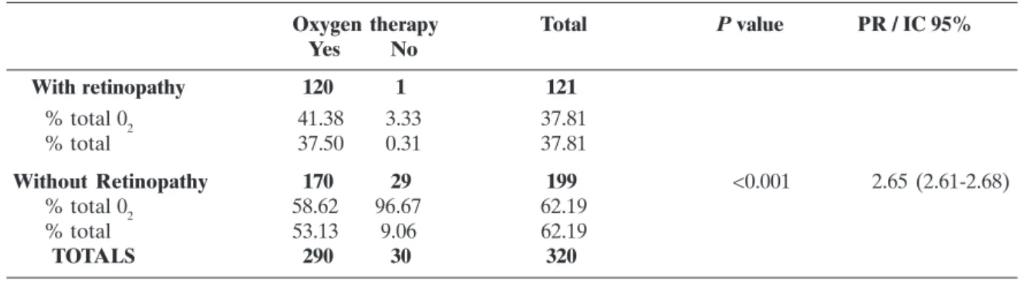
As for the aspects related to oxygen therapy, the prevalence ratio (PR) of ROP was 2.65, i.e. the prevalence of NBs who have used the oxygen therapy was 2.65 times higher than the prevalence of NBs who did not use it (Table 5).
In relation to the time of oxygen therapy, it was noted that the shorter the time of use of oxygen, the lower the chance of developing retinopathy (Table 6).
Oxygen therapy Total P value PR / IC 95% Yes No
With retinopathy 120 1 121
% total 02 41.38 3.33 37.81
% total 37.50 0.31 37.81
Without Retinopathy 170 29 199 <0.001 2.65 (2.61-2.68)
% total 02 58.62 96.67 62.19
% total 53.13 9.06 62.19
TOTALS 290 30 320
Fisher’s test.
Table 5
Oxygen therapy in relation to retinopathy of prematurity
Time Retinopathy Total P value Yes No
< 1 day 0 8 8 <0.001
% of the total time O2 0.00 100.00 100.00
% Total 0.00 4.06 4.06
1 a < 7 days 13 52 65
% of the total time O2 20.00 80.00 100.00
% Total 6.60 26.40 33.00
7 – 20 days 23 31 54
% of the total time O2 42.59 57.41 100.00
% Total 11.68 15.74 27.42
> 20 days 51 19 70
% of the total time O2 72.86 27.14 100.00
% Total 25.89 9.64 35.53
TOTAL 87 110 197
% do total tempo O2 44.16 55.84 100.00
% Total 44.16 55.84 100.00
Table 6
Retinopathy of Prematurity in relation to the time of use of oxygen
P value for the Chi-square test
DISCUSSION
Sight is one of the most important senses in the normal
physical and cognitive development of a child(2). The motor
development and communication skills are impaired in children with visual impairment because gestures and social behaviors
are learned by the sense of sight(2,13,14). This way, blindness and/
or low vision, besides affecting the physical, mental, economic
and cultural conditions, may change the whole dynamic of the
family and affect society as a whole(15). The present work aims at
determining the prevalence of ROP and its risk factors in the HRSJ, thus enabling the detection of possible severe cases and the prevention of the disease through a better understanding of this pathology.
found was higher than the one reported in the literature
(between 20 and 27.73%)(7,8,10). One possible explanation for
this high prevalence is that the HRSJ is a State reference to ROP, thus having many patients referred for treatment as well as a good screening of NBs. As in the HRSJ, we can also mention the high incidence of ROP (32%) at Hospital Regional ASA Sul, in Distrito Federal, which is the reference center in the Brazilian
Midwest(15). Recent data published in the city of Joinville(SC)(8)
and in the city of São Paulo(16) showed an overall prevalence of
ROP of 20 and 29.90%, respectively. In another study in Rio de Janeiro, Portes et al. described a prevalence of ROP of 27.81% at
Hospital Federal de Bonsucesso(17).
This study showed that the majority of NBs was diagnosed in stage 1 disease (17.81%), and only 9 NBs were diagnosed in stage 5 (2.81%), in which there is total retinal detachment; a similar result is found in the literature(7,10,17).
Only 1 patient was recorded with staging ROP 2, born with more than 37 weeks of gestational age, and stage 4B of ROP was not found in any NB. The same happened in the study carried put in Joinville(8).
In the present research, the factor gender showed no association (p = 0.993) to the disease in question, as well as in the
studies in Joinville(8) and São Paulo(16). There was a prevalence of
ROP of 37.79% in females, and of 37.84% in males, with no statistical difference.
Regarding the birth weight, we observed a prevalence of retinopathy in 83.33% of NBs with less than 1000 grams (extreme
low weight). Graziano et al.(16) similarly found in their study a
high prevalence of ROP (78.5%) among NBs with birth weight
lower than 1000 grams, and Fortes Filho et al.(18) found a
prevalence of 45.59%. In another paper, the authors drew attention to the high prevalence of the disease (78.5%) in the
group of children born with less than 1000 grams (18). It was
observed that NBs with very low weight (1000 g £ p < 1500 g) had a prevalence of ROP of 37.06%, and among those with low birth weight (1500 g £ p < 2500 g) a prevalence of 17.59%.
The same was found in relation to birth weight: the lower the gestational age the higher the risk of developing ROP, in this study having a ROP prevalence of 79.34% of NBs with less than 32 weeks of gestational age. Literature data confirm the data found, as they show that the occurrence of ROP is primarily
linked to low gestational age and birth weight(6,7,16,18,19).
From the analysis of the results we can see the combination of risk factors (oxygen therapy, mechanical ventilation, patent ductus arteriosus, perinatal asphyxia, respiratory distress syndrome, blood transfusion, intraventricular hemorrhage, sepsis, neonatal infection and hyaline membrane disease) included in this study with the presence of ROP, showing statistical significance to the study, which confirms the literature data(2,7,8,10).
On the other hand, no association between multiple pregnancy and sex was observed, suggesting that these variables may not be risk factors for the development of ROP.
Regarding oxygen therapy, the data presented here shows that the prevalence ratio (PR) of ROP was 2.65 times higher when compared to NBs who did not use it. From these results it is possible to suggest that the prevalence in the exposed patients is greater compared to the non-exposed ones. Another relevant observation is that the shorter the period of use of oxygen, the less chance of developing retinopathy. Other studies report oxygen therapy as an important risk factor for the development of retinopathy(7,8,16).
With the advance of neonatal intensive care units, increasing technological advancement in the area, there was an increase in NBs survival increasingly preterm and underweight, and hence the prevalence of ROP is becoming higher, thereby stimulating researches to allow pathophysiological knowledge, since this is not yet fully elucidated. Thus, it reinforces the need for research in this area, because blindness has achieved a significant number of children, representing a serious public health problem.
CONCLUSION
The clinical and epidemiological profile of the Preterm NBs
in the HRSJ can be defined.There was a significant prevalence
of ROP in preterm NBs, and among staging the stage 1 of ROP
presented a higher prevalence.The factors gender and multiple
pregnancy do not seem to be associated to the development of
ROP.And the risk factors oxygen therapy, mechanical ventilation,
patent ductus arteriosus, perinatal asphyxia, respiratory distress syndrome, blood transfusion, intraventricular hemorrhage, sepsis, neonatal infection and hyaline membrane disease seem to be associated regarding the development of ROP.
ACKNOWLEDGEMENTS
Dr. André Luis Freire Portes, head of the Department of Ophthalmology of Hospital Federal de Bonsucesso (RJ).
REFERENCES
1. Gilbert C, Rahi J, Eckstein M, O’Sullivan J, Foster A. Retinopathy of prematurity in middle-income countries. Lancet. 1997; 350 (9070):12-4.
2. Graziano RM, Leone CR. Problemas oftalmológicos mais freqüentes e desenvolvimento visual do pré-termo extremo. J Pedatr (Rio J). 2005; 81(1 Suppl):S95-100.
3. Moraes N, Bonomo P. Retinopatia da prematuridade: acompanhamento de 343 recém-nascidos pré-termo. Arq Bras Oftalmol. 1993; 56:192.
4. Tavano V, Nogueira R, Moraes N, Farah M. Associação entre retinopatia da prematuridade e hemorragia intraventricular em recém-nascidos de baixo peso. Arq Bras Oftalmol. 1996; 59:373. 5. Zin A, Florêncio T, Fortes Filho JB, Nakanami CR, Gianini N, Graziano RM, et al . Proposta de diretrizes brasileiras do exame e tratamento de retinopatia da prematuridade (ROP). Arq Bras Oftalmol. 2007; 70(5):875-83.
6. Sá LCF. Aspectos atuais da retinopatia da prematuridade. J Pediatr (Rio J). 1990; 66 (8/9): 220-4.
7. Fortes Filho JB, Eckert GU, Valiatti FB, Costa MC, Bonomo PP, Procianoy RS. Prevalência e fatores de risco para a retinopatia da prematuridade: estudo com 450 pré-termos de muito baixo peso. Rev Bras Oftalmol.2009; 68 (1):22-9.
8. Bonotto LB, Moreira AT, Carvalho DS. Prevalência de retinopatia da prematuridade em prematuros atendidos no período de 1992-1999 em Joinville (SC): avaliação de riscos associados – “screen-ing”. Arq Bras Oftalmol. 2007; 70 (1):55-61.
9. Kanski JJ, Menon J. Doenças vasculares retinianas. Oftalmologia clínica – uma abordagem sistemática. Rio de Janeiro: Elsevier; 2004. p. 438-86.
11. Lorena SH, Brito José MS. Estudo retrospectivo de crianças pré-termo no Ambulatório de Especialidades Jardim Peri-Peri. Arq Bras Oftalmol. 2009; 72 (3):360-4.
12. Fortes Filho JB. Revisão: Retinopatia da prematuridade. Rev Bras Oftalmol. 2006; 65(4):246-58.
13. Associação Médica Brasileira; Conselho Federal de Medicina. Projeto Diretrizes Retinopatia da prematuridade. São Paulo: Conselho Brasileiro de Oftalmologia e Sociedade Brasileira de Pediatria. Elaboração final em: 04 jul 2011. 18]. [Zin A, Uno F, Sociedade Brasileira de Retina e Vitreo, Sociedade Brasileira de Oftalmologia Pediatrica, Simoes R, organizadores ] [citado 2015 Dez 20]. Disponível em: http://www.projetodiretrizes.org.br/ diretrizes10/retinopatia_da_prematuridade.pdf
14. Endriss D, Ventura LM, Diniz JR, Celino AC, Toscano J. Doenças oculares em neonatos. Arq Bras Oftalmol. 2002; 65(5):551-5. 15. Souza RA, Santos PM, Santos RC. Retinopatia da prematuridade:
incidência, detecção e conduta em hospital de referência no Distrito Federal. Brasília (DF): Universidade de Brasília; FS 2010. 16. Graziano RM, Leone CR, Cunha SL, Pinheiro AC. Prevalência da retinopatia da prematuridade em recém-nascidos de muito baixo peso. J Pediatr (Rio J). 1997; 73(6): 377-82.
17. Portes AL, Barauna H, Jeveaux GC, Monteiro ML. Perfil Clínico e epidemiológico de recém-natos prematuros com muito baixo peso no Rio de Janeiro: estudo de 152 pacientes. Rev Bras. Oftalmol. 2010; 69(6):389-91.
Author corresponding:
Mara Barreto Theiss
E-mail: [email protected]
18. Fortes Filho JB, Lermann VL, Barros CK, Innocente C, Costa MC, Procianoy RS. Prevalência da retinopatia da prematuridade no centro de neonatologia do Hospital de Clínicas de Porto Alegre, Brazil. Revista HCPA 2006; 26 (2): 12-17.
19. Machado KC, Teixeira LL, Elpídio de Sá F. Perfil clínico dos recém-nascidos com retinopatia da prematuridade em um hospi-tal público do Ceará. RBPS 2008; 21(1): 47-54.
ERRATA
March / April 2016 , Vol. 75 ( 2 ) , pág.109-14
The original article “Epidemiologic profile of preterm
infants with retinopathy of prematurity in the Dr. Homero
Miranda Gomes Regional Hospital in San Jose,”
published in the March / April 2016 Journal of
Ophthalmology (Rev Bras Ophthalmol 2016;75 (2): 109-14), underwent correction in the names of the authors.
The names of the authors are correct, “Mara Barreto