Percutaneous Closure of a Patent Ductus Arteriosus
with the Cera
TM
PDA Occluder: Another Good
Option in the Toolbox
Francisco Chamié
1,2, Luiz Carlos Simões
1,3,
Daniel Silva Chamié de Queiroz
1,4, Renata Mattos
1,3ABSTRACT
Background: The percutaneous closure of a patent ductus
arteriosus (PDA) has been considered the treatment of choice by most authors, and several devices with different structural characteristics have been used. The initial experience with the novel CeraTM PDA Occluder is reported. Methods: From March of 2010 through December of 2011, patients weighing over 5 kg with a PDA diagnosed by transthoracic echocardiogram (TTE) with colour Doppler low mapping and no associated defects underwent the procedure. Follow-up was performed by TTE one, three, and six months after the procedure, and yearly thereafter. Results: Overall, 18 patients were referred for percutaneous occlusion; 61.2% were female. The mean age and weight were 13.7 ± 9.3 years and 42.9 ± 20.1 kg, respectively. Regarding morphology, 11 were type A, six were type E, and one had a residual postoperative defect. The mean diameter was 4.2 mm. Implantation was possible in all patients. Ten 6-4 mm, one 8-6 mm, three 10-8 mm, and four 12-10 mm devices were used. All defects were completely closed by the irst follow-up TTE. Deaths or complications were not observed in this series. Conclusions: The CeraTM prosthesis may be used for the occlusion of small or large defects, and delivers to excellent results in children and adults. The pro-cedure is easy, safe, has a high eficacy and low morbidity, and may be an excellent option for the percutaneous closure of a PDA. Due to its lexibility, oversized devices greater than 2 mm should be used.
DESCRIPTORS: Ductus arteriosus, patent. Heart catheterization. Prostheses and implants. Heart defects, congenital.
1 INTERCAT – Cardiologia Intervencionista – Rio de Janeiro, RJ, Brazil. 2 Hospital Federal dos Servidores do Estado – MS-RJ – Rio de Janeiro,
RJ, Brazil.
3 Instituto Nacional de Cardiologia – Laranjeiras – Rio de Janeiro,
RJ, Brazil.
4 University Hospitals at Case Medical Center – Cleveland, United States. Correspondence to: Francisco Chamié. Av. Borges de Medeiros, 3.501/103 – Lagoa – Rio de Janeiro, RJ, Brazil – CEP 22470-001
E-mail: [email protected]
Received: 1/9/2012 • Accepted: 3/5/2012
Original Article
RESUMO
Fechamento de Canais Arteriais com o Dispositivo CeraTM PDA Occluder: Mais uma
Boa Opção na Caixa de Ferramentas
Introdução: O fechamento percutâneo de persistência dos ca
-nais arteriais (PCA) tem sido considerado tratamento de escolha pela maioria dos autores, e diversos dispositivos com diferentes características estruturais têm sido utilizados. Apresentamos a experiência inicial do grupo com a nova pró tese CeraTM PDA Occluder. Métodos: Entre março de 2010 e dezembro de 2011 foram submetidos ao procedimento pacientes com mais de 5 kg de peso, com PCA diagnosticada por meio de ecocardio grama transtorácico com mapeamento de luxo em cores (ETT), sem defeitos associados. O seguimento foi feito com ETT no primeiro, no terceiro e no sexto meses subsequentes, e, a seguir, anualmente.
Resultados: No total, 18 pa cientes foram encaminhados para
oclusão percutânea, dos quais 61,2% eram do sexo feminino. As médias das idades e dos pesos foram, respectivamente, de 13,7 ± 9,3 anos e 42,9 ± 20,1 kg. Quanto à morfologia, 11 canais foram do tipo A, 6 foram do tipo E, e 1 pertuito residual após cirurgia. A média dos diâmetros foi de 4,2 mm. O implante foi possível em todos os casos. Foram utilizadas 10 próteses 6-4 mm, 1 prótese 8-6 mm, 3 próteses 10-8 mm e 4 próteses 12-10 mm. Todos os canais estavam completamente fechados por ocasião do primeiro ETT de controle. Não houve óbitos ou com plicações nesta casuís tica. Conclusões: A prótese CeraTM pode ser utilizada para o fechamento de canais de pequeno ou grande calibres com excelente resultado, em crianças e adultos. O procedimento é fácil, seguro, com alta eicácia e baixa morbidade, e pode ser excelente opção para o fechamento percutâneo de PCA. Suas características de lexibilidade sugerem que sejam utilizadas próteses superdimensionadas acima dos 2 mm habitualmente recomendados.
DESCRITORES: Permeabilidade do canal arterial. Cateterismo
car díaco. Próteses e implantes. Cardiopatias congênitas.
T
he percutaneous closure of a patent ductus arte-riosus (PDA) represents an established alternative to surgical ligation and has been deined as the treatment of choice by most authors. To this end, several devices have been used.1–15At the end of the 1990s, the first metal mesh prosthesis, the Amplatzer® Duct Occluder I (ADO I), capable of occluding larger diameter channels was developed as an alternative to embolisation coils.16 Universally used, it has occlusion rates of nearly 100%, with very low short- and long-term complica-tion rates.17–20 Despite the success, new devices with different structural characteristics have been produced.
The objective of the present study was to present an initial experience with a new prosthesis, the CeraTM PDA Occluder (Lifetech Scientiic Co. Ltd., Shenzhen, China), and to examine its role as another option for the occlu-sion of medium and large calibre PDA.
METHODS
Study design
This was a prospective, single-arm study performed at two centres. All patients underwent closure of PDA with the CeraTM prosthesis between March 2010 and December 2011. Characteristics of the device and the immediate results are described.
Selection criteria
All consecutive patients weighing more than 5 kg with PDA, and without any other associa ted defects that required surgical correction, were included in this study. Cases were chosen by transthoracic echocardiograms (TTEs) with colour low mapping. The dimensions and morphology of the defects did not constitute exclusion criteria.
The prosthesis
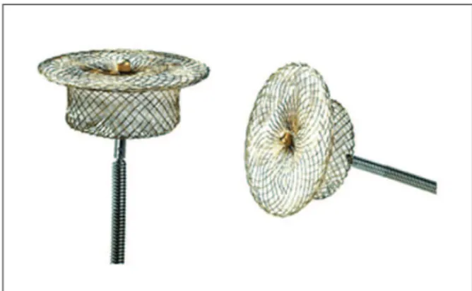
The CeraTM occluder is a self-expansible prosthesis with a nitinol fragmented cone and ceramic coating. The proximal (pulmonary) extremity has a female thread, measuring 2 mm less than the distal (aortic) extremity of the cone, and connects to the delivery system. There is a retention disc in the aortic extremity that measures 4 mm more than the distal extremity (Figure 1).
The device is available in 2-mm increments from 6 mm to 24 mm in diameter (distal extremity). The central portion measures 7 mm in 6- to 14-mm prostheses, 8 mm in 16- and 18-mm prostheses, 9 mm in 20- and 22-mm prostheses, and 10 mm in 24-mm prostheses.
The delivery system is composed of a 5 F to 12 F long and lexible sheath, a small loader of compatible size, a haemostatic valve, and a distal extremity with a metallic cable with threads.
Implant technique
The implant and follow-up protocols have been previously described and are the same protocols used for the ADO I prosthesis study.20
Statistical analysis
Continuous variables were expressed as mean and standard deviation, while categorical variables were expressed as numbers and percentages. The objective of this article was to present the initial experience with the new CeraTM prosthesis for the treatment of PDA in a single-arm registry; therefore, comparisons were not made.
RESULTS
Eighteen patients were referred for percutaneous closure with the CeraTM prosthesis; 61.2% were female. Their ages ranged from 1 to 33 years (13.7 ± 9.3 years), and their weights ranged from 10 kg to 72 kg (42.9 ± 20.1 kg).
Two patients had recent onset of exertional dyspnoea (cases 17 and 18). Regarding the morphology, 11 chan-nels were type A, six were type E,21 and the other was a residual channel after surgical ligation.
The smaller channel diameters, measured at the pulmonary extremity, ranged from 1 mm to 8.6 mm (4.2 ± 2.4 mm) (Table).
The systolic pulmonary pressure was higher than 30 mmHg in 50% (9/18) of patients and ranged from 18 mmHg to 45 mmHg (31 ± 7.9 mmHg).
Implantation of the device was possible in all cases. Ten 6-4 mm, one 8-6 mm, three 10-8 mm, and four 12-10 mm prostheses were used.
Figure 1 – Two details of the CeraTM PDA Occluder. The retention disc
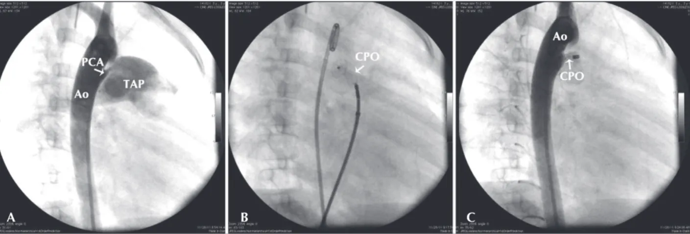
At the end of the procedure, two patients had minimal leakage of contrast inside the device; however, all defects were completely closed on the irst control TTE, which was performed within the irst week after the procedure (Figure 2). A gradient in the descending aorta or in the left branch of the pulmonary artery was not observed. In this initial series, complications were not observed.
DISCUSSION
The percutaneous closure of a PDA has been suc-cessfully performed through the use of several devices. The introduction of the ADO I prosthesis brought safety and ease of use, with excellent occlusion rates (99% to 100%) and a low incidence of complications (0% to 7%). This constituted an excellent alternative to per-cutaneous coil closure, despite the higher cost. The reported complications, in general, are minor and are usually observed in more severe patients and in patients with less body weight.22,23 There are no reports of late complications with this device.24
The Nit-Occlud® (PFM, Cologne, Germany) device was introduced as an intermediate device between the ADO I and coils. It utilises a controlled-release pre-molded coil that is shaped as an inverted double cone. It is capable of occluding intermediate size channels (< 6 mm) and costs less. With an occlusion rate of 91% to 100% and few minor complications (0% to 9%), it has been used in some centres, including in Brazil.25–28
The Amplatzer® Duct Occluder II (ADO II) was developed to occlude channels in smaller children by reducing the introducer proile. It maintained the high occlusion rate of existing devices. With two articulated disks at the extremities and a central portion, it closes channels of less than 12 mm in length and less than 5.5 mm in diameter. Occlusion rates were maintained at nearly 100%, without reports of signiicant com-plications.29–33 The report of a case in which residual low appeared 24 hours after the successful closure of a 3 mm channel due to kinking of the left disc in the ductal ampoule should be highlighted.34
TABLE
Characterisation of the Study Population
Case
No. ID Gender
Age (years)
Weight (kg)
Morphologic type
Diameter (mm)
Size of the
device* Results
1 JOC M 27 70 A 6 CPO 12 Closed
2 ASC F 17 56 A 5 CPO 10 Closed
3 JSO F 5 14 A 5 CPO 10 Closed
4 CGNA F 12 44 E 1 CPO 6 Closed
5 ACLO F 18 49 E 8.6 CPO12 Closed
6 GBB M 20 72 E 1 CPO 6 Closed
7 LHL F 20 55 A 1 CPO 6 Closed
8 JPAU M 1 10 A 1 CPO 6 Closed
9 DHDT M 14 73 E 1 CPO 6 Closed
10 KRC F 10 50 A 1 CPO 6 Closed
11 ACPO F 13 40 A 6 CPO 10 Closed
12 MRFS M 12 50 E 1 CPO 6 Closed
13 GRB F 8 30 PO 1 CPO 6 Closed
14 CVPS F 3 26 E 1 CPO 8 Closed
15 ALCA F 3 15 A 2.5 CPO 6 Closed
16 TAS M 2 12 A 2 CPO 6 Closed
17 SLRM M 33 57 B 8 CPO 12 Closed
18 LSG F 30 50 A 7 CPO 12 Closed
The design of the CeraTM prosthesis is very similar to that of the ADO I, but it has a more lexible nitinol mesh and a ceramic coating. This characteristic gives the CeraTM prosthesis the theoretical advantage of less nickel release on the days after the implant, although this has not been a problem reported with the ADO I.
Its great lexibility allows for the supersizing of the prosthesis according to the channel diameter without damaging neighbouring structures. Hence, the prosthesis is more stable (greater constriction in its central portion), and, as a consequence, it has a lower embolisation risk.
The long introducer allows passage through accen-tuated curves without kinks or breaks. Another advantage is the presence of a radiopaque mark in the distal end, which allows the surgeon to safely know the position of the long sheath during all the steps of the procedure.
CONCLUSIONS
In this initial experience, the CeraTM prosthesis could be used in small or large calibre channels. This device was easy to use, was extremely safe, had a low risk of morbidity, and was highly effective. The implant procedure is very similar to that of other existing nitinol mesh prostheses.
The author believe that its greater lexibility allows its diameter to be supersized more than 2 mm above the smaller channel diameter.
CONFLICTS OF INTEREST
Francisco Chamié is a Boynton technical consul-tant. The other authors declare no conlicts of interest.
REFERENCES
1. Bridges N, Perry S, Parness J, Keane J, Lock JE. Transcatheter closure of a large patent ductus arteriosus with the clamshell septal umbrella. J Am Coll Cardiol. 1991;18(5):1297-302. 2. Cambier PA, Kirby WC, Wortham DC, Moore JW. Percutaneous
closure of the small (less than 2.5 mm) patent ductus arteriosus using coil embolization. Am J Cardiol. 1992;69(8):815-6. 3. Chamié F, Pereira SJ, Sbafi F, Serra Junior AH, Athayde JG.
Fechamento de canal arterial com molas de Gianturco. Arq Bras Cardiol. 1996;67(1):23-7.
4. Grifka RG, Mullins CE, Gianturco C, Nihill MR, O’Laughlin MP, Slack MC, et al. New Gianturco-Grifka vascular occlu-sion device: initial studies in a canine model. Circulation. 1995;91(6):1840-6.
5. Lloyd TR, Fedderly R, Mendelsohn AM, Sandhu SK, Beekman RH 3rd. Transcatheter occlusion of patent ductus
arte-riosus with Gianturco coils. Circulation. 1993;88(4 Pt 1):1412-20. 6. Lloyd TR, Beekman RH 3rd, Moore JW, Hijazi ZM,
Hellenbrand WE, Sommer RJ, et al. The PDA Coil Registry Inves-tigators: 250 patient-years follow-up. Circulation. 1995;92:I380. 7. Porstmann W, Wierny L, Warnke H, Gertsberger G,
Romanuick PA. Catheter closure of patent ductus arteriosus: 62 cases treated without thoracotomy. Radiol Clin North Am. 1971;9(2):201-13.
8. Rao PS, Wilson AD, Sideris EB, Chopra PS. Transcatheter closure of patent ductus arteriosus with «buttoned» device: irst successful clinical application in a child. Am Heart J. 1991;121(6 Pt 1):1799-802.
9. Rashkind WJ, Cuaso CC. Transcatheter closure of a patent ductus arteriosus: successful use in a 3.5 kg infant. Pediatr Cardiol. 1979;1(1):3-7.
10. Rashkind WJ, Mullins CE, Hellenbrand WE, Tait MA. Non surgical closure of patent ductus arteriosus: clinical appli-cation of the Rashkind PDA occluder system. Circulation. 1987;75(3):583-92.
11. Tometzki AJP, Arnold R, Peart I, Sreeram N, Abdulhamed JM, Godman M, et al. Transcatheter occlusion of the patent ductus arteriosus with Cook detachable coils. Heart. 1996;76(6):531-5.
A B C
Figure 2 – Steps of the procedure. In A, descending aortography in the right anterior oblique incidence showing a conic patent ductus arteriosus (type A) with 2.5 mm in its smaller diameter, in the pulmonary entrance. In B, detail of the implant of a 6-mm (6-4 mm) CeraTM PDA Occluder, with the
12. Pauperio HM, Redinton AN, Rigby ML. Closing the patent arterial duct - plugs, umbrellas and coils. Cardiol Young. 1996; 6(2):252-4.
13. Arnoni DG, Peña JJS, Fontes VF, Braga SLN, Esteves CA, Ferreira WP, et al. Oclusão percutânea do canal arterial > 3 mm com auxílio do biótomo. Rev Bras Cardiol Invasiva. 2007;15(2):134-40.
14. Pedra CAC, Esteves CA, Braga SLN, Pedra SFR, Pontes Junior SC, Silva MAP, et al. Oclusão percutânea do pequeno canal arterial com molas de Gianturco: impacto da otimização da seleção das molas e dos pacientes e da não tolerância ao luxo residual signiicativo imediato nos resultados. Rev Bras Cardiol Invasiva. 2008;16(1):86-90.
15. Simões LC, Pedra CAC, Esteves CA, Camargo R, Braga SLN, Loureiro P, et al. Fechamento percutâneo do canal arterial com a prótese Amplatzer: experiência no Brasil. Arq Bras Cardiol. 2001;77(6):520-31.
16. Masura J, Walsh KP, Thanopoulos BV, Chan C, Bass JL, Goussous Y, et al. Catheter closure of moderate-to-large-sized patent ductus arteriosus using the new Amplatzer duct occluder: immediate and short-term results. J Am Coll Cardiol. 1998;31(4):878-82.
17. Faella HJ, Hijazi Z. Closure of the patent ductus arteriosus with the Amplatzer PDA device: immediate results of the interna-tional clinical trial. Catheter Cardiovasc Interv. 2000;51(1):50-4. 18. Pass RH, Hijazi Z, Hsu DT, Lewis V, Hellenbrand WE.
Multicenter USA Amplatzer patent ductus arteriosus occlusion device trial: initial and one-year results. J Am Coll Cardiol. 2004;44(3):513-9.
19. Pedra CAC, Esteves CA, Braga SLN, Kambara A, Fontes VF. Oclusão percutânea do canal arterial: estado da arte. Rev Bras Cardiol Invasiva. 1997;5(1):22-35.
20. Chamié F, Chamié D, Ramos S. Oclusão dos canais arteriais com prótese Amplatzer. Rev Bras Cardiol Invasiva. 2007;15(1): 15-24.
21. Krichenko A, Benson LN, Burrows P, Möes CAF, McLaughlin P, Freedom RM. Angiographic classiication of the isolated persistent ductus arteriosus and implications for percutaneous catheter occlusion. Am J Cardiol. 1989;63(12):877-80.
22. Al-Ata J, Ari AM, Hussain A, Kouatli AA, Jalal MO. The eficacy and safety of the Amplatzer ductal occluder in young children and infants. Cardiol Young. 2005;15(3):279-85.
23. Butera G, De Rosa G, Chessa M, Piazza L, Delogu A, Frigiola A, et al. Transcatheter closure of persistent ductus arteriosus with the Amplatzer duct occluder in very young symptomatic children. Heart. 2004;90(12):1467-70.
24. Masura J, Tittel P, Gavora P, Podnar T. Long-term outcome of transcatheter patent ductus arteriosus closure using Amplatzer duct occluders. Am Heart J. 2006;151(3):755e7-755e10. 25. Celiker A, Aypar E, Karagoz T, Dilber E, Ceviz N.
Transca-theter closure of patent ductus arteriosus with Nit-Occlud coils. Catheter Cardiovasc Interv. 2005;65(4):569-76. 26. Gamboa R, Mollón FP, Rios-Mendez RE, Arroyo G, Fogel A,
Villa DM. Patent ductus arteriosus closure using a new device: the Nit-Occlud device. Rev Esp Cardiol. 2007;60(4): 445-8.
27. Haddad JL, Lima Filho MO, Figueiredo GL, Nazzetta HE, Osterne ECV. Oclusão percutânea da persistência do canal arterial. Rev Bras Cardiol Invasiva. 2005;13(3):206-18. 28. Tometzki AJP, Chan K, De Giovanni JV, Houston A, Martin R,
Redel D, et al. Total UK multi-centre experience with a novel arterial occlusion device (Duct Occlud pfm). Heart. 1996;76(6):520-4.
29. Dua J, Chessa M, Piazza L, Negura D, Micheletti A, Bussadori C, et al. Initial experience with the new Amplatzer Duct Occluder II. J Invasive Cardiol. 2009;21(8):401-5.
30. Queiroz FJAC, Simões LC, Queiroz DSC, Ramos S, Silva JFA, Mattos R. Tratamento percutâneo do canal arterial com a prótese Amplatzer Duct Occluder II (ADO II): nova opção para um antigo defeito. Rev Bras Cardiol Invasiva. 2010;18(2):204-11. 31. Ribeiro MS, Pereira FL, Costa RN, Arruda A, Braga SLN,
Fontes VF, et al. Oclusão percutânea de defeitos cardíacos congênitos e estruturais com Amplatzer Duct Occluder II. Rev Bras Cardiol Invasiva. 2011;19(4):430-41.
32. Thanopoulos BV, Eleftherakis N, Tzannos K, Stefanadis C, Giannopoulos A. Further experience with catheter closure of patent ductus arteriosus using the new Amplatzer duct occluder in children. Am J Cardiol. 2010;105(7):1005-9. 33. Venczelova Z, Tittel P, Masura J. The new Amplatzer duct
occluder II: when is its use advantageous?. Cardiol Young. 2011;21(5):495-504.