TEMPOROMANDIBULAR JOINT DYSFUNCTION
AND ITS CORRELATION WITH AUDITORY TUBE
IN CLEFT PALATE PATIENTS
A relação entre disfunção temporomandibular e disfunção tubária
em pacientes com issura palatina
Flávio Ricardo Manzi (1), Priscila Dias Peyneau (2), Aline Lamas Lopes (3), Camila Lage Silveira (4),
Cíntia Santos Silva Machado (5), Camila Queiroz de Moraes Silveira Di Ninno (6)
1 Dental Surgeon, Assistant Professor of the Phonoaudiology
and Dentistry course from PUC Minas, Coordinator of the
Master course of Radiology from the Pontiical Catholic Uni -versity of Minas Gerais.
2 Dental Surgeon; Specialist in Radiology by Unigranrio.
Master in Dental Clinics with Emphasis on Radiology by the Catholic University of Minas Gerais.
3 Phonoaudiologist; graduated from the Pontiical Catholic
University of Minas Gerais.
4 Phonoaudiologist; graduated from the Pontiical Catholic
University of Minas Gerais.
5 Phonoaudiologist, Assistant Professor of the
Phonoau-diology course at Pontiical Catholic University of Minas
Gerais.
6 Phonoaudiologist, Assistant Professor of the
Phonoau-diology course at Pontiical Catholic University of Minas
Gerais.
Conlict of interest: non-existent
INTRODUCTION
Individuals with cleft palate have changes in the anatomy of the muscles attached to the median raphe of the palate, which causes the muscles responsible for their upper and lower movement lose
their ixation, therefore not exercising the adequate
traction on the velum1.
Casselbrant et al., in 1985, conducted a study in individuals with cleft palate, from computed tomog-raphy and image reconstruction in three dimen-sions, showed that the tensor and levator muscles of the soft palate function synergistically opening and dilating the Eustachian tube2. This study reveals
that the ibers of these muscles follow parallel to the
anatomy of the Eustachian tube and in the case of
ABSTRACT
Purpose: investigate the existence of dysfunctional temporomandibular disorders, and as a
predisposing factor to investigate tubal dysfunction in patients with cleft palate. Method: 10 individuals (20 temporomandibular joints), operated of cleft palate had been evaluated, that were in treatment in the Clinical Center of Fonoaudiologia of the PUC-MG, with age varying between 8 and 18 years, of
both gender, and that they presented history of middle otitis in irst infancy. The same ones had been submitted to the meatoscopia, imitanciometria, research of the tube function, examination of feel of
the face muscle and examinations of image (x-rays transcraniana in 6 positions and AP of Town).
Being compared for the Accurate Test of Fisher (p<0,05). Results: it was observed that the majority of the sample presented of auditory tube dysfunction, when analyzed for the test of the tube function corresponding 80% of the total. In relation to the joint to temporomandibular nine individuals (90%)
they had not presented dysfunction and only one individual (10%) got, observed in the examinations
of image, presence at the back of condyle process of the left side. Conclusions: as preliminary data, when analyzing the temporomandibular joint dysfunction and its correlation with auditory tube, the
Accurate Test of Fisher did not ind resulted signiicant, evidencing that the joint to temporomandibular of the cleft palate patients, does not suffer alterations proceeding from the ineficient functioning from
auditory tube.
individuals with cleft palate, fall into the area of cleft palate2.
The abnormal insertion of the levator muscles and tensor veli palatini, present in this type of cleft,
can vary from a mild degree to a more signiicant
degree one. In the presence of altered muscle palate may occur a reduced tubal functioning1.
The Eustachian tube, the structure that connects
the nasopharynx to the tympanic cavity of the
middle ear consists of three parts: cartilage, medial
and bone. The proximal portion also called cartilage ends in the nasopharynx. The bone or the distal
portion communicates with the anterior portion of the middle ear, since it is located in the petrous part of temporal bone. The medial portion refers to the part of the Eustachian tube that connects the carti-laginous portion of the bone3,4. Responsible for the
drainage and aeration of the tympanic cavity, the Eustachian tube allows the balance of air pressure in the middle ear, while it replenishes absolved
oxygen4. Also, it protects against middle ear
secre-tions, pathogens, pressure variations and sounds
from the nasal pharynx. The Eustachian tube also
has the function through the mucociliary system,
eliminating luids, debris, and foreign substances in
the middle ear4.
Individuals with cleft palate are then more prone to functional obstruction of the Eustachian tube, as the responsible muscles for opening and
dilation behave in this ineficient way5. The
persis-tence of negative pressure in the middle ear can be a triggering factor for sterile otitis media with effusion2,5. There is evidence that patients with cleft
lip and palate have several complaints about the ear, nose and throat5. The presence of serous otitis
media is considered almost universal in patients with cleft palate2,6-10.
Studies show changes in tympanometric curves of individuals with cleft palate. In these, it was possible to verify in most cases, tympanometric curve type B and C, suggesting the presence of hearing alterations resulting from problems in the middle ear5.
Temporomandibular disorders constitute of a set of signs and symptoms such as pain, presence of
crackles, clicking or even inlammation in the region
of the temporomandibular joint (TMJ)11,12. Cervical
tension and shoulder girdle also bind to this type of dysfunction13.
Joint changes present in temporomandibular disorders are described and related mainly in adult patients, probably due to the high incidence in this population11. However, these changes can also
occur in children. Generally the presence of joint in children is related to the history of tube dysfunction and otitis media in early childhood11.
The mandibular fossa during childhood and adolescence presents a framework called
tympa-nosquamous issure, which remains open until
the individual reaches the bone maturation12. It is
believed that the opening present in articular fossa
allows the passage of luid from the middle ear and
mastoid to the joint region of the temporomandibular joint11, and that there is a link between the
stomato-gnathic system and the auditory system, which can
be causes and consequences of temporomadibular
dysfunctions14,15.
As individuals with cleft palate exhibit signiicant
possibility of developing otitis media with effusion16,
the present study aimed to investigate the existence
of temporomandibular disorders, having as a predis-posing factor the tube dysfunction.
METHOD
We selected 28 individuals who operated cleft palate, aged from 4 to 20 years old, and were under-going treatment at the Phonoaudiology clinic at PUC Minas, and had a history of otitis media in early childhood. However, only 10 individuals agreed to
participate in all the steps required for this research. Thus, the sample consisted of 10 subjects, ive male and ive female, with age range from 8 to 18 years
old. So it was searched 20 temporomandibular joints and 20 tubal regions.
The sample subjects were assessed using the following tests:
– Meatoscopy: Through the TK otoscope it was investigated possible presence of wax plugs and/or foreign bodies in the ear canal;
– Immittanciometry: through AZ7 immittanciometer, it was assessed as the tympanum-ossicular system behaves at the sound;
– Research of the Tubal Function: Carried through the AZ7immittanciometer, this aimed to analyze the functioning of the Eustachian tube.
– Examination of the temporomandibular joints
and associated musculature: In this study it
was used the Speciic Protocol of Orofacial
Pain and Temporomandibular Disorders, based on research diagnostic criteria for temporomandibular joint disorders (RDC / TMD)17 and aims to investigate the presence
of muscle pain, stress points, popping and/or mandibular deviation.
– Imaging examinations: Through Oralix
equipment, Gendex-(70 kvp 10mA) and
Rotograph Dhabi Atlantis (80 Kvp 10mA), it was possible to perform transcranial radiography in
six positions and Town’s Ap, able to show the
By analyzing the relationship between tube dysfunction and temporomandibular dysfunction,
the Fisher exact test found no signiicant results,
showing that the temporomandibular joint of individuals with cleft palate does not change from
the ineficient functioning of the Eustachian tube.
DISCUSSION
It was observed in this study, that most of the sample had Eustachian tube dysfunction. This
inding is consistent with other studies that show the ineficiency of the tensor muscles and elevator of
the veil palate in patients with cleft palate, to play the role of opening and dilating the Eustachian tube1.
Study, using computed tomography and image reconstruction in three dimensions, showed that the anatomy of the Eustachian tube of individuals with cleft palate is different from tube anatomy of normal individuals. According to this work, the Eustachian
tube of the issured individuals, suffers a slight
deviation in its caudal portion, which may hinder its function3. This fact can be considered signiicant in
regard to the performance of the sample tube in the study of the tube function.
Some authors claim that the persistence of negative pressure within the middle ear due to
the ineficiency of the Eustachian tube function of
patients with cleft palate, can cause changes in the middle ear2,5. Studies have shown that the presence
of otitis media is almost universally considered This study was approved by the Ethics in Research Committee of PUC Minas CAAE 0219.0.213.000-05.
Data were tabulated and analyzed using the
Fisher exact test with a signiicance level of 5%.
RESULTS
In the studied sample, it was not veriied the presence of foreign body and or wax plug during
otoscopy, which made possible the realization of reliable audiological tests.
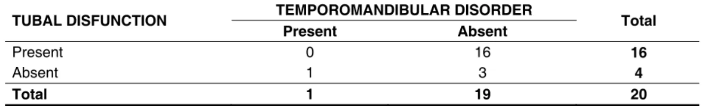
Considering the 10 analyzed patients, having as parameter 20 ATMs and 20 ears, it was observed
sixteen ears with Eustachian tube dysfunction and
absence of temporomandibular dysfunction, as it
was veriied temporomandibular dysfunction with
no tube dysfunction. Three ears and ATMs did not change (Table 1).
TUBAL DISFUNCTION TEMPOROMANDIBULAR DISORDER Total Present Absent
Present 0 16 16
Absent 1 3 4
Total 1 19 20
Table 1 - Relationship between temporomandibular disorders and tubal dysfunction
Fisher’s Exact Test. P = 0,2 (p< 0,05)
in this population. According to other authors, the incidence of otitis media in subjects with cleft palate, from 50% to 90% of the cases, this is 30 times more
frequent than in normal populations, as well as in
individuals with only lip issure6-10. Our work is in line
with these studies, as all individuals in the sample reported a history of otitis media in early childhood.
Audiological indings obtained from our sample,
showed that the vast majority of patients had changes in the middle ear, and found both tympanogram type
C and type B. This result correlates with the indings
in the literature since tympanometric studies claim that it can be seen in most cases of cleft palate, tympanogram type B or C, suggesting the presence of middle ear disease in these5.
Some authors claim that the growth and maturation is not complete until the second decade of life11-13. Another study shows that the mandibular
fossa during childhood and adolescence presents
a framework called tympanoescamosa issure,
which remains open until the individual reaches the maturation12. It is believed that this opening in the
fossa allows the passage of luid in the middle ear
and mastoid to the temporomandibular joint11.
Since individuals with cleft palate are more likely
to experience otitis media with effusion, and the vast
majority of this sample had abnormal middle ear, it
would be expected that these disorders present in
the temporomandibular region14. However, imaging
REFERENCES
1. Sheahan P, Miller I, Sheahan JN, Earley MJ, Blayney AW. Incidence and outcome of middle ear disease in cleft lip and/or cleft palate. Int J Pediat Otorhinolaryngol. 2003;67:785-93.
2. Casselbrant ML, Doyle WJ, Cantekin EL, Ingraham AS. Eustachian tube function in the rhesus monkey model of cleft palate. J Cleft Palate. 1985;22(3):185-91.
3. Arnold WH, Koch KHH. Morphology of the auditory tube and palatal muscles in case of bilateral cleft palate. Cleft Palate-Craniofac J. 2005;42(2):25-31.
4. Wood-Northem. Manual de Otorrinolaringologia. Barcelona: Edit Salvat; 1984.
disorders. This fact leads us to believe that this secretion in the middle ear of these individuals during infancy, had no way to drain
timpanoes-camosa issure.
Only one individual in our sample complained of pain in the neck, limitation of opening and popping
chewing. The imaging examination revealed a
posterior condyle of the left side. However, this individual did not show tube dysfunction, which leads us to suppose that the cause of temporoman-dibular dysfunction of this individual would possibly be associated with other aspects.
Temporomandibular dysfunction has as an etiology several factors such as: stress, psycho-logical disorders, harmful habits, malocclusion, among other causes13. One should not think of the
Eustachian tube dysfunction as a common cause of TMJ disorders, but as a possible predisposing factor for developing this.
CONCLUSION
From the results of this study, it was found that the tube dysfunction present in individuals with cleft palate is not a triggering factor for TMJ dysfunction.
RESUMO
Objetivo: investigar, em indivíduos com issura palatina, a existência de disfunções temporomandi
-bulares, tendo como fator predisponente a disfunção tubária. Método: foram avaliados 10 indivíduos
(20 articulações temporomandibulares), operados de issura de palato, que estavam em tratamento
no Centro Clínico de Fonoaudiologia da PUC Minas, com idade variando entre 8 e 18 anos, de ambos
os sexos, e que apresentavam história de otite média na primeira infância. Esses foram submetidos à
meatoscopia, imitanciometria, pesquisa da função tubária, exame de palpação da musculatura orofa
-cial e exames de imagem (radiograias transcraniana em seis posições e Ap de Town). Resultados:
foi observado que a maioria da amostra apresentava disfunção da tuba auditiva, correspondendo a 80% do total. Em relação à articulação temporomandibular nove indivíduos (90%) não apresentaram disfunção, em apenas um indivíduo (10%) foi veriicado presença de desordem temporomandibular.
Conclusão: ao analisar a relação entre a disfunção tubária e disfunção temporomandibular, não
foram observados resultados signiicantes de acordo com o Teste Exato de Fisher, evidenciando que a articulação temporomandibular dos issurados de palato, não sofre alterações provenientes do funcionamento ineiciente da tuba auditiva.
DESCRITORES: Fissura Palatina; Otite Média; Síndrome da Disfunção da Articulação
Temporomandibular; Tuba Auditiva
5. Filho OAC, Piazentin SHA. Aspectos otológicos.
In: Altmann EBC. Fissuras labiopalatinas. 4rd ed.
Carapicuba: Pró-Fono; 1997. p.485-98.
6. Paradise JL, Bluestone CD, Felder H. The unreversability of otitis media in 50 infants with cleft palate. Pediatr. 1969;44:35-42.
7. Potsic WP, Cohen, Randall P, Winschester. A retrospective study of hearing impairment in three groups of cleft palate patients. J Cleft Palate. 1979;16(2):56-62.
8. Stool SE, Randall P. Unexpected on disease
in infants with cleft palate. J Cleft Palate. 1968;4(1):99-105.
10. Annfrable M, Brandon GF, Theogarj DS. Velar closure and ear tubings as a primary procedure in the repair of cleft palates. Laryngosc. 1985;95(1):1044-9.
11. Bianchini JS, Amboni MP, Bóscolo FN, Lima JJG, Manzi FR. Repercussão da otite média nas
desordens craniomandibulares. In: 1º Encontro de
pesquisa PUC-MINAS; 2003. Belo Horizonte. 1º Encontro de pesquisa PUC-MINAS; 2003;1:69-79.
12. Keith DA, Glyman ML. Infratemporal space pathosis mimicking TMJ disorders. J Am Dent Assoc. 1991;122(11):59-61.
13. Bianchini EMG. Articulação temporomandibular e fonoaudiologia. In: Bianchini EMG. Articulação temporomandibular. Carapicuíba: Pró-Fono; 2000.
p. 34-43.
14. Barreto DC, Barbosa ARC, Frizzo ACF. Relação entre disfunção temporomandibular e alterações
auditivas. Rev CEFAC. 2010;12(6):1067-76.
15. Machado IM, Pialarissi PR, Minici TD, Rotondi
J, Ferreira LP. Relação dos sintomas otológicos
nas disfunções temporomandibulares. Arq.
Int. Otorrinolaringol./Intl Arch Otorhinolaryngol. 2010;14(3);274-9.
16. Marcusson A, List T, Paulin G, Dworkin S. Temporomandibular disorders in adults with repaired palate. Eur J Orthod. 2001;23(2):193-204. 17. Dworkin SF, LeResche L. Research diagnostic criteria for temporomandibular disorders: review,
criteria, examinations and speciications, critique. J
Craniomandib Disord 1992;6:301-55.
Received on: October 05, 2011 Accepted on: June 28, 2012
Mailing Address:
Flávio Ricardo Manzi
Av. Dom José Gaspar, 500 – Prédio 45 –
Clínica de Radiologia
Coração Eucarístico – Belo Horizonte – MG
CEP: 30535-610