249
Rev Bras Oftalmol. 2013; 72 (4): 249-52
O
RIGINALA
RTICLEThe authors declare no conflict of interest.
Recebido para publicação em 24/4/2012 - Aceito para publicação em 24/10/2012
Falha a longo prazo no tratamento do melanoma
de coróide com termoterapia transpupilar
Long-term failure after transpupillary
thermotherapy for small choroidal melanomas
Gabriela Soncini Pasetto
1, Henrique Pedroso de Freitas
1, Carina Graziottin Colossi
2, Manuel Augusto Pereira Vilela
3R
ESUMOObjetivo: Avaliar, retrospectivamente, num longo intervalo de tempo, os resultados da termoterapia transpupilar em casos selecio-nados do melanoma de coróide. Métodos: Foram identificados todos os casos com melanoma de coróide tratados com termoterapia transpupilar, como terapia única ou suplementar. Foram excluídos os casos com seguimento inferior a 60 meses, prontuários incompletos ou com o abandono do acompanhamento. Resultados: De um total de 18 olhos, 7 (38.9%) foram incluídos no estudo. Cinco (71,4%) pacientes foram tratados exclusivamente com termoterapia transpupilar, em 1 (14,2%) olho a crioterapia foi simul-tânea, 1 (14,2%) suplementou braquiterapia – placa episcleral. Três (42.8%) sofreram recorrência, 2 (28.5%) apresentaram metástases (um óbito), 2 foram enucleados. Complicações foram encontradas em 3 olhos: dobras maculares em 2, hemorragia vítrea em 1. Nos olhos enucleados, não havia extensão extraescleral, porém células interesclerais foram identificadas. Conclusão: Termoterapia transpupilar mostra um declínio em sua eficácia e conserva os riscos de metástases em períodos mais longos de acompanhamento, sendo necessária uma definição mais exata de seu papel nos melanomas de coróide.
Descritores: Melanoma/terapia; Coróide; Hipertermia induzida/métodos
A
BSTRACTObjective: To evaluate, retrospectively, in a long time interval, the results of transpupillary thermotherapy in selected cases of choroidal melanoma (CM). Methods: All patients with melanoma treated with transpupillary thermotherapy as sole therapy or supplement were identified. Cases with less than 60 months follow-up, incomplete records or abandonment of monitoring were excluded. Results: A total of 18 eyes, seven (38.9%) were included. Five (71,4%) patients were treated exclusively with transpupillary thermotherapy, in one (14,2%) eye criotherapy was used simultaneously, one (14,2%) was supplemented with brachytherapy – episcleral plate. Three (42.8%) had recurrence, two (28.5%) had metastases (one death), two eyes were enucleated. Complications were found in three eyes: two with macular folds, one with vitreous hemorrhage. In the enucleated eyes, extraescleral did not occur, however intrascleral tumoral cells were identified. Conclusion: Transpupillary thermotherapy shows a decline in its effectiveness and retains the risk of metastases in longer periods of follow-up, requiring a more exact definition of its role in choroidal melanoma.
Keywords: Melanoma/therapy; Choroidal; Hyperthermia, induced/methods
1Physicians at the Specialisation Course in Ophthalmology Professor Ivo Corrêa-Meyer. Porto Alegre/RS, Brazil. 2M.Sc., Preceptor at the Specialisation Course Professor Ivo Corrêa-Meyer. Porto Alegre/RS, Brazil.
3Ph.D., Professor of Ophthalmology, Federal University of Pelotas (UFPel), Pelotas/RS, Brazil.
250
I
NTRODUCTIONM
elanoma is the most frequent primary tumour of the eyeball. In its early stages it may be indistinguishable from a melanocytic nevus(1). It usually grows as aglobular mass protruding toward the vitreous cavity(1,2). Ocular
melanocytes are generally concentrated on the pigment epithelium, the uvea, and the conjunctival stroma. These cells have the same embryological origin of skin cells, i.e. the neural crest(3).
Specifically, choroidal melanoma (CM) occurs more frequently in the posterior pole and can present three different pigmentation patterns: amelanotic, densely pigmented, or mixed. In terms of appearance, the lesions may be nodular, multinodular, or diffuse(3).
For many years, enucleation was the treatment of choice for the condition. Currently, alternative therapeutic methods have been adopted that preserve the eyeball, and even its function, but without affecting the survival rate or the metastatic potential. These alternative therapeutic choices should take into account the patient’s overall clinical condition, the affected eye’s visual acuity and intraocular pressure, and the tumour’s size, location, growth pattern, and activity(4). Within certain limits, expectant
management is adequate; otherwise, therapeutic options include thermotherapy, brachytherapy, external radiotherapy, and surgical resection, among others.
Transpupillary thermotherapy (TTT) has emerged as a therapeutic option since 1998(1,2). In this technique, a laser beam
— infrared radiation — is applied through the pupil to raise the temperature and destroy tumour tissue through cytotoxicity(1,3).
The method can produce necrosis up to a maximum depth of 3.9 mm without affecting structures outside the treatment field. Its advantages include a limited field of action, the fact that it does not need to be conducted in an operating theatre, the preservation of the organ and its function (patients with extra-macular foci may retain good visual acuity), and good survival rates(1-4).
The aim of this study was to describe the long-term clinical course (60 months or more) of cases of CM treated with TTT, comparing them with different management alternatives.
M
ETHODSWe studied cases of CM treated with TTT that had a minimum follow-up time of 60 months. Inclusion criteria were
patients treated exclusively with TTT, patients who received “sandwich” therapy with TTT plus cryotherapy in the same surgical act, and patients who received TTT plus brachytherapy. Exclusion criteria were incomplete patient charts, a shorter follow-up period, and treatment dropouts.
The criteria and the technique adopted to indicate and perform TTT can be found in other sources(4); essentially, the method
was limited to tumours up to 4 mm thick and 12 mm in diameter on ultrasound. Treatment was administered using a slit lamp (Opto, Brazil) under peribulbar block. Each patient had their demographic data collected and underwent an assessment of corrected visual acuity using Snellen’s chart, assessment of extraocular movements, applanation tonometry, biomicroscopy, gonioscopy, and indirect binocular fundus examination. Retinography and fluorescein angiography were performed before the procedure and every 6 months afterwards. An oncological evaluation was performed every 6 months, with laboratory tests, ultrasound imaging, CT, and, in some cases, PET scan imaging. The study was approved by the Research Ethics Committee of the Federal University of Pelotas under number 00184/10.
R
ESULTSSeven eyes of patients (four women) with MC were identified using the set criteria. Mean age was 58.8 years, ranging from 45 to 72 years. Of these, 5 (71.4%) were treated with TTT only, 1 (14.2%) with TTT plus cryotherapy, and 1 (14.2%) with TTT plus episcleral plaque. Mean tumour thickness was 3.7 mm, with a diameter of 5.35 mm. The mean follow-up time was 75.4 months (Table 1).
Among patients treated exclusively with TTT (5 eyes), 3 ( 60%) progressed with tumour control, 1 (20%) with tumour growth compared to the original lesion, and 1 (20%) with metastasis. Among those with tumour control, one (20%) developed vitreous haemorrhage (with spontaneous clearing within 3 weeks) and 1 (20%) developed macular folds. The patient who progressed with tumour regrowth (20%) underwent enucleation, and the patient with metastasis (20%) died after a follow-up of 65 months.
Disease control was not achieved in the patient who underwent TTT plus cryotherapy (14.2%), with tumour regrowth and macular folds. Its final result was enucleation.
The patient treated with TTT plus episcleral plaque (14.2%) progressed with lung metastasis. This patient had been originally
Table 1
Summary of treated cases.
Rev Bras Oftalmol. 2013; 72 (4): 249-52
Pasetto GS, Freitas HP, Colossi CG, VilelaMAP
Patient Tumour Colour Site Treatament Course Complications Results
size(mm) (months)
Female, 49 years 2 x 20 Melanotic Post-equator TTT + 96 Phthisis Lung
episcleral plaque bulbi metastases
recent surgery
Female, 64 years 3.1 x 2.9 Amelanotic Nasal TTT 64 Controlled
Female, 45 years 3.2 x 2.0 Melanotic Macular TTT 65 Diffuse Death
metastases
Female, 72 years 4.0 x 4.1 Melanotic Equator TTT + 72 Folds, Enucleation
cryotherapy regrowt
Male, 65 years 3.8 x 3.2 Melanotic Macular TTT 65 Vitreous haemorrhage Controlled
Male, 56 years 3.0 x 2.1 Melanotic Post-equator TTT 82 Regrowt Enucleation
251
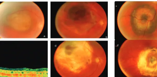
Figure 1:a. Submacular lesion, appearance before TTT; b. Submacular lesion, appearance after TTT; c. Optical coherence tomography (OCT), late; d. Submacular lesion, appearance after 6 months
Figure 2:a. Central melanoma with orange ring; b. Fluorescein angiography with mixed defects; c. Appearance after TTT; d. Optical coherence tomography (OCT) with fluid in the margins, before treatment
Figure 3: a. Diffuse melanoma before brachytherapy/episcleral plaque;
b. 60 days after brachytherapy; c. 130 days after brachytherapy and TTT; d. 150 days after two brachytherapy sessions
Figure 4:a. Appearance before TTT plus cryotherapy; b. Ultrasound imaging before TTT plus cryotherapy; c. After TTT, early; d. Late growth; e. Ultrasound imaging in the first 12 months; f. Histology after enucleation
Rev Bras Oftalmol. 2013; 72 (4): 249-52 Long-term failure in the treatment of small choroidal melanoma by transpupillary thermotherapy
treated with brachytherapy only; however, tumour recurrence was observed at the edges, leading to TTT. Treatment was then supplemented with brachytherapy and episcleral plaque. The patient underwent lobe resection on both lungs and is under oncological follow-up. She has been followed-up for 96 months and has progressed to phthisis bulbi.
Histological examination of enucleated eyes found no extrascleral extension, but tumour cells were found in the scleral stroma.
Of the three eyes successfully treated (all exclusively with TTT), two suffered a significant loss of central vision: one due to macular folds and the other due to the submacular location of the mass (Figures 1-4).
D
ISCUSSIONRecent study results have led to a more cautious, less enthusiastic approach towards TTT in the treatment of CM, mainly because of the detection of viable tumour cells in the
scleral parenchyma during follow-up. The depth to be reached can limit the method’s efficiency, either in primary applications or reapplications(5-12).
The choice to use TTT is based on the possibility of managing small lesions with a simple procedure that preserves the eyeball and, on certain cases, visual acuity. Early studies on TTT reported satisfactory and replicable results, but they had a short follow-up time(1-4). More recently, different publications
have supported a more cautious approach, and there is disagreement regarding the method’s results and complications. The detection of treatment failures (local or distant metastases) depends on the study’s follow-up period(6-11).
252
7. Fuisting B, Richard G. Transpupillary thermotherapy (TTT) - re-view of the clinically indication spectrum. Med Laser Applic. 2010;25(4):214-22. Edited By B.M. Stoffelns and S. Wolf 8. Win PH, Robertson DM, Buettner H, McCannel CA, Bennett
SR. Extended follow-up of small melanocytic choroidal tumors treated with transpupillary thermotherapy. Arch Ophthalmol. 2006;124(4):503-6.
9. Chojniak MM, Guia T, Uno F, Erwenne CM. Termoterapia transpupilar em melanoma maligno de coróide. Arq Bras Oftalmol. 2001;64(2):133-8.
10. Stoffelns BM, Schoepfer K, Vetter J, Mirshahi A, Elflein H. [Long-term follow-up 10 years after transpupillary thermotherapy (TTT) for small, posterior located malignant melanomas of the choroid]. Klin Monbl Augenheilkd. 2011;228(4):277-83. German. 11. Yarovoy AA, Magaramov DA, Bulgakova ES. Which choroidal
mela-noma should be treated with primary transpupillary thermotherapy? Our experience from 78 patients. Eur J Ophthalmol. 2010;20(1):186-93.
12. Bel DJ, Wilson MW. Choroidal melanoma: natural history and management options. Cancer Control. 2004;11(5):296-303. 13. The Collaborative Ocular Melanoma Study (COMS) randomized
trial of pre-enucleation radiation of large choroidal melanoma II: initial mortality findings. COMS report no. 10. Am J Ophthalmol. 1998;125(6):779-96. Comment in Am J Ophthalmol. 1998;125(6):865-7.
14. Mooy CM, De Jong PT. Prognostic parameters in uveal mela-noma: a review. Surv Ophthalmol. 1996;41(3):215-28.
15. Damato B, Coupland SE. A reappraisal of the significance of largest basal diameter of posterior uveal melanoma. Eye (Lond). 2009;23(12):2152-60; quiz 2161-2.
16. Seddon JM, Albert DM, Lavin PT, Robinson N. A prognostic fac-tor study of disease-free interval and survival following enucleation for uveal melanoma. Arch. Ophthalmol. 1983;101(12):1894-9. 17. Shields CL, Furuta M, Thangappan A, Nagori S, Mashayekhi A,
Lally DR, et al. Metastasis of uveal melanoma millimeter-by-millimeter in 8033 consecutive eyes. Arch Ophthalmol. 2009;127(8):989-98.
18. Zimmerman LE, McLean IW, Foster WD. Statistical analysis of follow- up data concerning uveal melanomas, and the influence of enucleation. Ophthalmology. 1980;87(6):557-64.
19. McLean IW, Foster WD, Zimmerman LE, Martin DG. Inferred natural history of uveal melanoma. Invest Ophthalmol Vis Sci. 1980;19(7):760-70.
Corresponding author:
Gabriela Soncini Pasetto
Rua Félix da Cunha 496 Bairro Floresta CEP: 90570-000 - Porto Alegre (RS), Brazil E-mail: [email protected]
Rev Bras Oftalmol. 2013; 72 (4): 249-52
Pasetto GS, Freitas HP, Colossi CG, VilelaMAP
The need for supplemental therapy is based mainly on the detection of tumour growth (in its thickness or margins) and subretinal fluid. Studies assessing the response to retreatment have also found divergent results, which may be due to different management strategies, but mainly due to differences in follow-up time.
Our study was limited due to its small, heterogeneous sample and its retrospective design. Still, we found a high frequency of tumour regrowth and metastasis.
Questions remain about the condition’s natural history and the influence of treatment on the prognosis of CM(12). Factors
that may predict a poor prognosis include: tumour size(13-15), old
age(16), tumour thickness(17), and involvement of the ciliary
body(18). It is believed that a small, light CM takes 7 years to grow
and another 4 years to develop metastases(12,18,19). The mortality
rate of uveal melanoma is 31% in 5 years, 45% in 15 years, 49% in 25 years, and 52% in 35 years(12).
C
ONCLUSIONTTT in tumours up to 4 x 12 mm shows low effectiveness and does not eliminate the risk of metastasis in longer follow-up periods. Its role in the treatment of CM needs to be more clearly defined.
R
EFERENCES1. Journeé-de Korver JG, Oosterhuis JA, de Wolff-Rouendaal D, Kemme H. Histopathological findings in human choroidal mela-nomas after transpupillary thermotherapy. Brit J Ophthalmol. 1997;81(3):234-9.
2. Oosterhuis JA, Journée-de Korver HG, Kakebeeke-Kemme HM, Bleeker JC. Transpupillary thermotherapy in choroidal melano-mas. Arch Ophthalmol. 1995;113(3):315-21.
3. Robertson DM, Buettner H, Bennett SR. Transpupillary thermo-therapy as primary treatment for small choroidal melanomas. Arch Ophthalmol. 1999;117(11):1512-9.
4. Shields CL, Shields JA, Perez N, Singh AD, Cater J. Primary transpupillary thermotherapy for small choroidal melanoma in 256 consecutive cases: outcomes and limitations. Ophthalmology. 2002;109(4):225-34.
5. Zaldivar RA, Aaberg TM, Sternberg P Jr, Waldron R, Grossniklaus HE. Clinicopathologic findings in choroidal melanomas after failed transpupillary thermotherapy. Am J Ophthalmol. 2003;135(5): 657-63.