49
Drusen-like beneath retinal deposits in type II
mesangiocapillary glomerulonephritis: a review
Depósitos retinianos como drusas na glomerulonefrite
tipo II mesangiocapilar: uma revisão
Miguel Hage Amaro
1, Cristina Muccioli
2,
Mario Martins do Santos Motta
3, Jorge Mitre
4,
João Jorge Nassaralla
5,
Luiz Augusto Siqueira Silva
6,
Teruo Aihara
71Instituto de Olhos e Laser de Belém (PA), Brazil; 2Universidade Federal de São Paulo (SP), Brazil;
3Professor, Universidade Federal do Estado do Rio de Janeiro (UNIRIO) – (RJ), Brazil; 4Faculdade de Medicina do ABC – Santo André (SP), Brazil;
5Instituto de Olhos de Goiânia – Goiânia (GO), Brazil; 6Oftalmologista – Belém (PA), Brazil;
7Santa Casa de Misericórdia de São Paulo (SP), Brazil.
Study carried out at Instituto de Olhos e Laser de Belém, (PA), Brazil
The authors declare no conflicts of interest
A
BSTRACTThe aim of this paper is to do a review of like beneath retinal deposits in type II mesangiocapillary glomerulonephritis. Drusen-like beneath retinal deposits in type II mesangiocapillary glomerulonephritis appear to develop at an early age, often second decade of life different of drusen from age-related macular degeneration (AMD).Long term follow-up of the cases in this disease shows in the most of them, no progression of the of drusen-like beneath retinal deposits in type II mesangiocapillary glomerulonefritis, the most of subjects retain good visual acuity and no specific treatment is indicated.
Keywords: Retinal drusen; Glomerulonephritis, membranoproliferative
R
ESUMOO objetivo deste artigo é fazer uma revisão dos trabalhos publicados sobre depósitos que se assemelham a drusas, na retina de pacientes com a glomerulonefrite mesangiocapilar tipo 2. Um trabalho da Universidade de Iowa sobre as características destes depósitos foi incapaz de diferenciá-lo das drusas retinianas. O acompanhamento a longo prazo destes depósitos parecem demonstrar que não há grande tendência a modificação e a maioria dos pacientes mantém boa visão.
Descritores: Drusas retinianas; Glomerulonefrite membranoproliferativa
U
PTADEA
RTICLEReceived for publication: 6/6/2014 - Accepted for publication: 13/12/2014
50
I
NTRODUCTIONT
he presence of drusen-like beneath retinal deposits in type II mesangiocapillary glomerulonephritis (MCGN) was first described in 1989 by Duvall-Young et al.(1,2) atthe Manchester Royal Infirmary Renal Unit analyzing subjects with biopsy proved glomerular disease.
They demonstrated the occurrence of deposits in the choriocapillaris and Bruch’s membrane which have the clinical appearance of retinal drusen but the histopathological characteristics of deposits in the glomerulus.
Hageman’s team(3) at the University of Iowa analyzing
eyes obtained from two human donors concluded that subretinal pigment epithelial deposits found are numerous and indistinguishable , both structurally and compositionally , from drusen in donors with AMD and exhibit sudanophilia , bind filipin, and react with antibodies directed against vitronecyin, complement C5 and C5b-9 complexes, TIMP-3 and amyloid P component. Reacted too with peanut agglutinin and antibodies directed against MHC class II antigens and IgG. They concluded that the ultrastructural characterstics of these deposits were also identical with those of AMD associated Drusen.
Type II mesangiocapillary glomerulonephritis is descriptively know as dense deposits disease(4).
Patients have C3 nephritic factor resulting in a loss of complement regulation systemically, characteristc histologically by the development histologically by development of electron dense material diffusely within the glomerular basement membrane accompanied by subendotelial and a subepithelial deposits.
Subjects with type II mesangiocapillary glomerulonephritis may present nephrotic syndrome or hematuria or proteinuria in the non-nephrotic range.
Spontaneous remission are rare, most of them developing chronic renal failure within 10 years of initial symptoms of the disease(5).
Type II mesangiocapillary glomerulonephritis may be associated with partial lipodystrophy (PLD).
Drusen-like beneath retinal deposits in type II mesangiocapillary glomerulonephritis appear to develop at an early age, often second decade of life different of drusen from age-related macular degeneration (AMD)(1,2).
Drusen can appears in younger age in individuals with other retinopathies, including, for example: individuals with pattern macular dystrophy(6,7), dominant drusen (Malattia Leventinese
or Doyne’s honeycomb retinal dystrophy)(8).
Recent genetic studies in patients with Drusen have found heterozygous Tyr402His AMD risk variant of Complement Factor H (CFH ) in five families of 30 probands. The association of the same variant in type II membranoproliferative
glomerulonephritis, wich also has drusen-like deposits as a feature(9).
Clinical data accumulated from a number of investigations(10-16) have revealed that patients with MPGN-II
frequently also develop drusen-like deposits beneath the RPE. The aim of this paper is to do a review, to show the 2 years follow up of drusen-like beneath retinal deposits in type II mesangiocapillary glomerulonephritis and to describe a multimodal analyses of a case.
In the Duvall-Young cases and others, none of patients had ocular symptoms. All the anterior segments appeared normal and no hypertensive retinopathy was detected.
Fluorescein angiography shows hyperfluorescent spots corresponding to scattered deposits posteriorly in the ocular fundus.
A clinical case of extensive drusen in type II mesangiocapillary glomerulonefritis was published and the optical coherence tomography(OCT) shows focal retinal pigment epithelial elevation(17).
Other report(18) a case of patient that had lost vision after
renal transplant from a possible occult choroidal neovascular membrane or posterior chorioretinopathy associated with organ transplantation as was described by Gass(19). Subretinal fluid(20)
and central serous chorioretinopathy(21) were related associated
with MPG type II.
For the researchers(17) who related the case, the extent of
ocular involvement did not consistently appear to be related to the renal involvement but was a relationship between the presence of ocular lesions and the duration of the disease. For the same authors(17) the fundus changes range from scattered
“small basal laminar drusen” to more extensive “soft” drusen involving the posterior pole that spread up to and beyond the equator often associated with pigmentary changes.
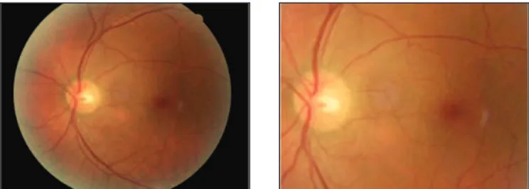
We described one case with scattered drusen-like deposits beneath the retina in posterior pole of the eye (unpublished report). The presentation of our case is similar a figure 2 page 902 case 18 as published by Duvall–Young et al.(1).
Our case show drusen-like deposits beneath retina, in the posterior pole, in both eyes,on colors photos and red-free 2 years
Figure 1: Colors photos of drusen-like beneath retinal deposits in right eye, 2 years ago
Figure 2: Color photos ofdrusen-like beneath retinal deposits in left eye, 2 years ago
Figure 3: Red-free photos showing drusen-like beneath retinal deposits in both eyes, 2 years ago
Rev Bras Oftalmol. 2015; 74 (1): 49-52
51
early (figure 1-3), no in peripheric retina but it was related(1,22) in
the follow-up 2 years later, colors photos (figures 4,5); fundus autofluorescence (figures 6,7), and spectral-domain OCT (figu-res 8,9) with no abnormalities.
In conclusion, long term follow-up of the cases in this disease shows in the most of them, no progression of the of Drusen-like beneath retinal deposits in type II mesangiocapillary glomerulonephritis(23), the most of subjects retain good visual
acuity and no specific treatment is indicated.
Acknowledgments
We would like to express our very great appreciation to Lawrence Yannuzzi, MD, Richard Spaide,MD and K. Bailey Freund,MD for their valuable and constructive analyses of the case cited in this review. Their willingness to give their time so generously has been very much appreciated.
R
EFERENCES1. Duvall-Young J, MacDonald MK, McKechnie N. Fundus changes in (type 2) glomerulonephritis simulating drusen: a histological report. Br J Ophthalmol. 1989; 73(4): 297"30.
2. Duvall-Young J, Short CD, Raines MF, Gokal R , Lawler L. Fundus changes in mesangiocapillary glomerulonephritis type 2: clinical and fluorescein angiographic findings. Br J Ophthalmol. 1989; 73(11):900"6.
3. Mullins RF, Aptsiauri N, Hageman GS. Structure and composition of drusen associated with glomerulonephritis: Implications for the role of comple-ment activation in drusen biogenesis. Eye (Lond). 2001;15(Pt 3):390–5. 4 . Appel GB, Cook HT, Hageman G, Jennette JC, Kashgarian M, Kirschfink M, et al. Membranoproliferative glomerulonephritis type II (dense deposit disease): an update. J Am Soc Nephrol. 2005 ;16(5):1392-403
5. Alpers CE. The kidney. In: Kumar V, Abbas AK, Fausto N. editors. Pathologic basis of disease. 7 th ed. Philadelphia: Elsevier Saunders; 2005. p.984-6 6. Gorin MB, Jackson KE, Ferrell RE, Sheffield VC, Jacobson SG, Gass
JD, et al. A peripherin/retinal degeneration slow mutation (Pro-210-Arg) associated with macular and peripheral retinal degeneration. Ophthalmology. 1995;102(2):246-55.
7. Piguet B, Héon E, Munier FL, Grounauer PA, Niemeyer G, Butler N, et al. Full characterization of the maculopathy associated with an Arg-172-Trp mutation in the RDS/peripherin gene. Ophthalmic Genet. 1996;17(4):175-86.
8. Stone EM, Lotery AJ, Munier FL, Héon E, Piguet B, Guymer RH, et al. A single EFEMP1 mutation associated with both Malattia Leventinese and Doyne honeycomb retinal dystrophy. Nat Genet. 1999;22(2):199-202. 9. Boon CJ, Klevering BJ, Hoyng CB, Zonneveld-Vrieling MN, Nabuurs
SB, Blokland E, Cremers FP, den Hollander AI. Basal laminar drusen caused by compound heterozygous variants in the CFH gene. Am J Hum Genet. 2008;82(2):516-23.
Figure 4: Colors photos of drusen-like beneath retinal deposits in right eye, 2 years later
Figure 5: Color photos ofdrusen-like beneath retinal deposits in left eye, 2 years later
Figure 6: Fundus autofluorescence with no abnormalities in left eye, 2 years later
Figure 7: Fundus Autofluorescence with no abnormalities in rigt eye, 2 years later
Figure 9: SD-OCT with no abnormalities in left eye , 2 years later
Rev Bras Oftalmol. 2015; 74 (1): 49-52 Drusen-like beneath retinal deposits in type II mesangiocapillary glomerulonephritis: a review
52
Correspondong author: Miguel Hage Amaro, MD
Travessa Quintino Bocaiúva, nº 516 Zip code: 66053-249 – Belém (PA), Brazil E-mail:[email protected] 10. Leys A, Proesmans W, Van Damme-Lombaerts R, Van Damme B.
Specific eye fundus lesions in type II membranoproliferative glom-erulonephritis. Pediatr Nephrol. 1991;5(2):189-92. Erratum in: Pediatr Nephrol 1991;5(3):364.
11. Kim DD, Mieler WF, Wolf MD. Posterior segment changes in membranoproliferative glomerulonephritis. Am J Ophthalmol. 1992 15;114(5):593-9.
12. Michielsen B, Leys A, Van Damme B, Missotten L. Fundus changes in chronic membranoproliferative glomerulonephritis type II. Doc Ophthalmol. 1990-1991;76(3):219-29.
13. Leys A, Vanrenterghem Y, Van Damme B, Snyers B, Pirson Y, Leys M. Fundus changes in membranoproliferative glomerulonephritis type II. A fluorescein angiographic study of 23 patients. Graefes Arch Clin Exp Ophthalmol. 1991;229(5):406-10.
14. Leys A, Vanrenterghem Y, Van Damme B, Snyers B, Pirson Y, Leys M. Sequential observation of fundus changes in patients with long stand-ing membranoproliferative glomerulonephritis type II (MPGN type II). Eur J Ophthalmol. 1991;1(1):17-22.
15. Leys A, Van Damme B, Verberckmoes R. Ocular complications of type 2 membranoproliferative glomerulonephritis. Nephrol Dial Transplant. 1996;11(1):211-4.
16. Leys A, Michielsen B, Leys M, Vanrenterghem Y, Missotten L, Van Damme B. Subretinal neovascular membranes associated with chronic membranoproliferative glomerulonephritis type II. Graefes Arch Clin Exp Ophthalmol. 1990;228(6):499-504.
17. Han DP, Sievers S. Extensive drusen in type I membranoproliferative glomerulonephritis. Arch Ophthalmol. 2009;127(4):577-9.
18. McAvoy CE, Silvestri G. Retinal changes associated with type 2 glom-erulonephritis. Eye (Lond). 2005;19(9):985-9.
19. Gass JD, Slamovits TL, Fuller DG, Gieser RG, Lean JS. Posterior chorioretinopathy and retinal detachment after organ transplanta-tion. Arch Ophthalmol. 1992;110(12):1717-22.
20. Polk TD, Kimura AE, Park D, Gass JD. Subretinal fluid associated with membranoproliferative glomerulonephritis type 2. Arch Ophthalmol. 1997;115(7):927-8.
21. Ulbig MR, Riordan-Eva P, Holz FG, Rees HC, Hamilton PA. Membranoproliferative glomerulonephritis type II associated with central serous retinopathy. Am J Ophthalmol. 1993;116(4):410-3. 22. Huang SJ, Costa DL, Gross NE, Yannuzzi LA. Peripheral drusen in
membranoproliferative glomerulonephritis type II. Retina. 2003;23(3):429-31.
23. D’souza Y, Short CD, McLeod D, Bonshek RE. Long-term follow-up of drusen-like lesions in patients with type II mesangiocapillary glom-erulonephritis. Br J Ophthalmol. 2008;92(7):950-3.
Rev Bras Oftalmol. 2015; 74 (1): 49-52