ABSTRACT
Head and neck reconstruction using
infrahyoid myocutaneous fl aps
Head and Neck Surgery Service, Department of Surgery, Faculdade de
Ciências Médicas da Universidade Estadual de Campinas (Unicamp),
Campinas, São Paulo, Brazil
INTRODUCTION
The use of myocutaneous fl aps is now a well-established alternative for reconstructing surgical defects in the head and neck.1-6 Despite
the evolution of free fl aps with microsurgical anastomoses, many axial pedicled fl aps are widely used because of their ease of harvesting, the reliability of the pedicle, the experience of most surgeons in using them and the non-availability of surgical teams able to perform free fl ap procedures.
First described by Wang et al. in 19861
and called the infrahyoid fl ap (IHF), these fl aps were proposed for the reconstruction of defects following resections of the oral tongue and, subsequently, for more complex defects of the oral cavity, oropharynx and facial skin. The fl ap is comprised of skin from the cervical region and the strap muscles, and its pedicle is based on the superior thyroid vessels.
OBJECTIVE
The objective of this article was to describe our experience with 14 patients with head and neck neoplasms, in whom infrahyoid myocu-taneous fl aps were used for reconstruction.
MATERIALS AND METHODS
Between January 1994 and December 2004, 14 patients with neoplasms of the head and neck underwent surgical resection of their lesions at the Head and Neck Surgery Service of the Department of Surgery, Uni-versidade Estadual de Campinas (Unicamp), Brazil. Patients with prior cervical surgical procedures were excluded from the study, as were patients with an ipsilateral scar on the fl ap area, advanced atherosclerosis that might have impaired the blood fl ow in the fl ap, or neck metastasis that required radical neck dissection (N2 or N3). Thirteen (92,8%) out of the 14 patients included over the period had squamous cell carcinoma (SCC) and one
(7.2%) had ameloblastoma. The disease stage was T3 in eight (61.5%) of the SCC cases and T4 in fi ve (38.5%). All of these patients had their surgical defects reconstructed using IHFs. Eleven of them were men and three were women, with ages ranging from 47 to 80 years (mean age of 66.4 years).
Excessively hairy skin was a relative con-traindication for fl ap use. Previous irradiation of the neck was also a contraindication for the use of IHFs, as stated by other authors.3,6
The infrahyoid strap muscles were used for this flap, including the sternohyoid, sternothyroid and the inferior belly of the omohyoid. The superior portion was vas-cularized by the superior thyroid artery and its branches, and the inferior third by the inferior thyroid artery. According to Eliachar et al.,2 this blood supply is segmental but
presents signifi cant anastomoses between the main superior and inferior branches, thereby permeating the entire musculature. Venous drainage takes place through the inferior thyroid vein, which drains directly into the internal jugular vein or through the anterior ipsilateral jugular vein (Figure 1).
According to Wang et al.,1 sensorimotor
innervation comes directly from branches of the cervical loops of C1 and C2 (upper half ) and C2 and C3 (lower half ), through nerve anastomoses.
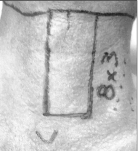
The fl ap was harvested by cutting the skin in a cranial-caudal direction, in a para-median or central location, depending on the site to be reconstructed (Figure 2). Care was taken not to go beyond the contralateral boundaries, thus avoiding damage to local micronutrition.
After the rectangular island of skin was constructed, in continuity with the sub-cutaneous tissue and the abovementioned muscles, the unit was separated by fi ne dis-section of the surgical capsule of the thyroid
Alfi o José Tincani André Del Negro Priscila Pereira Costa Araújo Hugo Kenzo Akashi Flávia da Silva Pinto Neves Antônio Santos Martins
ORIGINAL AR
TICLE
CONTEXT AND OBJECTIVE: The use of pedicled myocutaneous fl aps in head and neck reconstruc-tion is widely accepted. Here we describe our experience with infrahyoid fl aps (IHFs) employed to cover surgical defects in the oral cavity and oropharynx in patients with benign and malignant tumors. The aim was to evaluate the success rate for infrahyoid myocutaneous fl ap procedures performed at a single institution.
DESIGN AND SETTING: Retrospective study, at the Head and Neck Surgery Service, Unicamp.
METHODS: Fourteen IHFs were used to recon-struct surgical defects in eleven men (78.5%) and three women (21.5%) with a mean age of 66.4 years. The anterior fl oor of the mouth was reconstructed in nine patients (64.2%), the base of tongue in three (21.4%), the lateral fl oor in one (7.1%), and the retromolar area (7.1%) in one. Thirteen patients (92.8%) had squamous cell car-cinoma (SCC) and one (7.2%) ameloblastoma. The disease stage was T3 in eight (61.5%) of the SCC cases and T4 in fi ve (38.5%).
RESULTS: No patient presented total fl ap loss or fi stula. The most common complication was epidermolysis, which delayed the beginning of oral ingestion. The patients with SCC received postoperative radiotherapy without major conse-quences to the fl ap.
CONCLUSION: IHF is a safe and reliable proce-dure for reconstructing head and neck surgical defects. Due to its thinness and malleability, its use for oral cavity and oropharynx defects provides favorable cosmetic and functional outcomes. Complications, when present, are easy to manage.
KEY WORDS: Head and neck neoplasms. Surgi-cal fl aps. Reconstructive surgiSurgi-cal procedures. Neck muscles. Rehabilitation.
272
gland, in the plane of the superfi cial layer of the deep cervical fascia. Its size ranged from 10 to 14 centimeters,1,3 depending on
the defect to be corrected. The thyrohyoid muscle was preserved and remained in situ
in order to protect the superior laryngeal nerves and laryngeal vessels.4 As the fl ap
was harvested, even greater care was taken in dissecting the upper third. The vascular pedicle could be identifi ed easily and was clearly visible when dissection occurred in the medial-lateral direction. After identi-fying the pedicle, it was skeletonized by ligating its collateral branches (superior laryngeal artery, infrahyoid artery and cri-cothyroid artery, and their branches to the sternocleidomastoid muscle; and the collateral veins) and expanding its rotation arch,5 which could be up to 15 centimeters
in length.1 Special attention was given to the
dissection of the superior laryngeal nerve, which must always be preserved. As stated by Wang et al.,1 the collateral vessels should
be maintained whenever possible. The island of skin on the fl ap was sutu-red to the subjacent musculature to avoid sliding between its layers, which could injure the perforans vessels and thus compromise the microvasculature. The anterior jugular vein was ligated in its inferior and superior portions and the muscles were deinserted in this same direction, from the sternum toward the hyoid bone, where they were sec-tioned. The third branch of the cervical loop had to be sacrifi ced, thereby providing the fl ap with a broader rotation arch.
After adequate hemostasis, the skin of the donor area was closed primarily without diffi culties.
The fl ap was used to reconstruct defects in the fl oor of the mouth and the oropharynx. Nine cases were in the anterior fl oor of the mouth (all adjacent to or directly involving the jaw), one affected the lateral fl oor, three were at the base of tongue and one was in the retromolar area. All these lesions, except for one ameloblastoma, had fi rst been clinically staged as T3 or T4 (Table 1).
The variables analyzed were: size of the fl ap, stage of the tumor upon diagnosis, pri-mary site and functional results.
All patients (except the one case of ameloblastoma) received postoperative radiotherapy, with individualized doses for each case, ranging from 5000 to 7000 cGy. Supraomohyoid neck dissection was per-formed on all cases with malignant tumors (all N0 upon diagnosis), and the dissection was bilateral when the lesion was adjacent to or crossed the midline.
The fl ap was used with primary closing of the donor area in all cases. The skin paddle was rectangular and paramedian in all cases, as postulated by Wang et al.,1 with size
varying according to the area to be re cons-tructed (Table 1).
Motor innervation was preserved in all cases, and tracheostomy was performed in 13 patients.
Clinically palpable cervical lymph nodes located at levels I and II and no larger than two centimeters (N1) were considered for
Figure 1. Pedicle flap composed of infrahyoid strap muscles that was used for surgical reconstruction of the head and neck.
Table 1. Description of the series of cases of head and neck reconstruction using infrahyoid myocutaneous fl ap
Patient Tumor site Flap size (cm) Function Complications Oral diet (days) Feeding tube (days) Recurrence
1 FOM T3 8 x 4 Good No 6 7 No
2 FOM T4 8 x 5 Fair No 7 8 Yes
3 Tongue base T3 9 x 4 Fair Donor site dehiscence 10 12 Yes
4 RMT T4 8 x 4 Good No 5 6 No
5 FOM T4 8 x 3 Good Skin paddle necrosis 9 10 No
6 Tongue base T4 8 x 4 Fair Skin paddle necrosis 10 11 Yes
7 FOM T3 8 x 3 Good No 6 5 No
8 FOM T3 8 x 4 Good No 7 8 No
9 FOM T4 8 x 3 Good No 8 8 No
10 Tongue base T4 8 x 4 Fair Skin paddle necrosis 9 10 Yes
11 FOM T4 9 x 4 Good Post RT retraction 6 7 No
12 FOM T4 10 x 4 Good Skin paddle necrosis 9 10 No
13 FOM T4 10 x 4 Good No 7 8 No
14 Ameloblastoma 8 x 5 Good No 5 6 No
FOM = fl oor of the mouth; RMT = retromolar trigone; RT = radiotherapy.
Figure 2. The miocutaneous fl ap was harvested by cutting the skin in a cranial-caudal direction, in a paramedian or central location.
273
selective neck dissection, with no prejudice to the venous draining of the fl ap.
RESULTS
No case resulted in total loss of the fl ap and only four cases (28.5%) showed partial loss through epidermolysis, which did not affect the fi nal result. Damage to the cutaneous portion occurred within the fi rst three days following reconstruction and there was no loss of the muscular portion of the fl ap. There was one case of retraction of the fl ap caused by radiotherapy. However, no alteration of the functional results of the surgery was observed.
No cases of salivary fi stulas were seen, nor were there any early or late hemorrhagic or infectious complications.
Regarding the donor area, only one patient showed partial dehiscence of the suture line, which was resolved with local care and pro-duced very satisfactory fi nal aesthetic results.
The nasoenteral feeding tube was removed on average eight days after surgery and oral nu-trition was introduced on approximately the seventh day. The patients showed full capacity for oral ingestion of food on the twelfth day following surgery.
The patients with SCC received post-operative radiotherapy without major con-sequences to the fl ap. To date, four patients have shown local-regional recurrence, even after adjuvant treatment.
With regard to swallowing and speaking capacity, four patients required postoperative speech therapy. This evaluation was subjec-tive and based on the criteria of “good”, “intermediate” or “poor” capacity to produce phonemes, in comparison with the patient’s corresponding presurgery ability. The
evalua-tion was considered reliable since the patients did not present any complicating factors that might infl uence their speaking ability, such as radiotherapy or prior head or neck surgery. Nevertheless, both the anatomical location of the surgical defect and the harvesting of the fl ap negatively affected functional results.
DISCUSSION
A variety of fl aps are available for recon-structing surgical defects in the head and neck, including the pectoralis major, deltopectoral, trapezius and platysma, among others, and each has its own advantages and disadvantages. The ideal fl ap should be thin, pliable, hairless, easy to harvest and reliable and should have the capacity for implementation in a single-stage procedure.
Although the most commonly used fl ap is the pectoralis major, the IHF is thinner, lies close to the surgical defect and, based on our current experience, is very reliable. Other advantages are that it is easy and quick to harvest; there is no need to reposition the patient; there is primary closure of the donor area; and there are few or no cosmetic sequelae. The primary indications for using IHFs are intraoral, pharyngeal and parotid region skin defects.1-3,5,6
We believe that it is essential to suture the muscles to the skin, in order to attach them and thus prevent slipping. Likewise, there needs to be careful handling both in harvesting and elevation. These steps are likely to avoid the epidermolysis that occurred in several of our cases. Our success rate approached 100% and was comparable to larger series with simi-lar complications.1,3,5,6
As recommended by the majority of au-thors, only patients who had not undergone
previous cervical radiotherapy were selected for IHFs.3,6
In the cases with loss of the cutaneous portion of the fl ap, this probably occurred for the same reasons as described by Wang,1 that
is, because of a defi cit in venous drainage and consequent fl ap congestion.
Preserving the motor innervation in all cases provides greater symmetry, volume and mobility to the fl ap, thereby reducing atrophy and im-proving the functional results. This allowed for subsequent positive functional results regarding swallowing and speaking in our series.
Tumor recurrence was due to the ad-vanced stage of the disease at diagnosis, and this fi nding was similar to what has been described by other authors.4
CONCLUSION
The infrahyoid myocutaneous fl ap pro-cedure is simple for a head and neck surgeon familiar with the anatomy of this region to perform. In our experience, there were posi-tive aesthetic and functional results, and the complications were easy to manage. The patients were, however, carefully chosen so as to respect the contraindications.
Among the most important advantages of this type of fl ap are: the more appropriate thickness and coloring of the skin than in the material obtained from the usual fl aps (the type used here was thinner and more pliable than the skin of the thoracic wall or the forearm); the head and neck surgeon’s familiarity with the anatomical region; the proximity of the donor area to the reconstruction site; and ever-present possibility of primary closure. In addition, these fl aps present satisfactory cosmetic appearance and are well accepted by patients.
274
AUTHOR INFORMATION
Alfi o José Tincani, MD, PhD. Assistant professor, Head
and Neck Service, Department of Surgery, Faculdade de Ciências Médicas da Universidade Estadual de Campinas (Unicamp), Campinas, São Paulo, Brazil.
André Del Negro, MD. Attending surgeon, Head and
Neck Service, Department of Surgery, Faculdade de Ciências Médicas da Universidade Estadual de Campinas (Unicamp), Campinas, São Paulo, Brazil.
Priscila Pereira Costa Araújo, MD. Former resident,
Head and Neck Service, Department of Surgery, Faculdade de Ciências Médicas da Universidade Estadual de Campi-nas (Unicamp), CampiCampi-nas, São Paulo, Brazil.
Hugo Kenzo Akashi, MD. Resident, Head and Neck
Service, Department of Surgery, Faculdade de Ciências Médicas da Universidade Estadual de Campinas (Uni-camp), Campinas, São Paulo, Brazil.
Flavia Silva Pinto Neves, MD. Resident, Head and
Neck Service, Department of Surgery, Faculdade de Ciências Médicas da Universidade Estadual de Campinas (Unicamp), Campinas, São Paulo, Brazil.
Antônio Santos Martins, MD, PhD. Chief, Head and
Neck Service, Department of Surgery, Faculdade de Ciências Médicas da Universidade Estadual de Campinas (Unicamp), Campinas, São Paulo, Brazil.
Address for correspondence:
André Del Negro
Rua Governador Pedro de Toledo, 2157 — Apto. 141 Piracicaba (SP) — Brasil — CEP 13400-075 Tel. (+55 19) 3435-1898
E-mail: [email protected]
Copyright © 2006, Associação Paulista de Medicina
Resumo Reconstrução em cabeça e pescoço com retalho miocutâneo infrahióideo
CONTEXTO E OBJETIVO: O uso de retalhos miocutâneos pediculados para reconstrução cirúrgica na região de cabeça e pescoço (RCP) é consagrado. Apresentaremos a experiência com o uso do retalho infrahiódeo (RIH) em reconstrução de defeitos cirúrgicos na cavidade oral e orofaringe em portadores de tumores benignos e malignos. O objetivo foi avaliar o índice de sucesso do RIH em defeitos da cavidade oral em uma única instituição.
TIPO DE ESTUDO E LOCAL: Estudo retrospectivo, no Serviço de Cirurgia de Cabeça e Pescoço, Universidade Estadual de Campinas (Unicamp).
MÉTODOS: Foram utilizados 14 RIH para reconstrução em RCP em 11 homens (78,5%) e 3 mulheres (21,5%). Em nove (64,2%) pacientes, a reconstrução foi de assoalho oral anterior, três (21,4%) de base de língua, um (7,1%) de assoalho lateral e um de trígono retromolar (7,1%). O carcinoma espinocelular (CEC) foi neoplasia presente em 13 deles (92,8%) e um (7,2%) ameloblastoma de mandíbula. O estádio era T3 em oito (61,5%) e T4 em cinco (38,5%) dos casos de CEC.
RESULTADOS: Não houve caso de perda total do retalho ou fístula. A complicação mais comum foi epider-mólise, retardando o início da ingestão oral. Pacientes com CEC receberam radioterapia pós-operatória sem conseqüências para o retalho.
CONCLUSÃO:O RIH é um retalho seguro e confi ável para o cirurgião de cabeça e pescoço na reconstrução na RCP. Devido à espessura e maleabilidade, sua utilização para reconstrução de defeitos da cavidade oral e orofaringe proporciona bom aspecto cosmético e funcional. Complicações são de fácil manuseio.
PALAVRAS-CHAVE: Câncer de cabeça e pescoço. Retalhos cirúrgicos. Procedimentos cirúrgicos reconstru-tivos. Músculos do pescoço. Reabilitação.
1. Wang HS, Shen JW, Ma DB, Wang JD, Tian AL. The infrahyoid myocutaneous fl ap for reconstruction after resection of head and neck cancer. Cancer. 1986;57(3):663-8.
2. Eliachar I, Marcovich A, Har Shai Y, Lindenbaum E. Arterial blood supply to the infrahyoid muscles: an anatomical study. Head Neck Surg. 1984;7(1):8-14.
3. Dolivet G, Gangloff P, Sarini J, et al. Modifi cation of the infra hyoid musculo-cutaneous flap. Eur J Surg Oncol. 2005;31(3):294-8.
4. Zhao YF, Zhang WF, Zhao JH. Reconstruction of intraoral defects after cancer surgery using cervical pedicle fl aps. J Oral Maxillofac Surg. 2001;59(10):1142-6.
5. Rojananin S, Suphaphongs N, Ballantyne AJ. The infrahyoid musculocutaneous fl ap in head and neck reconstruction. Am J Surg. 1991;162(4):400-3.
6. Magrin J, Kowalski LP, Santo GE, Waksmann G, DiPaula RA. Infrahyoid myocutaneous fl ap in head and neck reconstruction. Head Neck. 1993;15(6):522-5.
Presented at the 20th Brazilian Congress of Head and Neck Surgery, Salvador, BA, Brazil, September 3-6, 2005.
Sources of funding:None
Confl ict of interest:None
Date of fi rst submission:December 23, 2005
Last received:September 14, 2006
Accepted:September 19, 2006
REFERENCES