Review article
general clinicians
Linfocitose monoclonal de células B: uma breve revisão para clínicos gerais
Daniel Mazza Matos
I, Roberto Passetto Falcão
I Department of Clinical Medicine and Center for Research on Cell-Based Therapy, Faculdade de Medicina de Ribeirão Preto,Universidade de São Paulo (FMRP/USP), São Paulo, Brazil
IMD, PhD. Hematologist, Department of Clinical Medicine, Faculdade de Medicina de Ribeirão Preto, Universidade de São Paulo (FMRP/USP), Ribeirão Preto, São Paulo, Brazil.
ABSTRACT
Monoclonal B-cell lymphocytosis (MBL) is a recently described medical condition that displays biological similarities to the most common subtype of adult leukemia in the Western world, i.e. chronic lymphocytic leukemia (CLL). Diagnostic criteria have been published with the aim of differentiating
between these two entities. The overall prevalence of MBL is at least 100 times higher than that of CLL, which indirectly suggests that MBL is not necessarily a pre-leukemic condition, although in some circumstances, CLL cases can really be preceded by MBL. In view of this high prevalence rate,
general clinicians and even non-hematological specialists have a high chance of being faced with individuals with MBL in their routine clinical practice. MBL is classiied as “clinical MBL”, “population-screening MBL” and “atypical MBL” and the clinical management of affected individuals depends
greatly on this differentiation. The present review provides a guide to diagnosing and following up MBL patients.
RESUMO
A linfocitose monoclonal de células B (LMB) é uma condição médica recentemente descrita que exibe similaridades biológicas com o mais comum subtipo de leucemia em adultos de países ocidentais, qual seja, a leucemia linfocítica crônica (LLC). Critérios diagnósticos foram publicados com o
intuito de separar as duas entidades. A prevalência global da LMB é pelo menos 100 vezes maior do que a da LLC, o que, indiretamente, sugere que a LMB não é necessariamente uma condição pré-leucêmica, embora, em algumas circunstâncias, casos de LLC possam realmente ser precedidos pela
LMB. Em virtude dessa alta taxa de prevalência, clínicos gerais e mesmo outros especialistas não hematologistas têm grande chance de deparar-se com casos de LMB em suas rotinas clínicas. A LMB é classiicada como “LMB clínica”, “LMB de screening populacional” e “LMB atípica”, sendo que o manuseio clínico dos indivíduos afetados depende substancialmente dessa diferenciação. A presente revisão fornece um guia para o diagnóstico e acompanhamento dos pacientes com LMB.
KEY WORDS:
B-Lymphocytes.
Leukemia, lymphocytic, chronic,
B-cell. Flow cytometry.
Epidemiology. Disease management.
PALAVRAS-CHAVES: Linfócitos B.
Leucemia linfocítica crônica de
células B. Citometria de luxo.
Epidemiologia. Gerenciamento clínico.
DEFINITION AND HISTORY
Over the last few years, the more widespread availability of au-tomated blood cell counters, associated with increasing technological advances in low cytometry, has made it possible to recognize very low levels of circulating monoclonal B-lymphocytes that are immunophe-notypically similar to chronic lymphocytic leukemia B-cells, in the peripheral blood of healthy subjects. Within this scenario, general cli-nicians and even non-hematological specialists have a high chance of coming across asymptomatic individuals with slight increases in abso-lute lymphocyte counts, composed of abnormal B-cell clones, in pe-ripheral blood, but without the diagnostic criteria for chronic lympho-cytic leukemia (CLL).1
he irst published paper to describe these abnormal B-cells dates back to 1991, consisting of a report from a health study conducted by the Centers for Disease Control (CDC) in the United States, which detected monoclonal B-cells in people living near hazardous waste sites. In 1995, a public health service workshop in the United States put forward recommendations for case deinitions and medical
fol-low-up.2 Subsequently, over the next ten years, an increasing number
of studies on diferent populations around the world, using diferent low cytometry approaches, recognized that CD5+ and CD5− mono-clonal B-lymphocytes could be found in individuals with no clinically apparent hematological disease.3
In 2005, the International Familial CLL Consortium proposed the term “monoclonal B-cell lymphocytosis” (MBL) to describe very low levels of circulating monoclonal B-cells that were identiied by means of immunophenotypic characterization, in the peripheral blood of ap-parently health subjects. Furthermore, diagnostic criteria for this con-dition were deined and, with little modiication, these continue to be used today (Chart 1).4
EPIDEMIOLOGY
(discussed later in “Biology and natural history of MBL”).5
More-over, the prevalence of MBL is age-related and population-related. Rawstron et al.6 showed that the overall prevalence of CLL-like MBL
among United Kingdom hospital outpatients without hematological or oncological diseases and with normal blood counts was 3.5%. he prevalence increased with age, from 2.1% in individuals between 40
and 60 years of age to 5.0% for individuals over 60 years of age. Ghia et al.7 studied individuals from a rural community referred for
rou-tine blood tests and showed that the overall prevalence was 5.1%. In a recent study conducted in a community in northern Italy, Dagk-lis et al.8 reported that the prevalence of CLL-like MBL and atypical
MBL was 6.3%. he prevalence in individuals over 60 years of age was 8.9%. Other published papers have found diferent prevalence rates going from as low as 0.14%9 to as high as 12.0%,10 but this
wide variation is probably related to the use of diferent low cytom-etry approaches, i.e. other than the most commonly used four-color low cytometry method. his hampers comparisons between these studies at the present time.
With regard to population subgroups, the highest prevalence of CLL-like MBL has been found in healthy irst-degree relatives of pa-tients with familial CLL, which is a condition characterized by the pres-ence of two or more individuals with the diagnosis of CLL inside the same family.11 hus, Rawstron et al.12 detected the presence of MBL in
eight out of 59 individuals (13.5%) pertaining to 21 families with CLL in the United Kingdom. he prevalence in another study on 9 families with CLL in the United States was 18%.13 hese studies clearly show
that MBL is more frequently observed in CLL kindreds than in individ-uals from the general population.
Lastly, using the worldwide four-color low cytometry approach, we have recently studied 167 healthy irst-degree relatives of patients with just one case of CLL inside the family (sporadic CLL). We found seven CLL-like MBL cases (overall prevalence of 4.1%). However, the prevalence of individuals over 60 years of age was 15.6%, which sug-gests that in older irst-degree relatives of patients with sporadic CLL, the risk of MBL detection is as high as in older irst-degree relatives from CLL families.14 Nonetheless, at this moment, there is no
evi-dence to suggest that searches for MBL should be conducted in irst-degree relatives of patients with the diagnosis of CLL. However, it is recommended that relatives who are potential donors for allogeneic stem cell transplantation should be screened for MBL, mainly because the transplanted monoclonal B-cells could interfere with the residual disease monitoring performed by low cytometry and, moreover, there is a possibility of expansion of the MBL clone in the recipient.4
Nev-ertheless, there is currently no consensus on whether or not matched relatives should be excluded, especially when there are no alternative donors inside the family.15
BIOLOGY AND NATURAL HISTORY OF MBL
With regard to the phenotype determined by low cytometry, monoclonal B-cell lymphocytosis has been classiied as MBL with CLL-like phenotype, MBL with atypical CLL phenotype and MBL with non-CLL phenotype (Chart 1). Moreover, MBL can also be
clas-siied as cases detected in clinical practice (clinical MBL), in which in-dividuals have absolute lymphocytosis, but with B-cell counts lower than 5 x 10⁹ cells/l, or otherwise,MBL cases accidentally found by
screening individuals with a completely normal blood count (popula-tion-screening MBL).15
Chart 1. Diagnostic criteria and subclassiication for monoclonal B-cell
lymphocytosis (adapted from Shanafelt et al.)15
DIAGNOSTIC CRITERIA
(1) Detection of a monoclonal B-cell population in the peripheral blood* by at least
one of following:
(a) Light-chain restriction: overall kappa:lambda ratio > 3:1 or < 0.3:1, or > 25% of B cells lacking or expressing low levels of surface immunoglobulin†
(b) Heavy-chain monoclonal immunoglobulin heavy chain variable region (IGHV) rearrangements
(2) Presence of a disease-speciic immunophenotype†,‡
(3) Absolute B-cell count < 5 x 10⁹ cells/l†
(4) No other features of a lymphoproliferative disorder:
(a) Normal physical examination (no lymphadenopathy or organomegaly)
(b) Absence of B-symptoms (fever, weight loss or nighttime sweating) attributable to a non-Hodgkin lymphoma
(5) No autoimmune or infectious disease
SUBCLASSIFICATION
(A) CLL-like phenotype:
(A.1) Coexpression of CD5 with CD19, CD20low and CD23†
(A.2) Light-chain restriction with low surface immunoglobulin expression (very small MBL clones may be oligoclonal and thus not light-chain restricted)†
(B) Atypical CLL phenotype:
(B.1) Coexpression of CD5 with CD19, but CD20strong or CD23 negative†
(B.2) Light-chain restriction with moderate/strong surface immunoglobulin expression†
(B.3) Exclude t(11;14) to rule out mantle cell lymphoma
(C) Non-CLL phenotype :
(C.1) CD5 negative†
(C.2) Expression of CD20†
(C.3) Light-chain restriction with moderate/strong surface immunoglobulin expression†
MBL with CLL-like phenotype accounts for most cases8 and
ex-hibits a population of monoclonal B-cells that, in addition to CD5 and CD23 positivity, show low expression of CD20 and CD79b antigens. his proile is phenotypically identical to CLL B-cells.16 Moreover, the
biological similarities of MBL with CLL-like and CLL phenotypes are demonstrable through other levels of scientiic evidence: irst, the pro-tein expression proile is almost identical between them and, second, some chromosomal abnormalities frequently found in CLL patients are also present in individuals with CLL-like MBL.16 However, despite this
biological relationship, a recent paper by Rossi et al.17 showed that MBL
and CLL are clinically diferent conditions, given that individuals with MBL have a more favorable clinical course than do patients with the di-agnosis of Rai 0 CLL.
With regard to clinical progression, Rawstron et al.18 followed up 185
subjects with clinical MBL for more than six years (median follow-up pe-riod of 6.7 years) and showed that CLL requiring treatment developed at a rate of 1.1% per year, mainly in subjects with clinical MBL, which is similar to the rate of progression to myeloma seen in patients with monoclonal gammopathy of undetermined signiicance. On the other hand, although limited data are available, progression among individuals with population-screening MBL is rarely seen in the practice of authors at diferent centers with wide experience of the management of MBL.18
Moreover, new studies are providing compelling evidence that not only the clinical characteristics but also the biology of population-screening MBL difer from that of clinical MBL. hus, Dagklis et al.8 have shown
that the usage of immunoglobulin heavy chain variable region (IGHV) is diferent between clinical and population-screening MBL, which means that the two conditions are probably biologically distinct entities.
Hence, it is possible to infer that individuals with population-screening MBL are unlikely to be at any substantially higher risk of developing CLL than observed among the general population.18 his
means that, in a general manner, MBL is not necessarily a pre-malig-nant condition. his conception is reinforced by studies showing that although MBL was originally thought to be exclusively monoclonal, both oligoclonal19 and polyclonal8,14 cases have been described. hus,
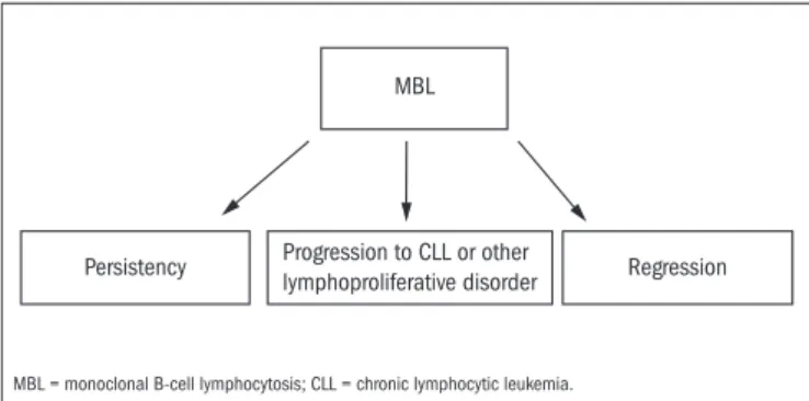
MBL can now be considered to be an entity with three possible difer-ent outcomes (Figure 1).
In this context, the distinction between a malignant and a premalig-nant condition is best made based on the individual’s risk of an adverse clinical outcome.20 Currently, only B-cell counts in peripheral blood can
be used to make a distinction among patients with monoclonal B-cells with regard to the necessity of starting some kind of therapy in the light of a risk of death. hus, at the present time, only limited data are available for ascertaining whether prognostic factors classically asso-ciated with CLL outcomes, as determined by low cytometry (CD38, CD49d and ZAP-70), cytogenetics (del 17 and del 11q) or molecular biology methods (IGHV rearrangement), can be used to predict the risk
of MBL progression.
DIAGNOSIS EVALUATION AND CLINICAL
MANAGEMENT
he diferential diagnosis between MBL, CLL and another related condition known as small lymphocytic lymphoma is based on periph-eral blood B-cell counts and physical examinations on patients. MBL is characterized by an absolute peripheral blood B-cell count lower than 5 x 10⁹ cells/l and absence of lymphadenopathy and
hepatosplenomeg-aly. Small lymphocytic lymphoma is also characterized by an absolute peripheral blood B-cell count lower than 5 x 10⁹ cells/l, but the
pres-ence of lymph nodes, liver or spleen enlargement is typical of this dis-ease. CLL is deined by the presence of more than 5 x 10⁹ B-cells/l in
pe-ripheral blood. Lymphadenopathy and hepatosplenomegaly are found to varying degrees in patients with CLL (Table 1).15 Patients with a
di-agnosis of CLL, small lymphocytic lymphoma, clinical CLL-like MBL or atypical/non-CLL-like MBL should undergo a complete evaluation by a hematologist.
he clinical management for individuals with MBL difers accord-ing to whether the patient presents population-screenaccord-ing MBL, clinical CLL-like MBL or atypical/non-CLL-like MBL. In practical terms, gen-eral clinicians should follow up individuals with population-screening MBL, given that these individuals have completely normal blood count and, in particular, that clinical experience has shown that progression in this group is very rare. For such individuals, close monitoring is not nec-essary and an annual examination with a complete blood count is suf-icient and appropriate.15
As previously stated, patients with clinical CLL-like MBL have a risk of progression to CLL requiring treatment that is 1.1% per year.18 Based
on this low risk, an annual follow-up with a complete blood count made by a hematologist is recommended. hese patients should be counseled to pay attention to speciic symptoms such as lymph node enlargement, nighttime sweating, extreme fatigue and weight loss, given that these clinical indings could be the irst evidence of disease progression.
MBL
Persistency Progression to CLL or other
lymphoproliferative disorder Regression
Figure 1. Possible outcomes from monoclonal B-cell lymphocytosis (MBL).
MBL = monoclonal B-cell lymphocytosis; CLL = chronic lymphocytic leukemia.
Table 1. Differential diagnosis: monoclonal B-cell lymphocytosis (MBL), small lymphocytic lymphoma (SLL) and chronic lymphocytic leukemia
(CLL) (adapted from Shanafelt et al. )15
Peripheral blood B-cells count < 5 x 10⁹/l
Lymphadenopathy or hepatosplenomegaly
MBL Yes No
SLL Yes Yes
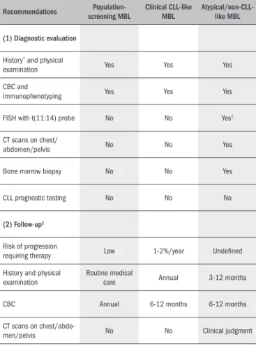
Patients with atypical or non-CLL-like MBL require a more thor-ough evaluation. In cases of lymph node enlargement found by com-puted tomography scans, these patients are best classiied as having a non-Hodgkin lymphoma subtype, which should be subclassiied based on complementary examinations (immunohistochemistry, cytogenetics etc). For such individuals, close clinical and laboratory monitoring is mandatory. Table 2 summarizes the recommendations for evaluation and follow-up of MBL in routine practice.
CONCLUSIONS
Monoclonal B-cell lymphocytosis is a very common clinical con-dition that is frequently found in asymptomatic subjects. It is biologi-cally related to chronic lymphocytic leukemia and, in some cases, can be found prior to the diagnosis of CLL, although, at the present time, MBL cannot necessarily be considered to be a pre-leukemic condition.
he prevalence of MBL is at least 100 times higher than that of CLL. hus, given that both availability of automated blood cell coun-ters and access to low cytometry tests are more widespread nowadays, it is not surprising that clinicians may unexpectedly ind MBL cases dur-ing their daily practice. Within this scenario, it is important to recognize situations in which patients with a diagnosis of MBL can be followed up in general clinicians’ oices, versus situations in which they must be referred to a hematologist.
For diagnostic purposes, monoclonal B-cell lymphocytosis should be distinguished from chronic lymphocytic leukemia and small lym-phocytic lymphoma. Moreover, it is essential to classify individual cases as “population-screening MBL” or “clinical MBL” and, furthermore, based on immunophenotyping, as “MBL with CLL-like phenotype”, “MBL with atypical CLL phenotype” or “MBL with non-CLL pheno-type”, given that the clinical management for these patients is strongly dependent on this classiication.
LITERATURE SEARCH
We conducted a search in the literature regarding MBL in the ive databases, using the term monoclonal B-cell lymphocytosis, and limit-ing the search to papers published in English over the last 20 years, in-cluding all types of articles (clinical trials, editorials, letters, meta-anal-yses, practical guidelines or randomized controlled trials). Articles with relevant information about the epidemiological, biological and clinical aspects of MBL have been included in the reference list of this paper. he results from the search are shown in Table 3.
REFERENCES
1. Redaelli A, Laskin BL, Stephens JM, Botteman MF, Pashos CL. The clinical and epidemio-logical burden of chronic lymphocytic leukaemia. Eur J Cancer Care (Engl). 2004;13(3): 279-87.
2. Vogt RF, Shim YK, Middleton DC, et al. Monoclonal B-cell lymphocytosis as a biomarker in environmental health studies. Br J Haematol. 2007;139(5):690-700.
3. Marti G, Abbasi F, Raveche E, et al. Overview of monoclonal B-cell lymphocytosis. Br J Hae-matol. 2007;139(5):701-8.
Table 2. Recommendations for evaluation and follow-up of monoclonal
B-cell lymphocytosis(MBL). Adapted from Shanafelt et al.15
Recommendations Population-screening MBL
Clinical CLL-like MBL
Atypical/non-CLL-like MBL
(1) Diagnostic evaluation
History* and physical
examination Yes Yes Yes
CBC and
immunophenotyping Yes Yes Yes
FISH with t(11;14) probe No No Yes†
CT scans on chest/
abdomen/pelvis No No Yes
Bone marrow biopsy No No Yes
CLL prognostic testing No No No
(2) Follow-up‡
Risk of progression
requiring therapy Low 1-2%/year Undeined
History and physical examination
Routine medical
care Annual 3-12 months
CBC Annual 6-12 months 6-12 months
CT scans on
chest/abdo-men/pelvis No No Clinical judgment
CLL, chronic lymphocytic leukemia; CBC, complete blood count; FISH, luorescence in situ hybridization; CT, computed tomography.
*Fever, nighttime sweating, weight loss and fatigue; †For patients with CD5+/CD23− MBL; ‡For the rare individuals fulilling the criteria for MBL who have an immunophenotype and cytogenetic evaluation suggestive of mantle-cell lymphoma, i.e. a “CD5+/CD23− MBL” case with the presence of t(11;14), or another aggressive non-Hodgkin lymphoma, the clinical follow-up should be done every 3-6 months with CT imaging at least every 6 months. For patients with MBL of atypical-CLL phenotype or non-CLL phenotype whose immunophenotype is consistent with a more indolent non-Hodgkin lymphoma subtype, follow-up every 6-12 months is recommended and the frequency of follow-up imaging requires clinical judgment.
Table 3. Database search results
Database Search limits Results
PubMed
Term: monoclonal B-cell lymphocytosis 19 reviews
Types of articles: all. 3 letters
Language: English 2 prospective cohort studies
Period: 1990-2010 1 editorial
SciElo Term: monoclonal B-cell lymphocytosis 1 review
Cochrane Library Term: monoclonal B-cell lymphocytosis None
Embase Term: monoclonal B-cell lymphocytosis None
4. Marti GE, Rawstron AC, Ghia P, et al. Diagnostic criteria for monoclonal B-cell lymphocytosis. Br J Haematol. 2005;130(3):325-32.
5. Ghia P, Prato G, Stella S, et al. Age-dependent accumulation of monoclonal CD4+ CD8+ double positive T lymphocytes in the peripheral blood of the elderly. Br J Haematol. 2007;139(5):780-90.
6. Rawstron AC, Green MJ, Kuzmicki A, et al. Monoclonal B lymphocytes with the characteristics of “indolent” chronic lymphocytic leukemia are present in 3.5% of adults with normal blood counts. Blood. 2002;100(2):635-9.
7. Ghia P, Prato G, Scielzo C, et al. Monoclonal CD5+ and CD5- B-lymphocyte expansions are frequent in the peripheral blood of the elderly. Blood. 2004;103(6):2337-42.
8. Dagklis A, Fazi C, Sala C, et al. The immunoglobulin gene repertoire of low-count chronic lym-phocytic leukemia (CLL)-like monoclonal B lymphocytosis is different from CLL: diagnostic implications for clinical monitoring. Blood. 2009;114(1):26-32.
9. Rachel JM, Zucker ML, Fox CM, et al. Monoclonal B-cell lymphocytosis in blood donors. Br J Haematol. 2007;139(5): 832-6.
10. Nieto WG, Almeida J, Romero A, et al. Increased frequency (12%) of circulating chronic lym-phocytic leukemia-like B-cell clones in healthy subjects using a highly sensitive multicolor low cytometry approach. Blood. 2009;114(1):33-7.
11. Caporaso N, Goldin L, Plass C, et al. Chronic lymphocytic leukaemia genetics overview. Br J Haematol. 2007;139(5):630-4.
12. Rawstron AC, Yuille MR, Fuller J, et al. Inherited predisposition to CLL is detectable as sub-clinical monoclonal B-lymphocytes expansion. Blood. 2002;100(7):2289-90. 13. Marti GE, Carter P, Abbasi F, et al. B-cell monoclonal lymphocytosis and B-cell
abnormali-ties in the setting of familial B-cell chronic lymphocytic leukemia. Cytometry B Clin Cytom. 2003;52(1):1-12.
14. Matos DM, Ismael SJ, Scrideli CA, et al. Monoclonal B-cell lymphocytosis in irst-degree rela-tives of patients with sporadic (non-familial) chronic lymphocytic leukaemia. Br J Haematol. 2009;147(3):339-46.
15. Shanafelt TD, Ghia P, Lanasa MC, Landgren O, Rawstron AC. Monoclonal B-cell lymphocytosis (MBL): biology, natural history and clinical management. Leukemia. 2010;24(3):512-20. 16. Rawstron AC, Bennett F, Hillmen P. The biological and clinical relationship between CD5+23+
monoclonal B-cell lymphocytosis and chronic lymphocytic leukaemia. Br J Haematol. 2007;139(5):724-9.
17. Rossi D, Sozzi E, Puma A, et al. The prognosis of clinical monoclonal B cell lymphocytosis differs from prognosis of Rai 0 chronic lymphocytic leukaemia and is recapitulated by bio-logical risk factors. Br J Haematol. 2009;146(1):64-75.
18. Rawstron AC, Bennett FL, O’Connor SJ, et al. Monoclonal B-cell lymphocytosis and chronic lymphocytic leukemia. N Engl J Med. 2008;359(6):575-83.
19. Lanasa MC, Allgood SD, Volkheimer AD, et al. Single-cell analysis reveals oligoclonality among ‘low count’ monoclonal B-cell lymphocytosis. Leukemia. 2010;24(1):133-40. 20. Shanafelt TD, Kay NE, Jenkins G, et al. B-cell count and survival: differentiating chronic
lymphocytic leukemia from monoclonal B-cell lymphocytosis based on clinical outcome. Blood. 2009;113(18):4188-96.
Sources of funding: This work was supported by a grant from Fundação de Amparo à Pes-quisa do Estado de São Paulo (Fapesp, grant numbers 05/59209-0 and 07/52462-7) and Conselho Nacional de Desenvolvimento Cientíico e Tecnológico (CNPq, grant number 472487/2006)
Conlict of interest: None
Date of irst submission: July 19, 2010
Last received: December 13, 2010
Accepted: January 31, 2011
Address for correspondence:
Roberto Passetto Falcão Departamento de Clínica Médica
Faculdade de Medicina de Ribeirão Preto (FMRP) Av. Bandeirantes, 3.900
Ribeirão Preto (SP) — Brasil CEP 14049-900
Tel. (+55 16) 3602-2336 Fax. (+55 16) 633-1144