Case report
literature review
Falência renal devida a amiloidose primária: um relato de caso e revisão da literatura
Ramon Andrade Bezerra de Mello
I, Dania Soia Neiva Marques Santos
II, Margarida Paula Rebelo Nunes Freitas-Silva
III,
Joaquim Aguiar Andrade
IVInternal Medicine Department of Hospital São João, Porto, Portugal
IMD. Doctoral student of Medicine and Molecular Oncology, School of Medicine, University of Porto, and Resident of Medical Oncology, “Francisco Gentil” Portuguese Institute of Oncology, Porto, Portugal. IIMD. Master’s degree student of Clinical Oncology, Abel Salazar Institute of Biomedical Sciences, University of Porto, Porto, Portugal, in association with Thomas Jefferson University, United States. IIIMD, MSc. Assistant professor, Department of Medicine, School of Medicine, University of Porto, and Specialist in Internal Medicine, Hospital São João, Porto, Portugal.
IVMD. Specialist in Clinical Hematology, Hospital São João, Porto, Portugal.
ABSTRACT
CONTEXT: Primary amyloidosis, also known as AL amyloidosis, is commonly caused by clonal expansion of plasma cells in the bone marrow, thereby segregating light chains of clonal immunoglobulin that settle in tissues in the form of insoluble amyloid ibrils. The aim of this study was to report a case of primary amyloidosis with renal failure, diagnosed in Hospital São João, Porto, Portugal, focusing on the diagnostic dificulties and presenting
a literature review.
CASE REPORT: A 68-year-old Caucasian man was admitted to the Internal Medicine Department of the hospital with a condition of anasarca and nephrotic syndrome. After performing a renal biopsy that tested positive using Congo red and immunohistochemistry, lambda light chain amyloidosis was diagnosed. This evolved into terminal renal disease, which led to hemodialysis and several episodes of urinary and catheter infections. He was
started on chemotherapy, consisting of bortezomib 0.7 mg/m2 and dexamethasone 40 mg in six cycles. This led to clinical improvement, stabilization of the illness and good tolerance of the treatment.
CONCLUSION: Amyloidosis is a rare entity that is dificult to diagnose. This is because of the unspeciic early clinical manifestations of the disease. The hypothesis of amyloidosis is only considered when speciic organ failure occurs. This case consisted of primary amyloidosis with involvement of
the kidneys as an initial presentation of the disease and its dificulties were shown, going from the clinical approach to the inal diagnosis.
RESUMO
CONTEXTO: A amiloidose primária, também conhecida como amiloidose AL, é geralmente causada pela expansão clonal de plasmócitos na medula óssea que segregam cadeias leves de imunoglobulina clonal, as quais se depositam nos tecidos na forma de ibrilas amiloides insolúveis. O objetivo
deste estudo é relatar um caso de amiloidose primária com acometimento renal diagnosticado no Hospital São João, Porto, Portugal, enfatizando as diiculdades do diagnóstico e apresentando uma revisão da literatura.
RELATO DO CASO: Homem de 68 anos, branco, foi admitido no Serviço de Medicina Interna do hospital com quadro de anasarca e síndrome nefrótica. Após realizar biópsia renal, que foi positiva para o vermelho congo e imunoistoquímica, foi diagnosticada amiloidose de cadeia leve
lambda. Evoluiu para doença renal terminal, o que levou a hemodiálise e tendo vários episódios de infecções urinárias e do cateter. Iniciou a quimioterapia com bortezimib, 0,7 mg/m2, e dexametasona, 40 mg em seis ciclos, levando a uma melhoria clínica, a estabilização da doença e boa
tolerância ao tratamento.
CONCLUSÃO: Amiloidose consiste em uma entidade rara e de difícil diagnóstico. Isso ocorre devido a manifestações clínicas da doença pouco especíicas, e esta hipótese só é considerada quando do acometimento de um órgão em particular. O caso em questão refere-se a uma apresentação da amiloidose primária com envolvimento renal, como apresentação clínica inicial da doença, e as diiculdades desde a abordagem clínica até o
diagnóstico inal.
KEY WORDS:
Amyloidosis. Amyloid.
Kidney failure. Nephrotic syndrome.
Congo red.
PALAVRAS-CHAVES: Amiloidose.
Amilóide. Insuiciência renal.
Síndrome nefrótica. Vermelho congo.
INTRODUCTION
Primary amyloidosis, also known as amyloid light (AL) chain disease, is most commonly caused by clonal expansion of plasma cells in the bone marrow, thereby segregating light chains of clonal immunoglobulin that settle in tissues in the form of insoluble amy-loid ibrils.1 he progressive accumulation of amyloid deposits in
normal tissues results in structural dysfunction, evolving into fail-ure of the afected organ, most commonly the kidney, heart, liv-er and pliv-eriphliv-eral nliv-ervous system. It may be associated with
multi-ple myeloma or other B-cell lymphoproliferative diseases, like non-Hodgkin lymphoma, Waldenstrom hypergammaglobulinemia or monoclonal gammopathy of undetermined signiicance (MGUS).1,2 If
untreated, AL amyloidosis has a mortality rate of approximately 80% over a two-year period. Meanwhile, treatments that suppress monoclo-nal light chain immunoglobulin frequently result in a clinical improve-ment, with stabilization or regression of the amyloid deposits, thus re-sulting in improvement and preservation of multiorganic function.2
the United States, a study showed that the estimated incidence was around 5.1 to 12.8 per million inhabitants per year. In another study conducted on 474 patients at the Mayo Clinic,3 it was estimated that
60% of the cases occurred over the age range from 50 to 70 years, and only 10% at ages below 50 years.4 his diagnosis occurs in about 2.5%
of all native renal biopsies and it is the cause of death of 1 in every 1,500 people in the United Kingdom.2,4 It is therefore an extremely
rare pathological condition and is often misdiagnosed. In some parts of the world, it leads to high morbidity and mortality rates when not treated adequately. he aim of this study was to report a case of prima-ry amyloidosis with renal failure that was diagnosed in Hospital São João, Porto, Portugal, focusing on the diagnostic diiculties and pre-senting a literature review.
CASE REPORT
A 68-year-old Caucasian man living in Porto, who had previous-ly been autonomous regarding his daiprevious-ly activities, was admitted to the Emergency Department of Hospital São João on August 4, 2009, pre-senting complaints of two months of severe dyspnea after minimal ef-fort, with anasarca, progressive asthenia and mild atypical thoracic pain in the left hemithorax. He had a previous medical history of chronic gastritis, systemic arterial hypertension (which had been diagnosed 15 years earlier and had been medicated and controlled), a difuse degener-ative osteomuscular pathological condition (with sporadic used of non-steroid anti-inlammatory drugs), sleep apnea corrected with surgery and benign prostatic hyperplasia. He was medicated with lisinopril in association with hydrochlorothiazide, lorazepam, inasteride, lansopra-zole and furosemide. While under observation, he seemed to be aware, well-oriented and cooperative in time and space, and was hemodynami-cally stable. Pulmonary auscultation reveled diminished bilateral breath-ing sounds, associated with difuse crackles (which were more accentu-ated in the expiratory phase), and also anasarca. An initial analytical assessment was made, which revealed renal failure and nephrotic pro-teinuria (Table 1). An electrocardiogram revealed poor progression of R waves. A chest roentgenogram presented a small bilateral pleural ef-fusion.
he patient was admitted to the Internal Medicine Department to evaluate the etiology of the nephrotic syndrome, among which the
possibilities were: lupus nephritis, amyloidosis, nephropathy associ-ated with human immunodeiciency virus (HIV), multiple myeloma or idiopathic origin. In the Internal Medicine ward, the patient still showed anasarca and now presented macroglossia (Figure 1). He was given diuretic therapy, without much response. Evaluations on au-to-antibodies and HIV serology were negative. He presented normal hepatic function and coagulation. A thoracic computed tomography (CT) scan was performed (Figure 2) to screen for adjacent neopla-sia, and this revealed an aerial bronchogram with condensation of the middle segment of the medial lobe. For this reason, he underwent bronchoscopy with bronchoalveolar lavage, which revealed Enter-obacter gergoviae that was sensitive to sulfamethoxazole/trimethoprim. He was on this antibiotic for 10 days.
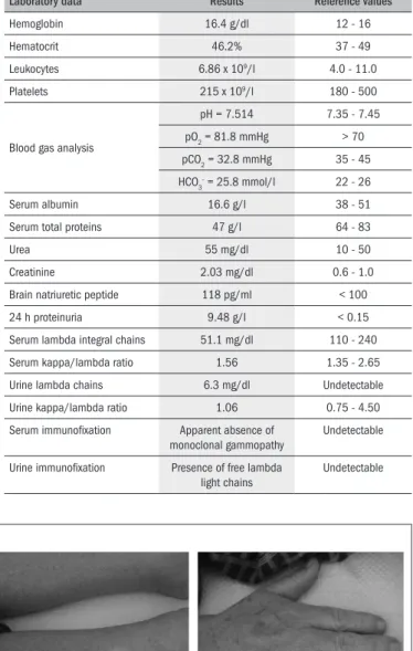
Table 1. Laboratory analysis carried out in August 2009
Laboratory data Results Reference values
Hemoglobin 16.4 g/dl 12 - 16
Hematocrit 46.2% 37 - 49
Leukocytes 6.86 x 10⁹/l 4.0 - 11.0
Platelets 215 x 109/l 180 - 500
Blood gas analysis
pH = 7.514 7.35 - 7.45
pO2 = 81.8 mmHg > 70
pCO2 = 32.8 mmHg 35 - 45
HCO
3
- = 25.8 mmol/l 22 - 26
Serum albumin 16.6 g/l 38 - 51
Serum total proteins 47 g/l 64 - 83
Urea 55 mg/dl 10 - 50
Creatinine 2.03 mg/dl 0.6 - 1.0
Brain natriuretic peptide 118 pg/ml < 100
24 h proteinuria 9.48 g/l < 0.15
Serum lambda integral chains 51.1 mg/dl 110 - 240
Serum kappa/lambda ratio 1.56 1.35 - 2.65
Urine lambda chains 6.3 mg/dl Undetectable
Urine kappa/lambda ratio 1.06 0.75 - 4.50
Serum immunoixation Apparent absence of
monoclonal gammopathy
Undetectable
Urine immunoixation Presence of free lambda
light chains
Undetectable
Figure 1. Clinical features: A and B show macroglossia; C shows the initial edemas; and D shows the upper extremities.
To screen for multiple myeloma, light chain and 24-hour urine immunoixation were performed, and this revealed free lambda light chains. A renal biopsy performed on August 7, 2009, revealed an im-age compatible with positive Congo red indings. Subsequently, under a polarizing optical microscope, apple-green birefringence was identi-ied (Figure 3), thereby diagnosing renal amyloidosis. he patient’s sta-tus improved and he was discharged with reinforcement of the diuretic therapy.
However, on September 1, 2009, the patient was admitted to the hospital again with an episode of kidney failure. An abdominal CT scan was performed to screen for another associated neoplasia, which turned out to be negative. He improved, and was discharged on September 15, 2009. Subsequent to this, he was again admitted, this time in the in-tensive care unit, with anasarca, severe renal failure and orthostatic hy-potension. He started hemodialysis on October 9, 2009, by means of a central venous line. His status evolved to urine and catheter infections, which were treated with adequate antibiotic therapy, with subsequent resolution of the inlammatory markers.
In the second week of October, the diagnosis of lambda light chain amyloidosis was conirmed through immunohistochemistry. he pa-tient was classiied as a high-risk case because he presented stage V renal failure and high basal brain natriuretic peptide at an age old-er than 65 years. He was started on chemothold-erapy with six cycles of bortezomib 0.7 mg/m2 and dexamethasone 40 mg, and his condition
improved, with good tolerance to the medication. After beginning the treatment, he also presented level IV+ hemiparesis in the left up-per arm, which improved after rehabilitation treatment and over the course of the chemotherapy cycles. Currently, the patient is stable and the disease has stagnated.
DISCUSSION
Amyloidosis is an extremely rare entity that is diicult to diagnose. his is because of the unspeciic early clinical manifestations of the
dis-Figure 3. A) Hematoxylin and eosin stain showing the kidney cortex with glomerulus (arrows), microtubules and arterioles; B) showing the detail of Congo red stain (arrow); C) showing Congo red stain (arrows) marking
A
B
C
approximate-ly 10% of all cases. It is considered to be a pathognomonic sign of the disease. However, other causes of macroglossia, like tongue cancer, hy-pothyroidism or vitamin B12 or folic acid deiciency also need to be investigated.2,4,5
Amyloidosis may be systemic or localized. he most common ways in which primary amyloidosis is presented are through nephrotic syndrome (which was initially observed in this case), peripheral neu-ropathy (which was also observed in this case), congestive cardiomyo-pathy and hepatomegaly. Fatigue and weight loss are very common, but usually they only occur when an organ is involved. his patient had complaints compatible with nephrotic syndrome over the two months prior to his irst visit to the Emergency Department. Some patients also present conditions related to multiple myeloma, which was not the case here. his association has a poor prognosis and thera-py is diicult. Renal and heart failure are the major causes of death.2,5,6
Almost every patient with AL amyloidosis has clonal B-cell dyscrasia, but monoclonal protein in the serum or urine is only identiied in 85% of the cases.4
Once a diagnosis of amyloidosis is suspected, it should be con-irmed through biopsy on the afected organ, with histopathological assessment using the Congo red technique. his technique was intro-duced by Bennhold in 1922; the amyloid substance has a red/brown coloration when observed in daylight, but the diagnosis is conirmed by the apple-green birefringence observed under a polarizing optical mi-croscope. Next, to diferentiate the type of amyloidosis, an immuno-histochemical test using monoclonal antibodies for the speciic case of light chains is performed.4,6-10 he presence of monoclonal light chains
in urine and serum is useful, but not always enough to diagnose system-ic disorders, like AL. For all patients, electrophoresis with serum (71% sensitivity) and urine (84% sensitivity) immunoixation needs to be per-formed, in order to attempt to reveal monoclonal light chains.11 In cases
where the results are initially negative, it may be necessary to redo the immunoixation.
Normally, all patients should undergo the serum-free light chain assay to assess disease evolution. Renal involvement is diagnosed through the evidence of renal amyloid deposits detected through bi-opsy, together with laboratory evidence of kidney dysfunction (24 hour proteinuria more than 0.5 g per day; mainly albumin). Heart involvement is identiied through biopsy, in the presence of clinical evidence and changes seen on echocardiogram (like ventricular walls thicker than 12 mm in the absence of hypertension or other potential causes of such thickening). Reduction of the ejection fraction occurs as a late event.9-14
hirty years ago, the treatment for this entity was mainly pallia-tive. New therapies directed towards stabilization of amyloid ibrils have substantially improved these patients’ quality of life. In patients with adequate criteria, stem cell transplantation has shown encourag-ing results in several recent studies, and the ive-year survival rate has been estimated to be approximately 60%.15,16 A systematic search in
major databases (Table 2) found some papers that showed the dii-culties in clinical approaches and the therapeutic implications of im-provements in clinical outcomes.17,18 Other studies have revealed that
despite the new therapeutic and clinical approaches, the survival rate remains unsatisfactory.19,20 he patient in the present report received a
regimen of high doses of steroids (dexamethasone, 40 mg) in associa-tion with a proteosome inhibitor (bortezomib, 0.7 mg/m2), and
de-spite its toxicity, it led the patient to a good response with good tol-erance, and an increase in the patient’s quality of life. Because he pre-sented stage V renal disease with hemodialysis support, age above 65 years and slightly increased basal brain natriuretic peptide, the patient did not fulill the criteria for stem cell transplantation and was clas-siied as high risk. Nonetheless, he beneited from the chemotherapy.
Table 2. Results from systematic search using description of the main clinical features observed in the patient of the present report, among papers indexed in PubMed, Lilacs (Literatura Latino-Americana e do Caribe em Ciências da Saúde), Embase, Scopus and Cochrane Library, from 1982 to 2010
Data Search strategy Results
PubMed Amyloidosis (MeSH) and kidney failure (MeSH) and nephrotic syndrome (MeSH) 57 manuscripts
8 reviews
27 case reports
1 comment/editorial
3 letters
4 comparative study
Lilacs Amyloidosis (DeCS) and kidney failure (DeCS) and nephrotic syndrome (DeCS) 1 case report
Embase “Amyloidosis” and “kidney failure” and “nephrotic syndrome” 161 manuscripts
6 reviews
11 case reports
3 letters
Scopus Amyloidosis (key words) and kidney failure (key words) and nephrotic syndrome (key words) 175 manuscripts
25 reviews
13 letters
1 note
4 short surveys
4 conference papers
Cochrane Amyloidosis 1 review
Currently, the patient’s quality of life is better and the disease has not progressed.
CONCLUSION
Amyloidosis is a disease with serious diagnostic diiculties because it commonly has unspeciic forms of presentation. In fact, many cases are underdiagnosed and this diagnosis is only considered when the pa-tient presents organ failure. herefore, this case alerts general practi-tioners to give more attention to this clinical entity and to the impor-tance of early diagnosis, particularly in patients with decompensated renal failure and severe clinical features of unknown cause that are dif-icult to control. his case consisted of primary amyloidosis, which is a rare entity, and showed involvement of the kidneys as an early presenta-tion of the disease, with diagnostic diiculties. Early diagnosis and treat-ment are helpful in stabilizing the disease, thereby improving the care and the outcome.
REFERENCES
1. Braunwald E, Hauser S, Jameson J, et al. Harrison’s principles of internal medicine. 17th ed. New York: McGraw-Hill; 2008. p. 2145-9.
2. D’Sa S, Abildgaard N, Tighe J, Shaw P, Hall-Craggs M. Guidelines for the use of imaging in the management of myeloma. Br J Haematol. 2007;137(1):49-63.
3. Kyle RA, Gertz MA. Primary systemic amyloidosis: clinical and laboratory features in 474 cases. Semin Hematol. 1995;32(1):45-59.
4. Lachmann HJ, Booth DR, Booth SE, et al. Misdiagnosis of hereditary amyloidosis as AL (primary) amyloidosis. N Eng J Med. 2002;346(23):1786-91.
5. Alambert CO, Sarpi MO, Dedivitis RA, et al. Macroglossia como primeira manifestação clíni-ca da amiloidose primária [Macroglossia as initial cliniclíni-cal manifestation of primary amyloi-dosis]. Rev Bras Reumatol. 2007;47(1):76-9.
6. Melo LV. Amiloidose sistêmica associada a mieloma múltiplo: relato de caso com amiloido-se cutânea exuberante. An Bras Dermatol. 1997;72(2):151-4.
7. Sipe JD, Cohen AS. Review: history of the amyloid ibril. J Struct Biol. 2000;130(2-3): 88-98. 8. Duston MA, Skinner M, Shirahama T, Cohen AS. Diagnosis of amyloidosis by abdominal fat
aspiration. Analysis of four years’ experience. Am J Med. 1987;82(3):412-4.
9. Sezer O, Eucker J, Jakob C, Possinger K. Diagnosis and treatment of AL amyloidosis. Clin Nephrol. 2000;53(6):417-23.
10. Lachmann HJ, Goodman HJ, Gilbertson JA, et al. Natural history and outcome in systemic AA amyloidosis. N Engl J Med. 2007;356(23):2361-71.
11. Katzmann JA, Dispenzieri A, Abraham RS, Kyle RA. Performance of free light chain assays in clinical practice. Blood (ASH Annual Meeting Abstracts). 2004;104:Abstract 757. Available from: http://abstracts.hematologylibrary.org/cgi/content/abstract/104/11/757. Acces-sed in 2011 (Jan 24).
12. Albright R, Brensilver J, Cortell S. Proteinuria in congestive heart failure. Am J Nephrol. 1983;3(5):272-5.
13. Falk RH, Skinner M. The systemic amyloidoses: an overview. Adv Intern Med. 2000;45: 107-37.
14. Hachulla E, Grateau G. Diagnostic tools for amyloidosis. Joint Bone Spine. 2002;69(6): 538-45.
15. Rajkumar SV, Gertz MA. Advances in the treatment of amyloidosis. N Engl J Med. 2007;356(23):2413-5.
16. Goodman HJ, Gillmore JD, Lachmann HJ, et al. Outcome of autologous stem cell transplan-tation for AL amyloidosis in the UK. Br J Haematol. 2006;134(4):417-25.
17. Mazuecos A, Araque A, Sánchez R, et al. Systemic amyloidosis secondary to pyonephrosis. Resolution after nephrectomy. Nephrol Dial Transplant. 1996;11(5):875-8.
18. Pozzi C, Locatelli F. The patient with insidious chronic renal failure and the patient with the nephrotic syndrome--two manifestations of a protean and not so rare disease. Nephrol Dial Transplant. 1996;11(9):1876-80.
19. Snanoudj R, Beaudreuil S, Arzouk N, et al. Recovery from pure red cell aplasia caused by anti-erythropoietin antibodies after kidney transplantation. Am J Transplant. 2004;4(2):274-7. 20. Gatica MA, Bertin CP, Tagle VR. Síndrome de lisis de glóbulos blancos después de un
tras-plante autólogo de células troncales hematopoyéticas en el tratamiento de la amiloidosis AL renal: Caso clínico [White blood cell lysis syndrome after autologous peripheral blood stem cell transplantation in the treatment of renal AL amyloidosis: Case report]. Rev Méd Chile. 2006;134(6):763-6.
Sources of funding: None
Conlict of interest: None
Date of irst submission: May 3, 2010
Last received: December 19, 2010
Accepted: January 31, 2011
Address for correspondence:
Ramon Andrade Bezerra de Mello
Faculdade de Medicina da Universidade do Porto Departamento de Medicina. Disciplina de Semiótica Clínica.
Alameda Professor Hernani Monteiro, s/no 4200-319, Porto, Portugal