Objective:To evaluate the impact of therapy on bone mineral density (BMD) and body composition in survivors of acute lymphoblastic leukemia (ALL) treated in accordance with Brazilian protocols by the Brazilian Cooperative Group of Treatment of Lymphoblastic Leukemia in Childhood (GBTLI) LLA-93 and LLA-99. Methods:A cross-sectional study with 101 patients was performed. BMD and body composition were evaluated using bone densitometry and were interpreted according to the age group and the reference population. Values between -1.1 and -1.9 in the group of children under 20 years were considered as risk group for low BMD z-scores. BMD values were compared to clinical characteristics, treatment received and body composition. A chi-square test, Fisher’s exact test, likelihood ratio and Student’s t-test were applied, with a 5% signiicance level.
Results:The patients presented a frequency of fractures of 2%, of osteonecrosis, 2%, and of low BMD, 2.9%. In the group of 79 patients under 20 years of age, three had low BMD. The 16 that presented risk for low BMD, demonstrated lower valutes in lumbar vertebrae L1–L4 (p=0.01) and whole body (p=0.005), and smaller values of lean body mass (p=0.03). In the group of 22 patients over 20 years of age, ten had osteopenia.
Conclusions: The low impact of treatment on BMD of this study conirms the concept that the bone mass gain occurs with increasing age and that the treatment does not inluence the process. The population at risk for low BMD values presented lower bone
Objetivo:Avaliar o impacto da terapia sobre a densidade mineral óssea (DMO) e composição corporal em sobreviventes da leucemia linfoide aguda (LLA), tratados de acordo com os protocolos brasileiros do Grupo Cooperativo Brasileiro de Tratamento de Leucemia Linfoide Aguda na Infância (GBTLI), LLA-93 e LLA-99. Métodos:Em estudo transversal com 101 pacientes, avaliaram-se a composição corporal e a DMO por meio da densitometria óssea, interpretando-a conforme a faixa etária e a população de referência. Foi considerado grupo de risco para baixa DMO valores de z-escore entre -1,1 e -1,9 no grupo dos menores de 20 anos. Compararam-se os valores da DMO com características clínicas, tratamento recebido e composição corporal. Foram utilizados os testes qui-quadrado, exato de Fisher, razão de verossimilhança e t de Student, com nível de signiicância de 5%.
Resultados:Foram encontradas 2% de fraturas, 2% de osteonecrose e 2,9% de baixa DMO. No grupo de pacientes com menos de 20 anos, três apresentaram baixa DMO. Os 16 pacientes com risco para baixa DMO exibiram menores valores em vértebras lombares L1–L4 (p=0,01), corpo total (p=0,005) e valores mais baixos de massa magra (p=0,03). No grupo de 22 pacientes com mais de 20 anos, dez demonstraram osteopenia. Conclusões: O baixo impacto do tratamento sobre a DMO neste estudo ratiica o conceito de que o ganho de massa óssea ocorre com o aumento da idade e que o tratamento não inluencia tal processo. A população de risco para baixa DMO demonstrou valores menores
ABSTRACT
RESUMO
*Corresponding author. E-mail: [email protected] (P.C.C. Molinari).
aInstitute of Pediatric Oncology, Child and Adolescent Cancer Support Group (IOP/GRAACC), Universidade Federal de São Paulo (UNIFESP), São Paulo, SP, Brazil.
bDepartment of Radiology, IOP/GRAACC/UNIFESP, São Paulo, SP, Brazil.
Received on February 25, 2016; approved on August 19, 2016; available online on January 27, 2017.
ASSESSMENT OF THE LATE EFFECTS ON BONES
AND ON BODY COMPOSITION OF CHILDREN AND
ADOLESCENTS TREATED FOR ACUTE LYMPHOCYTIC
LEUKEMIA ACCORDING TO BRAZILIAN PROTOCOLS
Avaliação dos efeitos ósseos tardios e composição corporal de
crianças e adolescentes tratados de leucemia linfoide aguda
segundo protocolos brasileiros
Poliana Cristina Carmona Molinari
a,*, Henrique Manoel Lederman
b,
79
INTRODUCTION
he therapeutic progress obtained in the treatment of children with acute lymphocytic leukemia (ALL) over the past 50 years has resulted in cure rates of around 80%, culminating in an impactful increase in the number of survivors. his population represents a group with increased risk for comorbidity related to treatment and the disease itself.1.2 Changes in bone metab-olism and body composition are considered important adverse late efects and represent a signiicant cause of morbidity in this population, through pain, fractures, decrease of bone mineral density (BMD) and chronic impairment of bone function.3.4 Exposure to corticosteroids, methotrexate, mercaptopurine and radiation, associated with low calcium intake, decreased physical activity, and obesity are some of the factors that lead to low BMD.5-7 Although patients treated for ALL can recover lost bone mass during the post-treatment period, a percent-age of them will not reach their maximum BMD acquisition potential, presenting signiicant bone deicit.1
Bone densitometry is the method of choice for evaluating bone mineral density and body composition. However, it must be performed and interpreted in accordance with the pediatric references for each population, whose norms have been previ-ously published.8-10
As such, the goal of this study was to evaluate the impact of chemotherapy and radiotherapy as adverse late efects of BMD in children and adolescents treated for ALL in accor-dance with Brazilian protocols, through the use of dual-energy X-ray absorptiometry (DXA).
METHOD
his is a cross-sectional retrospective study with a convenience sample that included 242 patients with ALL that had been treated in accordance with the Brazilian Cooperative Group of Treatment of Lymphoblastic Leukemia in Childhood (Grupo Cooperativo Brasileiro para Tratamento da LLA na Infância – GBTLI), protocols LLA-93 and LLA-99. hese patients were treated at the Institute of Pediatric Oncology (IOP) of the
Child and Adolescent Cancer Support Group (GRAACC) at the
Universidade Federal de São Paulo (UNIFESP), São Paulo (SP), Brazil.
he study included patients from both sexes who received such treatment from 1994 to 2006 and were over 5 years old at the time of data collection. At the time of the study, they were in regular clinical follow-up at the outpatient clinic and at pediatric endocrinology. All of the patients were in the irst complete clinical remission, which is deined as the lack of dis-ease in peripheral blood, in bone marrow, and in extramedullary compartments such as spinal luid or testicles. Patients in the following situations were excluded from the study: those with concurrent chronic conditions that could afect bone growth (including kidney or liver disease, as well as other immuno-logical and endocrinoimmuno-logical diseases); those who had not been under regular monitoring at the outclinics mentioned above for at least three years; those with physical disabilities; smok-ers and alcoholics; recipients of bone marrow or solid organ transplants; pregnant patients; those presenting recurrence of ALL during or after treatment; those with Down syndrome and those who had made use of exogenous growth hormones in the past two years. Finally, patients under the following conditions were removed from the study: those who did not return for the examinations or who decided to abandon the study and those who could not be reached by phone or by letter due to outdated contact information. his population was not statis-tically signiicant if compared to the population investigated. his study was approved by the Research Ethics Committee of the Medical School at the Universidade Federal de São Paulo (UNIFESP) under number 1.618/10. Patients and their par-ents (or legal guardians) were informed about the study and its goals, and agreed to sign an informed consent form and an agreement form.
Data collection took place between February 2011 and January 2013 and included the following information: demo-graphic data, previous history of the disease and of cancer treat-ment (including the patients’ risk rating and whether they had undergone cranial radiotherapy, accumulated doses of predni-sone, dexamethasone and chemotherapeutic agents), laboratory proile (creatinine serum, calcium, phosphorus, magnesium,
mass values and could beneit from a long-term monitoring for possible bone toxicity.
Keywords: Leukemia, lymphoblastic; Bone density/drug therapy; Bone mineral density/radiation efects.
de massa óssea, podendo beneiciar-se de um acompanhamento em longo prazo para uma possível toxicidade óssea.
80
alkaline phosphatase, thyroid and parathyroid hormones and vitamin D) and bone densitometry.
Demographic data included date of birth, current age and age at diagnosis, sex, race and time elapsed after treatment. Clinical characteristics related to the disease and treatment were obtained through a review of medical records, namely: symptoms of bone involvement at diagnosis; initial leukocyte count; immunophenotypical classiication of ALL; occurrence of central nervous system (CNS) involvement at diagnosis; chemotherapy protocol received; risk classiication; cumulative doses of prednisone, dexamethasone, methotrexate, alkylat-ing agents and mercaptopurine; need for prophylactic radio-therapy; presence of bone toxicity during treatment; and time outside of therapy.
Anthropometric and pubertal development data was obtained during the physical examination at the outpatient endocrinol-ogy clinic. A mechanical balance (Filizola®
) was used to measure weight, and height was measured using a wall-mounted stadi-ometer (Tonelli®
), allowing for the computation of the patients’ body mass index (BMI). Nutritional diagnosis was made based on the parameters of the World Health Organization (WHO), through BMI values.
BMD measurement was performed at the anteroposterior projection on the lumbar spine segment L1-L4, whole body and femur, through DXA, using the Lunar DPX (GE Lunar Corporation®
) instrument. All of the tests were analyzed by a single radiologist through the Encore software and interpreted according to age, sex and the software’s reference database. For patients under 20 years old, z-score levels under -2 standard deviations (SD) were considered as having low BMD values. Hypothetically, values between -1.9 and -1.1, conceptualized as normal by the literature, were considered as being at risk for low BMD. For patients above 20 years old, the T-score was calculated and compared with patterns of young adults. In this population, a T-score lower than -2.5 SD was considered as a case of osteoporosis. In the same fashion, a T-score between -1 and -2.5 SD indicated osteopenia. BMD values were matched against clinical characteristics and treatment, in addition to body composition.
Data was stored in spreadsheets, and all statistical analyses were performed through the use of the Statistical Package for Social Sciences (SPSS version 15.0) for Microsoft Windows. he following were treated as independent variables: gender, race, age at diagnosis, current age, time outside therapy, ini-tial leukocyte count, CNS iniltration at time of diagnosis, immunophenotyping, risk classiication, treatment received, cumulative doses of chemotherapy and corticosteroids, pubertal stage of Tanner, anthropometric data, body composition, and BMD values and their SD values. BMD classiication values
were treated as dependent variables: Normal BMD, low BMD and risk of low BMD for patients under 20 years; and normal BMD, osteopenia and osteoporosis for those over 20 years of age. Quantitative variables were expressed through arithme-tic means and SD values. he matching of BMD values with categorical variables was performed using the chi-square test, Fisher’s exact test and the likelihood ratio. he matching of BMD values with numerical variables was performed using
t-tests. he signiicance level was set at 5%.
RESULTS
Out of the 242 patients treated for ALL, 76 died due to various causes and 65 of them were excluded from the study due to one or more predetermined criteria. his led to a total of 101 patients, 59.4% of which were female and 77.2% were white. he mean age of the patients was 17.2±4.9 years. With regard to
character-istics at the time of diagnosis, the mean age was 5.2 ± 3.6 years,
and 94% of patients had an immunophenotypical classiication of precursor B-cell ALL. 78.2% of the patients presented an ini-tial leukocyte count lower than 50,000 cells/mm3, and CNS iniltration was diagnosed in nine patients.
he GBTLI LLA-93 protocol was used in 44 patients and the GBTLI LLA-99 in 57 patients, 54.5% of which were clas-siied as low risk. Treatment-related characteristics are shown in Table 1. All laboratory proiles showed values in agreement with the reference parameters for calcium, phosphorus, mag-nesium, serum creatinine and thyroid hormones; 16 patients presented alkaline phosphatase values above the reference for their age and gender; and 23 (23.4%) presented vitamin D values lower than 20 ng/dL.
With regard to the nutritional diagnosis, 22.8% of patients were considered overweight and 15.8% were considered obese, and there was no correlation between these conditions and previous exposure to radiation (p = 0.28). Body composition distribution showed that the lean mass levels and bone min-eral content were higher in males, and fat mass levels and fat percentages were higher in females. Patients who had spent more time from the end of their treatment up to the time of the study had higher values of lean body mass, fat mass and percentage of fat and bone mineral content.
During the research, we observed two patients with osteo-necrosis during treatment and two others who sufered a frac-ture of the forearm and tibia also in the same period.
81
the end of treatment up to the study. Two patients were classi-ied as having common ALL and one as pre-B ALL; two were treated with protocol GBTLI LLA-93 and one with GBTLI LLA-99; two were considered as having low risk of relapse and one with high risk; no patient had iniltration of the CNS. he mean dose accumulated from chemotherapeutic agents and corticosteroids was: prednisone 2240±1584 mg/m2, dexa-methasone 262±28 mg/m2, methotrexate 11713±1635 mg/m2, alkylating agents 2000 mg/m2 and 6-mercaptopurine (6-MP) of 32118-5007±5971 mg/m2; one patient was submitted to cranial radiotherapy with a dosage of 18 Gy. Two patients pre-sented low BMD at the spine segment L1-L4 and one whole body BMD. As for body composition, the mean bone mineral content (BMC) level was 1757±376 g, while the mean fat
per-centage was 28.8±15.3%. Average fat mass was 13.6±8.8 kg
and average lean mass was 30.6±3.3 kg.
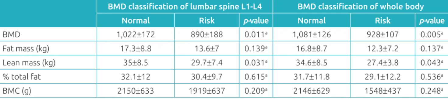
Comparing the normal BMD group with the group at risk for low BMD, there was no signiicant diference linked to gen-der, race, age at diagnosis, current age, time outside therapy and pubertal stage nor to characteristics of the disease such as immunophenotype, initial leukocyte count, involvement of the central nervous system, risk of recurrence, chemotherapy treat-ment received, accumulated doses of prednisone, dexameth-asone, methotrexate, alkylating agents, 6-MP and exposure to radiation therapy (Table 2). When matching lumbar spine segment L1-L4 and whole body BMD values with body com-position for both groups, a signiicant diference was found in lean mass, which was lower in the group at risk for low lum-bar spine L1-L4 and whole body BMD (p=0.043); no such
diference was found with relation to other body composition variables. (Table 3).
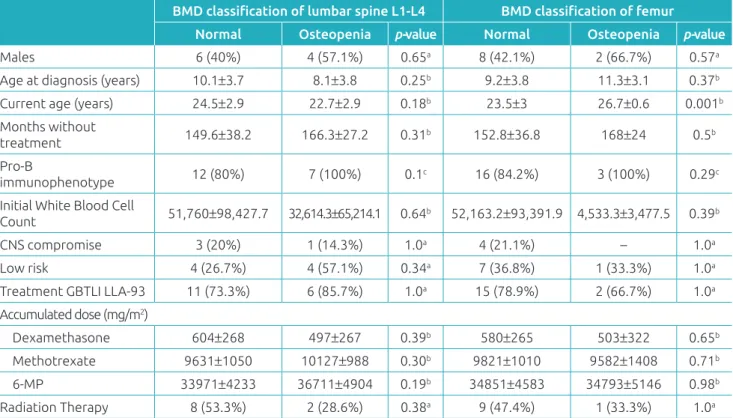
Of the 22 patients over the age of 20, eight (7.9%) had osteo-penia, and there was no diagnosis of osteoporosis. When clini-cal characteristics of the normal BMD group and those of the osteopenia group were compared, there was no signiicant dif-ference linked to gender, race, age at diagnosis, time without therapy, immunophenotype, initial leukocyte, involvement of the central nervous system, risk of recurrence, chemotherapy treatment received, accumulated doses by chemotherapeutic treatment of dexamethasone, methotrexate and 6-MP or com-pletion of radiation therapy (Table 4). he group that presented femoral BMD osteopenia was older than the group with nor-mal BMD (p=0.001) (Table 5).
DISCUSSION
he majority of patients in this study had normal bone min-eral density values in comparison to the reference population. his demonstrates that bone mass gain occurs with increasing age and that treatment does not inluence the process.9.11 In our research, three patients had low BMD. his inding varies 5–85% for populations of the same age and gender in several studies, and depends on the characteristics of the patients, the method used for BMD measurement and the way it is interpreted.12.13
he irst two years after the end of ALL treatment is the most critical period for bone loss, with recovery and progressive increase of bone mass taking place after that time.13 herefore, data suggests that this population had no risk for the loss of
Table 1 Characteristics of the treatment of lymphoblastic leukemia according to the protocols of the Brazilian
Cooperative Group of Treatment of Lymphoblastic Leukemia in Childhood (GBTLI), LLA-93 and LLA-99.
Standard Deviation GBTLI LLA-93 GBTLI LLA-99
n=44 % n=57 %
Sex: Male 17 38.6 24 42.1
Age at diagnosis (years)a 5.1±3.3 5.4±3.8
Months withouth treatmenta 154.6±26.5 89.9±20.8
Risk: low risk 24 54.5 31 54.4
Prophylactic radiotherapy (12 Gy) 17 38.6 1 1.7
Radiation therapy (24 Gy) 3 6.8 3 5.3
Accumulated dosesa
Prednisone 1,169±160
Dexamethasone 597±265 268±63
Methotrexate 10,373±331 9,879±2,150
Alkylating Agents – 6,197±4,623
6-mercaptopurine 36,145±4,936 32,898±539
82
BMD. Some studies showed similar results: the larger the elapsed time between the end of therapy and the younger the patient at the beginning of treatment, the greater the chance to recover the loss of BMD and acquire adequate bone mass.14.15 he clinical implication of low BMD in these patients is still uncertain, especially as it pertains to the risk of fractures.16
he frequency of low BMD in this study was 2%, compared to the prevalence of 11–18.5% of fractures in the literature.17.18 In the same fashion, a lower prevalence of osteonecrosis was found in our study.19.20 hese results are attributed to possible subdiagnoses and the absence of active search for osteonecro-sis in asymptomatic patients.
Table 2 Rating distribution of the bone mineral density of whole body and lumbar spine L1-L4 of normal and risk
groups of patients younger than 20 years old, regarding clinical features and related to treatment. Lumbar spine L1-L4 BMD Whole body BMD
Normal Risk p-value Normal Risk p-value
Males 25 (40.3%) 5 (33.3%) 0.83a 28 (38.9%) 3 (50%) 0.67c
Age at diagnosis (years) 4.0±2.6 4.4±2.6 0.62b 4.3±2.6 2.7±1.0 0.54b Current age (years) 15.0±2.9 15.5±3.4 0.61c 15.1±3.0 14.7±3.1 0.72c Months without treatment 107±33 111±44 0.68b 106±33 120±44 0.94a Pubertal stage: pubertal 57 (91.9%) 13 (86.7%) 0.61c 66 (91.6%) 5 (83.3%) 0.44c Pro-B
immunophenotype 60 (96.7%) 14 (93.3%) 0.56d 69 (95.8%) 6 (100%) 0.49d Initial White Blood Cell
Count 33,049±47,097 33,767±53,261 0.95b 28,532±41,732 54,717±63,659 0.39b
CNS compromised 5 (8.1%) – 0.57c 5 (6.9%) – 1.0c
Low risk 36 (58.1%) 9 (60%) 1.0a 43 (59.7%) 4 (66.7%) 1.0c
Treatment
GBTLI LLA-93 20 (32.2%) 6 (40%)
0.79a 23 (31.9%) 3 (50%) 0.39c
GBTLI LLA-99 42 (67.7%) 9 (60%) 49 (68.1%) 3 (50%)
Accumulated dose (mg/m2)
Prednisone 1147±121 1244±247 0.27b 1154±136 1307±323 0.27b
Dexamethasone 358±202 416±290 0.47b 353±201 446±333 0.69b
MTX 10197±1911 9842±765 0.26b 10141±1817 10656±1443 0.25b
Alkylates 6162±4635 4044±4057 0.19b 5943±4600 2000 0.19b
6-MP 34091±3461 33447±3070 0.51b 34122±3423 33338±3702 0.65b Radiation Therapy 10 (16.1%) 4 (26.7%) 0.45c 11 (15.3%) 2 (33.3%) 0.26a BMD: bone mineral density; CNS: central nervous system; GBTLI LLA: Brazilian Cooperative Group of Treatment of Lymphoblastic Leukemia in Childhood; MTX: methotrexate; 6-MP: 6-mercaptopurine; achi-square test; bt test; cFishers exact test; dlikelihood ratio test; p<0.05.
Table 3 Rating distribution of the bone mineral density of whole body and lumbar spine L1-L4 and body composition
in relation to normal and risk groups in patients younger than 20 years old.
BMD classiication of lumbar spine L1-L4 BMD classiication of whole body
Normal Risk p-value Normal Risk p-value
BMD 1,022±172 890±188 0.011a 1,081±126 928±107 0.005a
Fat mass (kg) 17.3±8.8 13.6±7 0.139a 16.8±8.7 12.3±7.2 0.137a Lean mass (kg) 35±8.5 29.7±7.4 0.031a 34.6±8.5 27.4±3.8 0.043a % total fat 32.1±12 30.4±9.7 0.615a 31.7±11.8 29.1±12.2 0.536a
BMC (g) 2150±633 1919±637 0.209a 2146±629 1548±437 0.248a
at test; BMD: bone mineral density; p<0.05; BMC: bone mineral content; normal: values of z-score less than or equal to -2; risk: values of z-score
83
Table 4 Distribution of the classiication of bone mineral density of the whole body and femoral head of normal and
osteopenia groups in patients aged 20 years or more in relation to clinical characteristics and related to treatment.
BMD classiication of lumbar spine L1-L4 BMD classiication of femur
Normal Osteopenia p-value Normal Osteopenia p-value
Males 6 (40%) 4 (57.1%) 0.65a 8 (42.1%) 2 (66.7%) 0.57a
Age at diagnosis (years) 10.1±3.7 8.1±3.8 0.25b 9.2±3.8 11.3±3.1 0.37b Current age (years) 24.5±2.9 22.7±2.9 0.18b 23.5±3 26.7±0.6 0.001b Months without
treatment 149.6±38.2 166.3±27.2 0.31b 152.8±36.8 168±24 0.5b
Pro-B
immunophenotype 12 (80%) 7 (100%) 0.1c 16 (84.2%) 3 (100%) 0.29c
Initial White Blood Cell
Count 51,760±98,427.7 32,614.3±65,214.1 0.64b 52,163.2±93,391.9 4,533.3±3,477.5 0.39b
CNS compromise 3 (20%) 1 (14.3%) 1.0a 4 (21.1%) – 1.0a
Low risk 4 (26.7%) 4 (57.1%) 0.34a 7 (36.8%) 1 (33.3%) 1.0a
Treatment GBTLI LLA-93 11 (73.3%) 6 (85.7%) 1.0a 15 (78.9%) 2 (66.7%) 1.0a Accumulated dose (mg/m2)
Dexamethasone 604±268 497±267 0.39b 580±265 503±322 0.65b
Methotrexate 9631±1050 10127±988 0.30b 9821±1010 9582±1408 0.71b 6-MP 33971±4233 36711±4904 0.19b 34851±4583 34793±5146 0.98b
Radiation Therapy 8 (53.3%) 2 (28.6%) 0.38a 9 (47.4%) 1 (33.3%) 1.0a
BMD: bone mineral density;p<0.05; ALL: acute lymphocytic leukemia; CNS: central nervous system; GBTLI LLA: Brazilian Cooperative Group of Treatment of Lymphoblastic Leukemia in Childhood; 6-MP: 6-mercaptopurine; aFishers exact test; bt test; clikelihood ratio test.
Table 5 Distribution of the classiication of bone mineral density of the lumbar spine L1-L4 and femur and body
composition in relation to normal and osteopenia groups in patients aged 20 years or more.
BMD classiication of lumbar spine L1-L4 BMD classiication of femur
Normal Osteopenia p-value Normal Osteopenia p-value
BMD CL L1-L4 1,243±71 1,051±29 <0.001a – – –
Femoral BMD – – – 1.1±0.1 0.9±0.1 0.48a
Fat mass (kg) 28.0±15.5 17.9±12.8 0.15a 25.0±16.0 23.3±10.9 0.86a Lean mass (kg) 41.7±9.1 42.2±8.9 0.89a 41.6±8.6 43.8±1.8 0.69a
% total fat 37.6±13.9 28±15.7 0.15a 34.7±15.8 33.7±5.0 0.91a
BMC (g) 2741±345 2458±420 0.10a 2652±364 2656±601 0.98a
BMD: bone mineral density; CL L1-L4: lumber spine, vertebrae L1-L4; BMC: bone mineral content; at test; p<0.05. Vitamin D levels were below normal in 23 patients
(23.4%). Some analyses with Brazilian children and adoles-cents showed that 60-70% of them had insuicient levels of vitamin D, with inadequate intake of this vitamin and cal-cium. herefore, the lack of these nutrients may not be related to the treatment received.21-24 However, some studies have shown that ALL treatment increases the incidence of vita-min D deiciency, which potentially increases the risk of low bone mineral density.25
Although current densitometry standards deine low BMD (for people under 20), as that which presents z-scores under
84
With regard to to body composition, in that same group, there was also a diference between the amount of lean mass and lumbar spine and whole body BMD. Another study with a similar population also found this diference.27 his reinforces the fact that lean mass is one of the most important predic-tive factors of bone mass.16 ALL treatment may cause changes in body composition, such as obesity, decrease in lean body mass, growth deicits and changes in BMD. he relationship between bone mass, anthropometric data and lean mass may vary according to age, gender and growth rate, as well as nutri-tional status, often measured by the BMI, which is also a deter-minant of bone mass.16
We also analysed patients older than 20 years of age in con-trast with the group with osteopenia and with the group with normal lumbar spine and femoral BMD. here were no difer-ences found related to the disease or to the accumulated doses of corticosteroids and chemotherapy agents related to lumbar spine L1-L4 and femoral head BMD. Only the average lumbar spine L1-L4 BMD was signiicantly lower in the osteopenia group in comparison to the normal BMD group. his may be due to the fact that the lumbar spine vertebrae, which are more sensitive to toxic bone factors, are composed of trabecular bone, more metabolically active than cortical bone. he basis for the establishment of bone mass in adults is initiated in childhood and adolescence. Bone metabolism and BMD development are inluenced by countless factors. Any interference in this pro-cess causes a deicit in the acquisition of bone mass, impairing the acquisition of peak bone mass, an important determinant of the risk of fractures in adulthood.28
For the irst time, the bone toxicity of patients treated for ALL was analysed in accordance with two Brazilian protocols coming from a single institution. Understanding the reality of Brazilian patients treated for childhood ALL with regard to late bone toxicity allows for the development of a longitudinal follow-up focused on this population, with its peculiar charac-teristics, providing good opportunity for future interventions. In Brazil, so far, there is no national guideline for tracing late efects of antineoplastic treatment, or their impact on BMD. Such a guideline would provide subsidies for the creation of risk scores based on characteristics of the Brazilian ALL survivor
population, with respect to the treatment received and the bone development commonly associated with our population. his research had some limitations: there was a signiicant loss of patients - 53% died and the rest were excluded by crite-ria), with a decrease in the study sample and a possible impact on the results presented. As this was a cross-sectional study, it was not possible to verify the association of cause and efect between the bone toxicity and ALL treatment. Furthermore, the software used had no reference data based on the Brazilian pediatric population with speciic BMD values for compari-son. Finally, there was no healthy children population available as a control for data comparison, and thus we were not able to determine whether there were other factors that inluence bone toxicity other than exposure to treatment.
It can be concluded that the impact of the treatment in accor-dance with Brazilian protocols GBTLI LLA-93 and LLA-99 on survivors’ BMD in the long term was low, and 2.9% of patients had low BMD.
hrough this study, it was possible to deine a risk group for low BMD, made up of 15.8% of the patients, whose values of whole body and lumbar spine L1-L4 BMD were signiicantly lower than the population studied, values that are in fact con-sidered normal (below 2 SD) in the literature. his group may be beneit from preventive actions against bone mass loss and the development of protocols for longitudinal follow-up and detection of bone toxicity. In the population studied, the occur-rence of fractures (2%) and osteonecrosis (2%) was uncommon, however no active search for osteonecrosis was performed on asymptomatic patients. Lean mass was related to whole body and lumbar spine L1-L4 BMD values in patients under 20 years of age. he higher the values of lean mass, the greater the BMD. he present study has suggested a positive association between BMD and lean mass, conirming its importance in the development of bone mass.
Funding
his study did not receive funding.
Conflict of interests
he authors declare no conlict of interests.
REFERENCES
1. Wasilewski-Masker K, Kaste SC, Hudson MM, Esiashvili N, Mattano LA, Meacham LR, et al. Bone mineral density deficits in survivors of childhood cancer: long-term follow-up guidelines and review of the literature. Pediatrics. 2008;121:e705-13.
85 3. Haddy TB, Mosher RB, Reaman GH. Osteoporosis in survivors
of acute lymphoblastic leukemia. Oncologist. 2001;6:278-85.
4. Davies JH, Evans BA, Jenney ME, Gregory JW. Skeletal morbidity in childhood acute lymphoblastic leukaemia. Clin Endocrinol (Oxf). 2005;63:1-9.
5. Tragiannidis A, Dokos CH, Sidi V, Papageorgiou T, Koliouskas D, Karamouzis M, et al. Alterations of bone mineral metabolism of children with diferent cell lineage types of acute lymphoblastic leukaemia under chemotherapy. Hippokratia. 2011;15:43-7.
6. Kohler JA, Moon RJ, Sands R, Doherty LJ, Taylor PA, Cooper C, et al. Selective reduction in trabecular volumetric bone mineral density during treatment for childhood acute lymphoblastic leukemia. Bone. 2012;51:765-70.
7. Watsky MA, Carbone LD, An Q, Cheng C, Lovorn EA, Hudson MM, et al. Bone turnover in long-term survivors of childhood acute lymphoblastic leukemia. Pediatr Blood Cancer. 2014;61:1451-6.
8. Gordon CM, Leonard MB, Zemel BF, International Society for Clinical Densitometry. 2013 Pediatric Position Development Conference: executive summary and reflection. J Clin Densitom. 2014;17:219-24.
9. Ma NS, Gordon CM. Pediatric osteoporosis: where are we now? J Pediatr. 2012;161:983-90.
10. Bianchi ML, Baim S, Bishop NJ, Gordon CM, Hans DB, Langman CB, et al. Oicial positions of the International Society for Clinical Densitometry (ISCD) on DXA evaluation in children and adolescents. Pediatr Nephrol. 2010;25:37-47.
11. Silva CC, Goldberg TB, Nga HS, Kurokawa CS, Capela RC, Teixeira AS, et al. Impact of skeletal maturation on bone metabolism biomarkers and bone mineral density in healthy Brazilian male adolescents. J Pediatr (Rio J). 2011;87:450-6.
12. Kaste SC, Rai SN, Fleming K, McCammon EA, Tylavsky FA, Danish RK, et al. Changes in bone mineral density in survivors of childhood acute lymphoblastic leukemia. Pediatr Blood Cancer. 2006;46:77-87.
13. Muszynska-Roslan K, Konstantynowicz J, Krawczuk-Rybak M, Protas P. Body composition and bone mass in survivors of childhood cancer. Pediatr Blood Cancer. 2007;48:200-4.
14. Mandel K, Atkinson S, Barr RD, Pencharz P. Skeletal morbidity in childhood acute lymphoblastic leukemia. J Clin Oncol. 2004;22:1215-21.
15. Marinovic D, Dorgeret S, Lescoeur B, Alberti C, Noel M, Czernichow P, et al. Improvement in bone mineral density and body composition in survivors of childhood acute lymphoblastic leukemia: a 1-year prospective study. Pediatrics. 2005;116:e102-8.
16. Chaiban J, Muwakkit S, Arabi A, Jomaa L, Daouk LO, El-Rassi R, et al. Modeling pathways for low bone mass in children with malignancies. J Clin Densitom. 2009;12:441-9.
17. Högler W, Wehl G, van Staa T, Meister B, Klein-Franke A, Kropshofer G. Incidence of skeletal complications during treatment of childhood acute lymphoblastic leukemia: comparison of fracture risk with the General Practice Research Database. Pediatr Blood Cancer. 2007;48:21-7.
18. Rayar MS, Nayiager T, Webber CE, Barr RD, Athale UH. Predictors of bony morbidity in children with acute lymphoblastic leukemia. Pediatr Blood Cancer. 2012;59:77-82.
19. te Winkel ML, de Muinck Keizer-Schrama SM, de Jonge R, van Beek RD, van der Sluis IM, Hop WC, et al. Germline variation in the MTHFR and MTRR genes determines the nadir of bone density in pediatric acute lymphoblastic leukemia: a prospective study. Bone. 2011;48:571-7.
20. Strauss AJ, Su JT, Dalton VM, Gelber RD, Sallan SE, Silverman LB. Bony morbidity in children treated for acute lymphoblastic leukemia. J Clin Oncol. 2001;19:3066-72.
21. Peters BS, dos Santos LC, Fisberg M, Wood RJ, Martini LA. Prevalence of vitamin D insuiciency in Brazilian adolescents. Ann Nutr Metab. 2009;54:15-21.
22. Oliveira RM, Novaes JF, Azeredo LM, Cândido AP, Leite IC. Association of vitamin D insuiciency with adiposity and metabolic disorders in Brazilian adolescents. Public Health Nutr. 2014;17:787-94.
23. Bueno MB, Fisberg RM, Maximino P, Rodrigues GP, Fisberg M. Nutritional risk among Brazilian children 2 to 6 years old: a multicenter study. Nutrition. 2013:29:405-10.
24. Martini LA, Verly Jr. E, Marchioni DM, Fisberg RM. Prevalence and correlates of calcium and vitamin D status adequacy in adolescents, adults, and elderly from the Health Survey-São Paulo. Nutrition. 2013;29:845-50.
25. Reisi N, Iravani P, Raeissi P, Kelishadi R. Vitamin D and bone mineral status in the long-term survivors of childhood acute lymphoblastic leukemia. Int J Prev Med. 2015;6:87.
26. Jarfelt M, Fors H, Lannering B, Bjarnason R. Bone mineral density and bone turnover in young adult survivors of childhood acute lymphoblastic leukaemia. Eur J Endocrinol. 2006;154:303-9.
27. Siviero-Miachon AA, Spinola-Castro AM, Lee ML, Andreoni S, Geloneze B, Lederman H, et al. Cranial radiotherapy predisposes to abdominal adiposity in survivors of childhood acute lymphocytic leukemia. Radiat Oncol. 2013;8:39.
28. Borges JL, Brandão CM. Low bone mass in children and adolescents. Arq Bras Endocrinol Metab. 2006;50:775-82.