ABSTRACT
http://dx.doi.org/10.1590/1678-775720160139
The effect of
hippophae rham noides
extract on
oral mucositis induced in rats with methotrexate
Ozan KUDUBAN1, Muhammed Recai MAZLUMOGLU2, Selma Denktas KUDUBAN3, Ertugrul ERHAN41LKDO&(7ø15,
Osman KUKULA6, Oguzhan YARALI7, Ferda Keskin CIMEN8, Murat CANKAYA9
1- Erzurum Education and Research Hospital, Ear Nose and Throat Head&Neck Surgery Clinic, Erzurum, Turkey. 2- Hinis State Hospital, Department of Otorhinolaryngology, Erzurum, Turkey.
3- Palandoken State Hospital, Department of Plastic Reconstructive and Esthetic Surgery, Erzurum, Turkey. 4- Erzincan University, Faculty of Medicine, Department of Otorhinolaryngology, Erzincan, Turkey. 5- Erzincan University, Faculty of Medicine, Department of Pharmacology, Erzincan, Turkey. 6- Ondokuzmayis University, Faculty of Medicine, Department of Pharmacology, Samsun, Turkey. 7- Erzurum Training and Research Hospital, Department of Medical Genetics, Erzurum, Turkey. 8- Mengucek Gazi Education and Research Hospital, Department of Pathology, Erzincan, Turkey. 9- Erzincan University, Faculty of Arts and Sciences, Department of Biology, Erzincan, Turkey.
Corresponding address: Nihal Cetin - Department of Pharmacology, Faculty of Medicine - Erzincan University, 24030 - Erzincan - Turkey - Phone: +90 5556307638 - Fax: +90 446 2261819 - e-mail: [email protected]
6XEPLWWHG0DUFK0RGL¿FDWLRQ0D\$FFHSWHG0D\
O
bjective: To investigate the effect of HRE (Hippophae rham noides extract) on oralmucositis induced in rats with MTX. Material and Methods: Experimental animals were divided into groups as healthy (HG), HRE+MTX (HMTX), and control group, which received MTX (MTXC). HMTX group received 50 mg/kg HRE while MTXC and HG groups received equivolume distilled water with gavage once a day. After one hour of HRE and distilled water administration, HMTX and MTXC groups received a single dose of oral MTX 5 mg/
TNF-D
animals receiving MTX, compared with HG and HMTX groups; however, these parameters were lower in the cheek and low lip tissue, and a milder damage ocurred in these tissues, compared with the tongue tissue in MTXC group. No histopathologic damage was observed in the cheek, lower lip, and tongue tissues of the rats treated with HRE. Conclusion: This
synthetic drugs for prophylaxis of oral mucositis developed due to MTX.
Ke yw or ds: Gene expression. Hippophae rham noides. Methotrexate. Oral mucositis. Rats.
I N TROD UCTI ON
Methotrexate (MTX) is an antiproliferative folic acid antagonist used in the treatment of various
and high doses are used to treat malignancies2.
However, high-dose MTX cause serious adverse effects29. MTX and some antineoplastic drugs
destruct the proliferating cells of the mucosal layer, creating damage in the oral tissues14. This side
effect, known as oral mucositis, occurs in 40% of patients receiving chemotherapy12. Mucositis
reportedly consists of steps beginning with the formation of reactive oxygen species (ROS),
mucosal damage, infection, and cell death21. Oral
mucositis causes either discontinuation or reduced chemotherapy dose19,28. Therefore, numerous
studies have been conducted about protection against mucositis, but there are no specific medications in clinical practice for the treatment of mucositis. MTX has been reported to decrease
levels of myeloperoxidase (MPO), malondialdehyde
system1,16. This information from the literature
indicates that MTX may also damage the oral
antiulcer, and antimicrobial effects, and inhibiting the production of proinflammatory cytokines.
Hippophae rham noides fruit extract (HRE), tested in this study against MTX oral mucositis, showed antioxidant, antiulcerogenic, antiinflammatory, antimicrobial, and proinflammatory cytokine antagonist properties18,27,31. Hippophae rham noides
L. plant, member of the El eag n aceae family, contains carotenoids (D
C, tocopherol, tocotrienol, folic acid, tannin, and fatty acids18,31. No studies on the protective effect
of HRE against oral mucositis induced by MTX were found in the literature screening. Therefore, the objective of this study is to investigate and evaluate the effect of HRE on oral mucositis induced in rats with MTX through biochemical, gene expression, and histopathologic examinations.
M ATERI AL AN D M ETH OD S
An im a ls
Experimental animals were obtained from Atatürk University Medical Experimental Application and Research Center. A total of 21 rats weighing 224-232 g were used. Before the experiment, animals were doused and fed as groups (n-7) in the pharmacology laboratory at normal room temperature (22°C). The study was conducted in Ataturk University Experimental Studies and Research Center, Erzurum. The experimental procedure was approved by the Committee for Animal Research of Ataturk University, Erzurum. This study was carried out in accordance with international guidelines on the ethical use of animals. (Ethics Committee Number: 23.10.2015/168).
Ch e m ica l a ge n t s
The chemical agents used in the experiment
Thiopental sodium from I.E. Ulagay-Turkey, and
Hippophea rham noides extract from Karen Bilim-Turkey.
Th e e x pe r im e n t
Experimental animals were divided into groups as healthy (HG), HRE+MTX (HMTX), and control group, which received MTX (MTXC). HMTX group of rats (n-7) was given 50 mg/kg HRE while MTXC (n-(n-7) and HG (n-7) groups were given equivolume distilled water with gavage once a day. After one hour of HRE and distilled water administration, HMTX and MTXC groups received a single dose of oral MTX 5 mg/kg. This procedure was repeated for one month. At the
high-dose anesthesia. Then the amounts of MDA
and tGSH were determined in the removed cheek,
TNF-D gene expressions were measured, and all the tissues were histopathologically studied.
Bioch e m ica l a n a lyse s
M D A a n a lysis
et al.22 (1979), MDA forms a pink complex with
thiobarbituric acid (TBA) at 95°C, which can be measured using spectrophotometry at a wavelength of 532 nm22. The volume of 0.1 mL homogenat
was added to a solution containing 0.1 mL of 8.1% sodium dodecyl sulphate (SDS), 1.5 mL of 20% acetic acid (Merck, Darmstadt, Hessen, Germany) 1.5 mL of 0.9% TBA (Sigma-Aldrich, Steinheim, Nordrhein-Westfalen, Germany), and 0.3 mL dH2O.
The mixture was incubated at 95°C for 1 h. Upon cooling, 5 mL of n-butanol: pyridine (v/v, 15:1, Merck, Darmstadt, Hessen, Germany) was added. The mixture was vortexed for 1 min and centrifuged for 30 min at 4000 rpm. The absorbance of the 0.15 mL supernatant was measured at 532 nm by spectrophotometry. The Standard curve was obtained by using 1,1,3,3-tetramethoxypro pane (Sigma-Aldrich, Steinheim, Nordrhein-Westfalen, Germany).
t GSH a n a lysis
al.25 (1968), DTNB [5,5’-dithiobis (2-nitrobenzoic
and DTNB is reduced easily by sulfhydryl groups. The yellow color produced during the reduction is measured by spectrophotometry at 412 nm25.
For measurement, cocktail solution [5.85 mL 100 mM Na-Fosfat buffer, 2.8 mL 1 mM DTNB (Sigma-Aldrich, Steinheim, Nordrhein-Westfalen, Germany), 3.75 mL 1 mM NADPH (Sigma-Aldrich, Steinheim, Nordrhein-Westfalen, Germany), and 80
Steinheim, Nordrhein-Westfalen, Germany)] was prepared. Before measurement, 0.1 mL metaphosphoric acid (Sigma-Aldrich, Steinheim, Nordrhein-Westfalen, Germany) was added onto 0.1 mL homogenate and centrifuged for 2 min at 2000 rpm for deproteinization. The volume of
of supernatant. The Standard curve was obtained using GSSG (Sigma-Aldrich, Steinheim, Nordrhein-Westfalen, Germany).
*HQHH[SUHVVLRQRI,/ǃDQG71)D
the isolated RNA were assessed with a nucleic acid measurement device (Maestro, Nano, Nucleotest Bio
samples were stored at -80°C.
cDNA synthesis: cDNA was synthesized from the isolated RNA samples using the Transcriptor First Strand cDNA synthesis kit (Roche Diagnostics GmbH, Mannheim, Germany). For each subject, 1
2
combined and incubated in Thermal Cycler for 10
were incubated for 10 min at 25°C, 30 min at 55°C, 5 min at 85°C, then held at 4°C.
Quantitative gene expression evaluation by real-time polymerase chain reaction (RT-qPCR): For each cDNA sample, gene expression of both MPO and reference gene (G6PD) was analyzed using the Roche LightCycler 480 II Real-Time PCR instrument (Roche Diagnostics GmbH, Meinheim, Germany).
Master (Roche Diagnostics GmbH, Mannheim,
Ready single assay - Roche Diagnostics GmbH, Mannheim, Germany). Cycle conditions of the relative quantitative PCR (qPCR) were preincubated
cycles of 95°C for 10 s, 6°C for 30 s, 72°C for 1 s, followed by cooling at 40°C for 30 s. qPCR
using the LightCycler 480 Software, Version 1.5 (Roche Diagnostics GmbH, Mannheim, Germany). Relative quantitative amounts were calculated by dividing the target genes by the expression level of the reference gene. Reference gene was used for normalization of target gene expression.
Figure 1- a: The effects of methotrexate on malondialdehyde (MDA) levels in the cheek mucosal tissues; b: The effects of PHWKRWUH[DWHRQW*6+OHYHOVLQWKHFKHHNPXFRVDOWLVVXHVF7KHHIIHFWVRIPHWKRWUH[DWHRQ,/ȕJHQHH[SUHVVLRQOHYHO in the cheek mucosal tissues. *p<0.0001
H ist opa t h ologic e x a m in a t ion
The cheek mucosa, lower lip, and tongue
10% formalin solution for 24 h. Following routine
μm thick sections and stained with hematoxylin and eosin (H&E). All the sections were evaluated by a pathologist who was blinded to the treatment protocols under optic microscope (BX-52; Olympus, Tokyo, Japan).
St a t ist ica l a n a lysis
Experimental results were expressed as
the difference between the groups was determined using one-way ANOVA test followed by LSD test. All statistical analyses were performed using the SPSS Statistics Version 22 statistical software, and
RESULTS
Bioch e m ica l r e su lt s
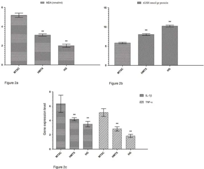
Figure 1a shows that the amounts of MDA were determined as 4.71±0.66, 2.61±0.34, and 1.61±0.30 μmol/gr protein, and the amounts of tGSH were found as 5.55±0.90, 7.65±0.32, and 8.26±0.51 nmol/gr protein in the cheek mucosal tissues of MTXC, HMTX, and HG groups; respectively (Figure 1b). MDA values were found as 5.18±0.55, 3.11±0.29, and 2.00±0.41 (Figure 2a) and, tGSH values were found as 5.83±0.49, 8.05±0.53, and 10.25±0.59 nmol/gr protein in the lower lip tissues of MTXC, HMTX, and HG groups; respectively (Figure 2b). The amounts of MDA were measured as 9.23±0.46, 3.31±0.47, and 2.80±0.40 μmol/ gr protein in the tongue tissues of MTXC, HMTX, and HG groups (Figure 3a). Whereas, the amount of tGSH were found as 1.78±0.33, 6.81±0.37, and 7.38±0.46 nmol/gr protein in the tongue tissues of these groups (Figure 3b).
Ge n e e x pr e ssion r e su lt s
expression in the cheek tissue of rats to 8.16±1.72. This rate was found as 5.33±0.81 and 4.00±0.89 in HMTX and HG groups. Again, MTX raised TNF-D gene expression in the cheek tissue of rats to 7.13±0.40, while HRE reduced TNF-D gene expression to 3.05±0.24. This rate was measured
expression in the lower lip tissue was found as 6.33±1.21, 4.18±0.21, and 3.50±0.38, and TNF-D gene expression was measured as 5.11±0.54, 2.83±0.30, and 1.85±0.22 in MTXC, HMTX, and HG,
D gene expressions in the tongue tissue compared with HMTX and HG.
IL-gene expression was measured as 10.11±1.12, 2.73±0.39, and 2.00±0.58 in MTXC, HMTX, and HG; respectively (Figure 3c).
H ist opa t h ologic r e su lt s
Examinations under optic microscope revealed a normal histopathologic appearance of the cheek, lower lip, and tongue tissues of the healthy group (Figure 4a, 4b, and 4c).
In Figure 4d, dilated congested blood vessels (arrow) and edema (star) were distinguished in the cheek mucosal tissue of MTXC group, which received MTX, while there were mild edema (star) and dilated congested blood vessels (arrow) observed also in the lower lip mucosa of this group (Figure 4e). However, besides dilated congested blood vessels (Figure 4f),
muscle layer (arrow) were also monitored in the tongue mucosal tissue of MTXC group (Figure 4g). Additionally, dilated congested proliferated blood vessels (Figure 4h) and marked edema (star) were found in the tongue tissue of MTXC group (Figure
lower lip, and tongue tissues of the animals treated with HRE (Figure 4j, 4k, 4l).
Figure 3- a: The effects of methotrexate on malondialdehyde (MDA) levels in the tongue tissues; b: The effects of PHWKRWUH[DWHRQW*6+OHYHOVLQWKHWRQJXHWLVVXHVF7KHHIIHFWVRIPHWKRWUH[DWHRQ,/ȕJHQHH[SUHVVLRQOHYHOLQWKH tongue tissues. *p<0.0001
D I SCUSSI ON
In this study, the effect of HRE on oral mucositis induced in rats with MTX was investigated and evaluated through biochemical, gene expression, and histopathologic examinations. Experimental results showed that the amount of MDA was increased and the amount of tGSH was decreased in the cheek, lower lip, and tongue tissues of the D
in the tissues in which MDA was significantly increased. MDA is known to be an oxidant and GSH an antioxidant parameter17. Therefore, increased
MDA and decreased tGSH amounts indicate the development of oxidative stress. MTX has been experimentally demonstrated to increase the tissue levels of MDA, which is a marker of lipid peroxidation to decrease the level of tGSH15,20. HRE,
which is used against the toxicity of MTX, was found
decrease of tGSH in the cheek, lower lip, and tongue tissues. This demonstrates that HRE protects the tissues against oxidative stress. Fruits of Hippophae
r h am n oides contain numerous phytochemicals, such as vitamins A, E, C, carotens, fatty acids, and
10,18,31. These antioxidant phytochemicals
are used to treat cancer, ulcer, liver, and skin diseases32. Many researchers have reported that
lipid peroxidation products are increased, and the plasma level of vitamin E is decreased in the tissues of cancer patients due to chemotherapy30. Lipid
peroxidation products increased with chemotherapy have been reported to decrease free radical scavengers antioxidant vitamins, such as vitamins A, E, and C4,26. In another in v iv o and in v it r o
study, the use of vitamins E, A, and C against oxidative stress caused by chemotherapy has been found to enhance the therapeutic effect and also protect normal cells against apoptosis5. It has been
reported in a cell culture and animal model that the combinations of vitamins A, B6, B12, C, D, and E
of chemotherapy prolonged survival time and increased response to treatment26. Furthermore,
it has been argued in a study evaluating
risk-and multivitamin combinations that vitamins had Figure 4- a: Normal histopathologic appearance of the rat cheek tissue of Healthy Group (HG) (HE&40); b: Normal histopathologic appearance of the rat cheek tissue of HG (HE&20); c: Normal histopathologic appearance of the rat cheek tissue of HG (HE&40); d: Dilated congested blood vessels (arrow) and edema (star) are distinguished in the cheek mucosa of MTXC group given methotrexate (MTX) (HEx40); e: Mild edema (star) and dilated congested blood vessels (arrow) are monitored in the lip mucosa of MTXC group given MTX (HEx40); f: Dilated congested blood vessels are seen in the tongue mucosa of MTXC group given MTX (HE&100); g: Fibroblastic proliferation (star) and fat cells replaced muscle layer are monitored in the tongue mucosal tissue of MTXC group given MTX (arrow) (HE&40); h: Dilated, congested, proliferated blood vessels (arrow) are seen in the tongue mucosa of MTXC group given MTX (HE&100); i: Edema (star) and dilated congested blood vessels (arrow) are monitored in the tongue mucosal tissue of MTXC group given MTX (HE&100); j:
Appearance of the cheek tissue of HMTX group treated with Hippophae rhamnoides extract (HRE) resembling normal
protective effect in cancer patients at risk6. This
information from the literature indicates that our experimental results were in parallel with previous studies.
D gene expressions in the cheek, lower lip, and tongue
MTX, compared with healthy and HRE groups.
D, were elevated in the oral tissue with mucositis developed due to chemotherapy8. Another study shows that the
D were increased and
tissue with MTX3 D
No information was found in the literature about D in MTX oral mucositis. However, Çakir, et al.7 (2015) reported that MTX
gene expressions in the kidney tissue. In our
D gene expressions. Vitamin C, alpha-tocopherol, and beta-carotens, also found in HRE, are known to have ihibitor
23. Again,
palmitic, oleic, and linoleic fatty acids appear to
and TNF-D13.
Macroscopically, no mucosal ulcerations were found in the cheek, lower lip, and tongue tissues of the animals given MTX. In addition, we found histopathological signs including mild edema and dilated congested blood vessels in the cheek and lower lip tissues, which have lower levels of D than the tongue tissue. However, marked congested blood vessels,
layer, proliferated blood vessels, and edema were observed in the tongue mucosal tissue.
cheek, lower lip, and tongue tissues of the healthy and HMTX groups, suggesting that histopathologic results were consistent with biochemical and gene expression results. Vascular congestion,
11.
Nothing was found in the literature about excessive fat accumulation in the muscle layer due to MTX. However, several studies associate the accumulation
this is a pathologic event9. Edema caused by MTX in
the cheek, lower lip, and tongue tissue might be a result of vascular changes. In the literature, edema
vascular changes24. In conclusion, in this study we
demonstrated with biochemical, gene expression,
damage in the cheek and lower lip tissues and more severe damage in the tongue tissue of rats.
HRE was found to protect the cheek, lower lip, and tongue tissue against the toxic effect of MTX.
the prophylaxis of oral damage due to MTX.
ACKN OW LED GM EN TS
We would like to thank Dr. Halis Suleyman.
D I SCLOSURE
The authors declare no potential conflict of interests regarding the authorship and/or publication of this article.
REFEREN CES
1- Alamir I, Boukhettala N, Aziz M, Breuillé D, Déchelotte P,
chemotherapy-induced intestinal mucositis. Clin Exp Immunol. 2010;162(2):298-305.
2- Altindag Ö, Küçükoglu B. Intoxication due to high dose methotrexate in a patient with rheumatoid arthritis: a case report. Turk J Rheumatology. 2011;26(1):58-61.
3- Araújo AA, Borba PB, Souza FH, Nogueira AC, Saldanha TS, Araújo TE, et al. In a methotrexate-induced model of intestinal mucositis, olmesartan reduced inflammation and induced enteropathy characterized by severe diarrhea, weight loss, and reduced sucrose activity. Biol Pharm Bull. 2015;38(5):746-52. 4- Bairati I, Meyer F, Gélinas M, Fortin A, Nabid A, Brochet F, et al. A randomized trial of antioxidant vitamins to prevent second primary cancers in head and neck cancer patients. J Natl Cancer Inst. 2005;97(7):481-8.
5- Blumenthal RD, Lew W, Reising A, Soyne D, Osorio L, Ying Z, et al. Anti-oxidant vitamins reduce normal tissue toxicity induced by radio-immunotherapy. Int J Cancer. 2000;86(2):276-80. 6- Borek C. Dietary antioxidants and human cancer. Integr Cancer Ther. 2004;3(4):333-41.
effect of alpha lipoic acid on rat kidneys in methotrexate induced oxidative injury. Eur Rev Med Pharmacol Sci. 2015;19(11):2132-9. 8- Chang C-T, Hsiang CY, Ho TY, Wu CZ, Hong HH, Huang YF.
induced oral mucositis through transcriptomic analysis. PloS One. 2015;10(8):e0135102.
9- Cilla M, Peña E, Martínez MA. Mathematical modelling of atheroma plaque formation and development in coronary arteries. J R Soc Interface. 2014;11(90):20130866.
10- Fatima T, Kesari V, Watt I, Wishart D, Todd JF, Schroeder WR,
vitamin C and tocopherol biosynthesis genes in the antioxidant-rich sea buckthorn (Hippophae rham noides L.). Phytochemistry. 2015;118:181-91.
11- Freire MR, Freitas R, Colombo F, Valença A, Marques AM, Sarmento VA. LED and laser photobiomodulation in the prevention and treatment of oral mucositis: experimental study in hamsters. Clin Oral Investig. 2014;18(3):1005-13.
12- Hejna M, Köstler W, Raderer M, Steger G, Brodowicz T, Scheithauer W, et al. Decrease of duration and symptoms in chemotherapy-induced oral mucositis by topical GM-CSF: results of a prospective randomised trial. Eur J Cancer. 2001;37(16):1994-2002.
13- Hua KF, Hsu HY, Su YC, Lin IF, Yang SS, Chen YM, et al. Study
Zost era j aponica. J Agric Food Chem. 2006;54(2):306-11. 14- Ilgenli T, Ören H, Uysal K. The acute effects of chemotherapy upon the oral cavity: prevention and management. Turk J Cancer. 2001;31:93-105.
prevents methotrexate-induced hepatorenal oxidative injury in rats. J Pineal Res. 2003;34(4):282-7.
Amelioration of methotrexate-induced enteritis by melatonin in rats. Cell Biochem Funct. 2004;22(3):169-78.
17- Kisaoglu A, Borekci B, Yapca OE, Bilen H, Suleyman H. Tissue damage and oxidant/antioxidant balance. Eurasian J Med 2013;45(1):47-9.
18- Kwon DJ, Bae YS, Ju SM, Goh AR, Choi SY, Park J. Casuarinin suppresses TNF-D-induced ICAM-1 expression via blockade of
2011;409(4):780-5.
19- McGowan D. Chemotherapy-induced oral dysfunction: a literature review. Br J Nurs. 2008;17(22):1422-6.
20- Miyazono Y, Gao F, Horie T. Oxidative stress contributes to methotrexate-induced small intestinal toxicity in rats. Scand J Gastroenterol. 2004;39(11):1119-27.
21- Niscola P, Romani C, Cupelli L, Scaramucci L, Tendas A, Dentamaro T, et al. Mucositis in patients with hematologic malignancies: an overview. Haematologica. 2007;92(2):222-31. 22- Ohkawa H, Ohishi N, Yagi K. Assay for lipid peroxides in animal tissues by thiobarbituric acid reaction. Anal Biochem.1979;95(2):351-8.
23- Oliveira BF, Veloso CA, Nogueira-Machado JA, Moraes EN, Santos RR, Cintra MT, et al. Ascorbic acid, alpha-tocopherol,
cytokines in mononuclear cells of Alzheimer's disease patients. Nutr Neurosci. 2012;15(6):244-51.
24- Scully C, Bagan JV. Adverse drug reactions in the orofacial region. Crit Rev Oral Biol Med. 2004;15(4):221-39.
25- Sedlak J, Lindsay RH. Estimation of total, protein-bound, and nonprotein sulfhydryl groups in tissue with Ellman's reagent. Anal Biochem. 1968;25:192-205.
26- Simone CB 2nd, Simone NL, Simone V, Simone CB. Antioxidants
and other nutrients do not interfere with chemotherapy or radiation therapy and can increase kill and increase survival, part 1. Altern Ther Health Med. 2007;13(1):22-8.
27- Süleyman H, Demirezer L, Büyükokuroglu M, Akcay M, Gepdiremen A, Banoglu Z, et al. Antiulcerogenic effect of
Hippophae rham noides L. Phytother Res. 2001;15(7):625-7. 28- Thomson PJ, Greenwood M, Meechan JG. General medicine and surgery for dental practitioners. Part 6 - cancer, radiotherapy and chemotherapy. Br Dent J. 2010;209(2):65-8.
29- Vanhoecke B, Bateman E, Mayo B, Vanlancker E, Stringer A, Thorpe D, et al. Dark Agouti rat model of chemotherapy-induced mucositis: establishment and current state of the art. Exp Biol Med. 2015;240(6):725-41.
30- Weijl N, Cleton F, Osanto S. Free radicals and antioxidants in chemotherapyinduced toxicity. Cancer Treat Rev. 1997;23(4):209-40.
31- Yilmaz I, Demiryilmaz I, Sener E, Cetin N, Ucuncu Y, Altuner D, et al. The effect of Hippophae r ham noides extract on oxidative damage on rat's gastric tissue depending on co-implementation of methotrexate and indomethacin. Lat Am J Pharm. 2014;33(3):453-8.