Ethyl-2-cyanoacrylate as a sealant after partial cecum resection in
Ethyl-2-cyanoacrylate as a sealant after partial cecum resection in
Ethyl-2-cyanoacrylate as a sealant after partial cecum resection in
Ethyl-2-cyanoacrylate as a sealant after partial cecum resection in
Ethyl-2-cyanoacrylate as a sealant after partial cecum resection in
rattus norvegicus albinus
rattus norvegicus albinus
rattus norvegicus albinus
rattus norvegicus albinus
rattus norvegicus albinus
O etil-2-cianoacrilato como selante após ressecção parcial de ceco em
O etil-2-cianoacrilato como selante após ressecção parcial de ceco em
O etil-2-cianoacrilato como selante após ressecção parcial de ceco em
O etil-2-cianoacrilato como selante após ressecção parcial de ceco em
O etil-2-cianoacrilato como selante após ressecção parcial de ceco em rattus
rattus
rattus
rattus
rattus
norvegicus albinus
norvegicus albinus
norvegicus albinus
norvegicus albinus
norvegicus albinus
ADILSON GOMES FAION, ACBC-MG1; AUGUSTO DIOGO FILHO, TCBC-MG2;TANIA MACHADODE ALCANTARA3;
TACIANA FERNANDES ARAUJO FERREIRA4
A B S T R A C T A B S T R A C T A B S T R A C T A B S T R A C T A B S T R A C T
Objective: Objective: Objective: Objective:
Objective: To evaluate the use of ethyl-2-cyanoacrylate in the treatment of an injury caused in a partially excluded segment of the mouse gut: the cecum. Methods:Methods:Methods:Methods:Methods: We used 45 male Wistar rats, divided into three equal groups; in all there was performed a partial resection of the cecum. The groups were designated as Group 1: the lesion was treated with application of ethyl-2-cyanoacrylate, Group 2: suture and application of ethyl-2-cyanoacrylate, Group 3: purse-string suture. The animals were monitored postoperatively and half of each group was necropsied 14 days after the procedure, the remaining on the 28th. They were subjected to macroscopic
evaluation, had cecal samples collected for histological examination and the findings were submitted to statistical analysis. Results:Results:Results:Results:Results: Weight gain after the experiment was different among groups (p=0.028). The presence of microabscesses was higher at 28 days postoperatively in group 2 when compared to group 3 (p=0.003). The collagen deposition on the 28th postoperative day was greater in group 1 (p=0.036) and intensity of inflammation at the 14th postoperative day was greater in group 1 (p=0.045). In the other parameters there was no statistical difference.Conclusion:Conclusion:Conclusion:Conclusion:Conclusion: The use of ethyl-2-cyanoacrylate was effective in the treatment of cecal stump exclusion of rats as for macroscopic and microscopic findings and postoperative outcome.
Key words: Key words: Key words: Key words:
Key words: Cyanoacrylate. Wound Healing. Colon. Rats. Adhesives.
Work performed at the Laboratory of experimental surgery and surgical technique of the Faculty of Medicine, Federal University of Uberlandia (UFU). Dissertation submitted to the Graduate Program in Health Sciences, Faculty of Medicine, Federal University of Uberlandia as partial requirement for obtaining a master’s degree.
1. Master’s Degree, Health Sciences, Faculty of Medicine, Federal University of Uberlandia, Uberlandia, Minas Gerais, Brazil. 2. Assistant Professor, Department of Surgery, Faculty of Medicine, Federal University of Uberlandia, Uberlandia, Minas Gerais, Brazil. 3. Associate Professor, Department of Clinical Medicine, Federal University of Uberlandia, Uberlandia, Minas Gerais, Brazil. 4. Undergraduate Research Fellow of the CNPq, Uberlândia, Brazil.
INTRODUCTION
INTRODUCTION
INTRODUCTION
INTRODUCTION
INTRODUCTION
T
he healing process and the means used to obtain acceleration and more efficient results are a constant concern in the medical field.Among the devices studied as alternative pro-cesses to the commonly used in medicine, synthetic adhesives are described and used since the mid-fifties of last century. Among these, there are several formulations based on cyanoacrylate, such as ethyl-2-cyanoacrylate, displaying positive features such as easy application, fast polymerization and low toxicity, among others, thus being a well cited substance1.
Typically, these compounds have been used as supplements to suturing processes, the report of their use alone being rare and, when applied, displaying questionable results1.
Souza and Oliveira2 concluded that the
ethyl-cyanoacrylate was better tolerated in the closure of the
rats’ skin, without inducing necrosis, allergic reactions or infections, presenting several advantages over octyl-cyanoacrylate.
Besides their use as adhesives for closing hollow viscera surgical wounds, cyanoacrylate has also been described in the experimental treatment of fistulas. Recent research has cited theie use in the treatment of digestive systemfistulas3.
The creation of an experimental model using ethyl-2-cyanoacrylate as a treatment for a terminal, infected, digestive tract segment, for example, the cecum of rats subjected to cecotomia, will deliver contributions to the establishment of alternatives in the healing process and control of fistulas of the digestive system.
rimental Surgery, Faculty of Medicine, Federal University of Uberlandia, which followed the ethical principles of animal experimentation and was approved by the Ethics Committee on Use of Animals (CEUA) of UFU protocol 088/08.
S a m p l e S a m p l e S a m p l e S a m p l e S a m p l e
To perform the experiment we used 45 male rats (Rattus norvegicus albinos) of Wistar strain, with a mean age of 120 days and weighing between 135g and 337g. The animals were kept in appropriate cages, were exposed to light in a regular cycle of 12 hours and fed specific indus-trial diets and water ad libitum at all stages of the experiment. We equally and randomly divided them to each experimental group and a raffle was subsequently held for distribution into three groups with 15 animals each: Group 1: cecal stump closure by exclusive application of ethyl-2-cyanoacrylate; Group 2: cecal stump closure with a 4-0 cotton needled suture stitch and application of ethyl-2-cyanoacrylate; and Group 3: cecal stump closure with 4-0 cotton purse-string needle suture.
Procedures Procedures Procedures Procedures Procedures
The surgical team was composed of four surgeons, an experimental laboratory technician and an undergraduate research scholar. We also had guidance from a veterinarian.
The animals fasted for 6 hours, were weighed and then anesthetized with intraperitoneal injection of 10% ketamine hydrochloride (Ketalar®, Agener Union 100mg/ ml), in dosages of 30mg per kilogram of weight, and 2% xylazine hydrochloride (“Calmiun®” Agener Union 20mg/ ml) at a dosage of 10mg per kilogram. Then the animals were sent to one of the teams. The rats were marked with a code of rings of different colors on their tails, the correct identification. Data were recorded on specific charts for each animal.
The animals were all operated on the same day. After the surgical procedure, the animals were placed in individual cages to recover from anesthesia for two hours. They were later transferred to cages in groups of three with food and water ad libitum.
Technique Technique Technique Technique Technique
The animals were prepared by a laboratory technician who fixed them with adhesive tape in a proper bulkhead in supine position, subjecting them to shaving of the abdominal wall. Later, they were randomly distributed to each of the surgical teams.
We proceeded with antisepsis using an alcohol solution of 1% polyvinylpyrrolidone-iodine and placement of sterile drapes.
Then it was sectioned at 0.5 cm from the clip and its proximal portion resected, respecting the 1 cm margin from the implantation of ileum and colon.
After that, we protected the base of the cecum with gauze and irrigated the exposed area by means of a syringe mounted needle under pressure with saline 0.9%. Finally, there was scrapping of the cecal stump mucosa with a scalpel blade until active bleeding from the edges started (Figures 1 and 2).
Treatment of the cecal stump was performed according to the groups: Group 1 - Application of ethyl-2-cyanoacrylate, using the pour spout of the tube homogeneously in the cecal stump. The stump was kept tractioned for three minutes, followed by the release of the clip and repositioning inside abdominal cavity (Figu-res 3 and 4); Group 2 - Approximation of the edges with a central caecal extramucosal 4-0 cotton stitch and then ethyl-2-cyanoacrylate. The stump was also kept tractioned for three minutes and released into the abdo-minal cavity; Group 3 - Making of purse-string suture at the base of the cecum with a 4-0 cotton suture and submucosal cecal stump due invagination. The stump was kept tractioned for three minutes and released into the abdominal cavity.
After local review, we proceeded to the closure of the abdominal wall using the same technique in all groups: simple 4-0 chrome catgut stitches in the peritoneum-aponeurosis, and separated 4-0 mononylon for the skin.
In the immediate postoperative period, animals received an intraperitoneal injection that contained 0.1 ml/ 100 g of dipyrone and 0.1 ml/100 g of sodium diclofenac.
Necropsies Necropsies Necropsies Necropsies Necropsies
The 45 animals were autopsied in the following order: 24 on the 14th postoperative day, after a lethal dose
of anesthetic, eight animals in each group. The other (21 animals, seven from each group) on day 28 after surgery, also undergoing the same procedure. The dose was calculated to be equivalent to twice the therapeutic dose used in anesthesia.
Animals were weighed approximately 30 minutes before being subjected to a lethal anesthetic dose. The autopsy was performed by one researcher involved in the experiment. He had no information about which procedure the animal had undergone. Thus, we followed a schedule of evaluation, as will be described below.
presence and degree of adhesion formation, presence of nodules, enlarged lymph nodes, fistulae, abscesses, among others. The grading of adhesions adopted was the one proposed and modified by Diogo Filho4.
Then we resected the cecal stump including 0.5 cm of ileum and 0.5 cm of the distal colon. Thus, the specimen comprised the cecal stump treated by one of the approaches already described.
Histological analysis Histological analysis Histological analysis Histological analysis Histological analysis
The excised postsurgical anatomical parts were fixed in 10% formalin solution for at least 48 hours and
sent to the pathology laboratory of the Hospital de Clíni-cas of UFU. They were opened in the largest diameter, the surgical site being identified with the aid of the research team; at least five longitudinal sections of this area were performed. They were then duly processed and dehydrated in increasing ethanol solutions, cleared in xylene and embedded in paraffin. The blocks were sectioned on microtome. We obtained sections of 3 µm thick, which were mounted on slides and stained with hematoxylin-eosin.
The slides were examined by a pathologist blinded to which group each animal belonged. We used
Figure 4 Figure 4 Figure 4 Figure 4
Figure 4 – Final appearance of the cecal stump treated with ethyl-2-cyanoacrylate adhesive, prior to clips removal. Figure 1
Figure 1 Figure 1 Figure 1
Figure 1 – Cecal exposure and ligation of the mesenteric vessels.
Figure 2 Figure 2 Figure 2 Figure 2
Figure 2 – Cecal edges scarification.
Figure 3 Figure 3 Figure 3 Figure 3
a control in comparison with the surgically manipulated cecal stump.
The morphological study of the cecal stump focusing on the healing process assessed the presence or absence of: reepithelialization, abscesses and granulomas, whether foreign body (FB) or epithelioid (E) types. Collagen deposition, fibroblast proliferation, neovascularization and inflammation were also observed. To assess these parameters, we used a scoring system from zero to three proposed by Durmus et al.5.
Statistical analysis Statistical analysis Statistical analysis Statistical analysis Statistical analysis
The results were statistically analyzed with BioEstat, version 5.0. For comparison between groups, we used analysis of variance (ANOVA). We also used descriptive statistics to compare the macroscopic findings in the analysis and postoperative findings using bimodal analysis of variance, Kruskal-Wallis, Mann Whitney, Student T, Tukey and Chi-square tests. Differences were considered significant for values corresponding to p <0.05.
The groups were compared in the 14th and 28th
postoperative days, as well as their respective results between the 14th and 28th postoperative days.
RESULTS
RESULTS
RESULTS
RESULTS
RESULTS
Animals were assessed daily, with notes Animals were assessed daily, with notes Animals were assessed daily, with notes Animals were assessed daily, with notes Animals were assessed daily, with notes of all events.
of all events.of all events. of all events. of all events.
Five animals, three belonging to group 2 and two belonging to group 3, died on the second day after surgery. At necropsy, there was intrabdominal bleeding in all cases, without other probable cause of death. One animal in group 3 died on the third day after surgery. Peritonitis by cecal stump dehiscence was identified at autopsy. These animals were excluded from the study and randomly reset to the corresponding groups.
Preoperative weight Preoperative weight Preoperative weight Preoperative weight Preoperative weight
The average weight for animals in groups 1, 2 and 3 before surgery were not statistically significant.
Weight postoperative day and autopsy Weight postoperative day and autopsy Weight postoperative day and autopsy Weight postoperative day and autopsy Weight postoperative day and autopsy Animals were weighed approximately 30 minutes before being subjected to a lethal dose of anesthetic. There was no statistical difference in the groups on the 14th and 28th postoperative days (PO). There was
also no statistical difference (Student t test) in the comparison between the corresponding groups in the 14th
and 28th postoperative day.
weight of 32g. Several loops’ adhesions in the surgical site were noted in its autopsy, giving an appearance of semi-intestinal obstruction.
The comparison of changes in animal weights between groups G1, G2, G3 and preoperatively, with the weights on the 28th postoperative day showed significant
difference (p = 0.028). There were also differences between G1 and G2 when using the Tukey test, but not between G1 and G3 and between G2 and G3. Based on the Student t test for comparison within the same group in the 14th and
28th postoperative day, it was found that animals in G1
gained weight on both occasions, however, without statistical difference (p = 0.0946). In G2 animals gained weight with statistical difference (p <0.0001), the same as in G3 (p = 0.0018). The analysis of animal weight gains, comparing preoperative with 14th and 28th POs is shown in
table 1.
Grading of adhesions Grading of adhesions Grading of adhesions Grading of adhesions Grading of adhesions
The macroscopic evaluation of adhesions, by the time of autopsies, was represented by degrees, as reported in the methods. Most animals had adhesions degrees 2 and 3. An animal of G1 (14th PO) had no adhesions (grade
0), and one animal in G3 (28th PO) had grade 4 adhesions.
No animal in this study, presented grade 5adhesions. In the POs 14 and 28 there was no statistical difference between G1 and G2, between G1 and G3 and between G2 and G3. When comparing the same group on the 14th and 28th postoperative days, there were no
statistically significant differences. We also compared the three groups in the POs (14th and 28th) and did not observe
any significant differences.
Presence of other microscopic changes Presence of other microscopic changes Presence of other microscopic changes Presence of other microscopic changes Presence of other microscopic changes Other gross changes were observed in only six animals, one in G1, three in G2 and two in G3. They showed granulomas in the region of the parietal peritoneum and also mesenteric lymphadenopathy. There were no abscesses, fistulas, or other changes in any ani-mal (Table 2)
Microscopic analysis Microscopic analysis Microscopic analysis Microscopic analysis Microscopic analysis
In one animal belonging to the G3, which had its autopsy performed on the 28th postoperative day,
histological analysis was not possible due to difficulties in identifying the surgical site under the microscope.
Cecal wall thickness in the area of surgical Cecal wall thickness in the area of surgical Cecal wall thickness in the area of surgical Cecal wall thickness in the area of surgical Cecal wall thickness in the area of surgical manipulation
manipulationmanipulation manipulationmanipulation
For an animal of G1 (28th PO) and two from G3
this analysis was not possible because of the position of the intestinal loops on the slide, hampering the measurement of their thickness.
The results showed no significant difference when comparing the groups on the 14th and 28th POs. When
comparing the groups two by two and at the evaluation between the groups in the 14th and 28th postoperative day,
there was no statistical difference either.
Thickness of surgical manipulation-free Thickness of surgical manipulation-free Thickness of surgical manipulation-free Thickness of surgical manipulation-free Thickness of surgical manipulation-free Cecal wall
Cecal wall Cecal wall Cecal wall Cecal wall
The results showed that when comparing the groups at the 14th postoperative day there was no significant
difference. Comparing the groups two by two, between G1 and G2 and between G1 and G3 showed no significant difference, the opposite occurring to G2 and G3 (p = 0.0233), with statistical difference in the 14th postoperative
day. Likewise, in applying the analysis of variance, when comparing the groups on the 28th postoperative day, there
was no significant difference (p = 0.2801). When comparing the groups two by two and evaluating the groups in the 14th and 28th postoperative day, there was no statistical
difference.
Epithelialization of the area of surgical Epithelialization of the area of surgical Epithelialization of the area of surgical Epithelialization of the area of surgical Epithelialization of the area of surgical manipulation
manipulation manipulation manipulation manipulation
All animals showed partial or complete reepithelialization in the cecal stump. On the 14th
postoperative day, epithelialization was incomplete in 75% of cases in each group. On the 28th postoperative day, the
incomplete reepithelialization was 71.4%, 85.7% and 57.1% for groups 1, 2 and 3, respectively. We used the binomial test for comparing two proportions.
Incomplete reepithelialization predominated in all groups, both on the 14th and 28th postoperative days.
There was no statistical difference between groups.
Microabscesses at the site of cecal Microabscesses at the site of cecal Microabscesses at the site of cecal Microabscesses at the site of cecal Microabscesses at the site of cecal surgical manipulation
surgical manipulation surgical manipulation surgical manipulation surgical manipulation
In all groups, most animals necropsied at 14th
PO showed microabscesses in the area of surgical manipulation. For those animals necropsied on the 28th
postoperative day, only in G2 microabscesses predominated, and none of the animals in G3 showed that complication. Binomial test was performed and showed statistical difference between G2 and G3 on the 28th postoperative day. The other groups showed no
significant difference.
Collagen deposition at the site of cecal Collagen deposition at the site of cecal Collagen deposition at the site of cecal Collagen deposition at the site of cecal Collagen deposition at the site of cecal surgical manipulation
surgical manipulation surgical manipulation surgical manipulation surgical manipulation
There was a tendency for greater deposition of collagen in all groups. Most animals had grade 2 classification of collagen deposition. Only one animal in group 3, on the 28th postoperative day, had a grade 1
collagen deposition and in group 1, on the 28th PO, there
was a slight predominance of grade 3 (Table 3). When comparing both groups, we found no statistical difference in the 14th postoperative day. On the 28th PO there was
also no statistical difference between groups. When comparing the groups in the 14th and 28th PO, there was
no statistical difference. However, using the Kruskal-Wallis test for the 28th PO there was statistically significant
difference between groups G1 and G3 (p = 0.0368), with more collagen deposition in G1. For the 14th postoperative
day, using this same test, no difference was found between groups.
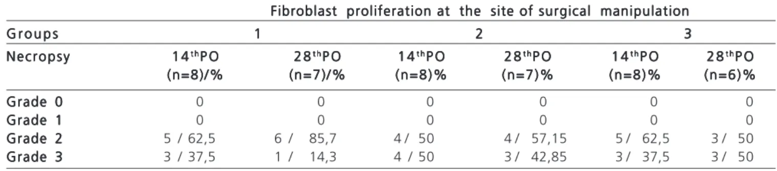
Fibroblast proliferation at the site of Fibroblast proliferation at the site of Fibroblast proliferation at the site of Fibroblast proliferation at the site of Fibroblast proliferation at the site of surgical manipulation
surgical manipulation surgical manipulation surgical manipulation surgical manipulation
All animals showed fibroblastic proliferation at the surgical site, and, generally, in each group, a predominance of grade 2, except for G2 and G3 on the 14th PO and on the 28th PO, in which half the animals had
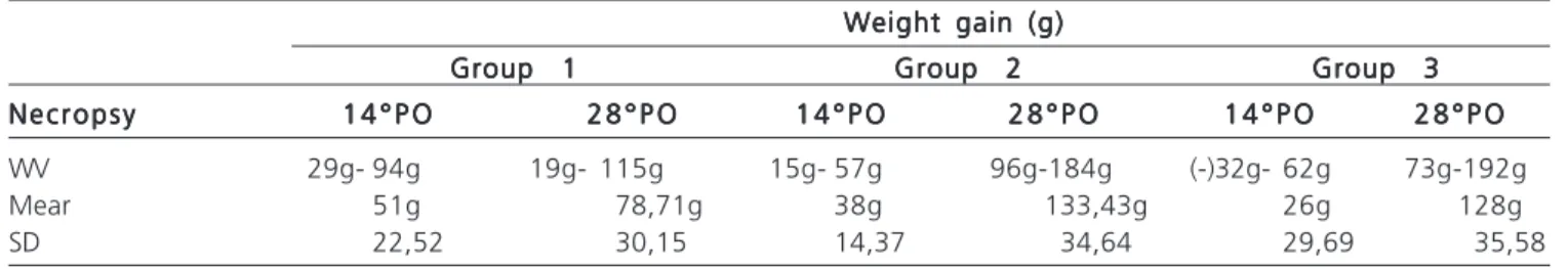
Table 1 Table 1 Table 1 Table 1
Table 1 – Weight gain of animals in each experimental group.
Weight gain (g) Weight gain (g) Weight gain (g) Weight gain (g) Weight gain (g) Group 1
Group 1Group 1
Group 1Group 1 Group 2Group 2Group 2Group 2Group 2 Group 3Group 3Group 3Group 3Group 3 N e c r o p s y
N e c r o p s y N e c r o p s y N e c r o p s y
N e c r o p s y 1 4 ° P O1 4 ° P O1 4 ° P O1 4 ° P O1 4 ° P O 2 8 ° P O2 8 ° P O2 8 ° P O2 8 ° P O2 8 ° P O 1 4 ° P O1 4 ° P O1 4 ° P O1 4 ° P O1 4 ° P O 2 8 ° P O2 8 ° P O2 8 ° P O2 8 ° P O2 8 ° P O 1 4 ° P O1 4 ° P O1 4 ° P O1 4 ° P O1 4 ° P O 2 8 ° P O2 8 ° P O2 8 ° P O2 8 ° P O2 8 ° P O
WV 29g- 94g 19g- 115g 15g- 57g 96g-184g (-)32g- 62g 73g-192g
Mear 51g 78,71g 38g 133,43g 26g 128g
SD 22,52 30,15 14,37 34,64 29,69 35,58
WV: weight variation; SD: standard deviation; PO: postoperative; (-): negative value.
Table 2 Table 2 Table 2 Table 2
Table 2 – Distribution of granulomas in groups 1, 2 and 3 in 14th e 28th POs.
Presence of nodules Presence of nodulesPresence of nodules Presence of nodulesPresence of nodules N e c r o p s y
N e c r o p s y N e c r o p s y N e c r o p s y
N e c r o p s y 1414141414ththththth PO (n/%) PO (n/%) PO (n/%) PO (n/%) PO (n/%) 2828282828ththththth PO (n/%) PO (n/%) PO (n/%) PO (n/%) PO (n/%)
Group 1 (n=8) 0 1* 14,3
Group 2 (n=8) 2 25 1* 14,3
Group 3 (n=8) 2 25 0*
PO: postoperative
there was also no difference.
Neovascularization at the site of cecal Neovascularization at the site of cecal Neovascularization at the site of cecal Neovascularization at the site of cecal Neovascularization at the site of cecal surgical manipulation
surgical manipulationsurgical manipulation surgical manipulation surgical manipulation
All groups showed neovascularization in the region of cecal surgical manipulation, predominantly, in general, those classified as grade 3. By means of the Kruskal-Wallis test, there was no difference between groups, both for animals on the 14th PO and on the 28th.
Likewise, no difference was observed among animals of the same group necropsied at different times in the 14th
and 28th POs. When comparing the groups two by two,
there was no statistical significance on the 14th and 28th
POs.
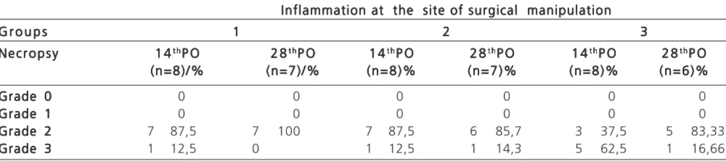
Intensity of inflammation at the site of Intensity of inflammation at the site of Intensity of inflammation at the site of Intensity of inflammation at the site of Intensity of inflammation at the site of cecal surgical manipulation
cecal surgical manipulationcecal surgical manipulation cecal surgical manipulation cecal surgical manipulation
The vast majority of animals showed a tendency to grade 2 inflammation at the site of surgical manipulation, as 100% of the animals in G1 sacrificed on 28th PO were so
classified and only in G3 animals sacrificed on the 14th
However, when using the Kruskal-Wallis test to compare the animals necropsied on the 14th postoperative
day, there was statistical difference (p = 0.0454), and this was between G1 and G3, with greater inflammation in G3 and between G2 and G3, with more inflammation in G3 (Table 5).
Presence and type of microgranulomas at Presence and type of microgranulomas at Presence and type of microgranulomas at Presence and type of microgranulomas at Presence and type of microgranulomas at the site of cecal surgical manipulation
the site of cecal surgical manipulationthe site of cecal surgical manipulation the site of cecal surgical manipulationthe site of cecal surgical manipulation
In one animal in group 3, sacrificed at the 28th
day, we could not perform this analysis due to difficulties in the visualization site
All others showed granuloma formation in the anastomosed site, being of both types – epithelioid and foreign body.
DISCUSSION
DISCUSSION
DISCUSSION
DISCUSSION
DISCUSSION
Among the various types of cyanoacrylates available, we chose ethyl-2-cyanoacrylate because it is a
Table 3 Table 3 Table 3 Table 3
Table 3 – Classification of collagen deposition at the site of surgical manipulation.
Collagen deposition at the site of surgical manipulation Collagen deposition at the site of surgical manipulationCollagen deposition at the site of surgical manipulation Collagen deposition at the site of surgical manipulation Collagen deposition at the site of surgical manipulation G r o u p s
G r o u p sG r o u p s G r o u p s
G r o u p s 11111 22222 33333
N e c r o p s y N e c r o p s yN e c r o p s y N e c r o p s y
N e c r o p s y 1 41 41 41 41 4t ht ht ht ht hP OP OP OP OP O 2 82 82 82 82 8t ht ht ht ht hP OP OP OP OP O 1 41 41 41 41 4t ht ht ht ht hP OP OP OP OP O 2 82 82 82 82 8t ht ht ht ht hP OP OP OP OP O 1 41 41 41 41 4t ht ht ht ht hP OP OP OP OP O 2 82 82 82 82 8t ht ht ht ht hP OP OP OP OP O
(n=8)/% (n=8)/%(n=8)/%
(n=8)/%(n=8)/% (n=7)/%(n=7)/%(n=7)/%(n=7)/%(n=7)/% (n=8)%(n=8)%(n=8)%(n=8)%(n=8)% (n=7)%(n=7)%(n=7)%(n=7)%(n=7)% (n=8)%(n=8)%(n=8)%(n=8)%(n=8)% (n=6)%(n=6)%(n=6)%(n=6)%(n=6)% Grade 0
Grade 0Grade 0 Grade 0
Grade 0 0 0 0 0 0 0
Grade 1 Grade 1Grade 1 Grade 1
Grade 1 0 0 0 0 0 1 / 16,66
Grade 2 Grade 2Grade 2 Grade 2
Grade 2 7 / 87,5 3 / 42,85 6 / 75 6 / 85,7 8 / 100 5 / 83,33
Grade 3 Grade 3Grade 3 Grade 3
Grade 3 1 / 12,5 4 / 57,15 2 / 25 1 / 14,3 0 0
PO: postoperative.
Table 4 Table 4 Table 4 Table 4
Table 4 – Classification of fibroblast proliferation at the site of surgical manipulation.
Fibroblast proliferation at the site of surgical manipulation Fibroblast proliferation at the site of surgical manipulation Fibroblast proliferation at the site of surgical manipulation Fibroblast proliferation at the site of surgical manipulation Fibroblast proliferation at the site of surgical manipulation G r o u p s
G r o u p sG r o u p s G r o u p s
G r o u p s 11111 22222 33333
N e c r o p s y N e c r o p s yN e c r o p s y N e c r o p s y
N e c r o p s y 1 41 41 41 41 4t ht ht ht ht hP OP OP OP OP O 2 82 82 82 82 8t ht ht ht ht hP OP OP OP OP O 1 41 41 41 41 4t ht ht ht ht hP OP OP OP OP O 2 82 82 82 82 8t ht ht ht ht hP OP OP OP OP O 1 41 41 41 41 4t ht ht ht ht hP OP OP OP OP O 2 82 82 82 82 8t ht ht ht ht hP OP OP OP OP O
(n=8)/% (n=8)/%(n=8)/%
(n=8)/%(n=8)/% (n=7)/%(n=7)/%(n=7)/%(n=7)/%(n=7)/% (n=8)%(n=8)%(n=8)%(n=8)%(n=8)% (n=7)%(n=7)%(n=7)%(n=7)%(n=7)% (n=8)%(n=8)%(n=8)%(n=8)%(n=8)% (n=6)%(n=6)%(n=6)%(n=6)%(n=6)% Grade 0
Grade 0Grade 0 Grade 0
Grade 0 0 0 0 0 0 0
Grade 1 Grade 1Grade 1 Grade 1
Grade 1 0 0 0 0 0 0
Grade 2 Grade 2Grade 2 Grade 2
Grade 2 5 / 62,5 6 / 85,7 4 / 50 4 / 57,15 5 / 62,5 3 / 50
Grade 3 Grade 3Grade 3 Grade 3
Grade 3 3 / 37,5 1 / 14,3 4 / 50 3 / 42,85 3 / 37,5 3 / 50
readily accessible, effective and relatively less toxic substance than others of the same class6,7. Corroborating
with our choice, Souza et al.2 stated the superiority of this
compound over octyl-2-cyanoacrylate and conventional sutures in the synthesis of rat skin. The same studies report that the ethyl-2-cyanoacrylate is better tolerated, with less infection, necrosis or allergic reactions in treated animals, and lower costs.
The species and number of animals used in each group were similar to or higher than those found in some studies2,8, besides having been previously determined by
statistical data.
The weight displayed by the animals immediately before the autopsy results did not differ between groups when the animals’ post-experimental weight gain in the 14th postoperative day was evaluated.
It was found that the animals of G1 had a greater mean weight gain than the G2 and G3, but without statistical significance (p = 0.1544). In the 28th PO we found that
the animals of G1 had a lower weight gain than G2 and G3, with statistical difference (p = 0.0128 and p = 0.0422, respectively). This lower weight gain in G1 animals may be related to tissue toxicity caused by different cyanoacrylate monomers6,7. That said, it opens room for
further studies to examine if indeed the ethyl-2-cyanoacrylate is a product with some degree of toxicity to animals. Moreover, the prolonged healing process could be an aggravating factor that would hamper weight gain in G1. However, the histological parameters evaluated showed no significant differences between groups when compared on the 14th and 28th postoperative
days. This may mean that the association of octyl-2-cyanoacrylate with conventional sutures in the jejunum of rabbits was similar to one that used only convencio-nal suture9. In this study, we found no evidence to explain
this fact in the evaluated microscopic and macroscopic parameters.
The analysis of the degree of peritoneal adhesion in different groups and postoperative moments revealed no statistically significant difference, which may indicate that both the cyanoacrylate and the gold standard technique of invaginating the cecal stump have similar tissue reactions. Moreover, we can state that the tissue reaction was minimal
for all groups, given the minimal macroscopic changes observed, which was also homogeneously distributed in the sample groups. These findings are controversial in the literature: Matera et al.10, in a comparative study between
the isobutyl-2-cyanoacrylate and n-butyl-2-cyanoacrylate, found that both compounds caused a similar degree of adhesions.
Amaral et al. 9 evaluated jejunal anastomosis in
rabbits using synthetic (octyl-2-cyanoacrylate) and biological (fibrin) adhesives associated with multiple conventional sutures and found that the rate of adhesions did not differ between groups.
According to Von Bahten et al.11, who studied
splenic wound treatment using octyl-2-cyanoacrylate and poliglecaprone suture, the animals treated with adhesive showed less adhesion.
Souza et al.1 observed prevalence of fibrosis in
the small intestine and colon anastomoses using methyl-2-cyanoacrylate compared to 6-0 silk.
Of the seven parameters used in microscopic analysis, in four statistical differences were not detected (re-epithelialization of cecal manipulation area, fibroblast proliferation, neovascularization and presence and type of granuloma).
The thickness of the intestinal wall in the area of cecal surgical manipulation presented greater than the thickness in an area free from manipulation, which was expected by surgical damage caused at the site. In all three groups there were no statistical differences in the cecal wall thickness at the site of surgical manipulation, independent of time after surgery. This showed that this is an early step in the healing process and a similar intensity of aggressive factors (suture, ethyl-2-cyanoacrylate and the combination of both.), unlike shown by Souza et al.1, who
found a stronger reaction with thickening and stenosis with the use of synthetic adhesive (methyl-2-cyanoacrylate) compared with 6-0 silk.
Animals from G3 presented with differences in relation to others on the normal intestinal wall thickness, which can be attributed to edema and the preparation of slides for histological processing.
Regarding the presence of microabscesses in the long term (animals necropsied at 28th PO), they were more
Tabela 5 Tabela 5 Tabela 5 Tabela 5
-Tabela 5 - Classification of Inflammation at the site of surgical manipulation.
Inflammation at the site of surgical manipulation Inflammation at the site of surgical manipulation Inflammation at the site of surgical manipulation Inflammation at the site of surgical manipulation Inflammation at the site of surgical manipulation G r o u p s
G r o u p s G r o u p s G r o u p s
G r o u p s 11111 22222 33333
N e c r o p s y N e c r o p s y N e c r o p s y N e c r o p s y
N e c r o p s y 1 41 41 41 41 4t ht ht ht ht hP OP OP OP OP O 2 82 82 82 82 8t ht ht ht ht hP OP OP OP OP O 1 41 41 41 41 4t ht ht ht ht hP OP OP OP OP O 2 82 82 82 82 8t ht ht ht ht hP OP OP OP OP O 1 41 41 41 41 4t ht ht ht ht hP OP OP OP OP O 2 82 82 82 82 8t ht ht ht ht hP OP OP OP OP O
(n=8)/% (n=8)/%(n=8)/% (n=8)/%
(n=8)/% (n=7)/%(n=7)/%(n=7)/%(n=7)/%(n=7)/% (n=8)%(n=8)%(n=8)%(n=8)%(n=8)% (n=7)%(n=7)%(n=7)%(n=7)%(n=7)% (n=8)%(n=8)%(n=8)%(n=8)%(n=8)% (n=6)%(n=6)%(n=6)%(n=6)%(n=6)% Grade 0
Grade 0 Grade 0 Grade 0
Grade 0 0 0 0 0 0 0
Grade 1 Grade 1 Grade 1 Grade 1
Grade 1 0 0 0 0 0 0
Grade 2 Grade 2 Grade 2 Grade 2
Grade 2 7 87,5 7 100 7 87,5 6 85,7 3 37,5 5 83,33
Grade 3 Grade 3 Grade 3 Grade 3
material. Conventional sutures complemented by biological or synthetic adhesives in rabbits’ enteric anastomoses showed no difference as for the presence of abscesses9. Therefore, this assessment should be
reviewed in similar studies. Still from this analysis we have that the use of suture material (G3 animals) determined lower inflammatory reaction than the cyanoacrylate, as the animals in this group had a lower incidence of microabscesses in relation to others. In the study by Duarte et al.12 it was found an abscess in the
group treated with cyanoacrylate and one in the group treated with polyglactin, which determined the occurrence of these reactions to processes inherent to the characteristics of the material or to the procedure, with no difference between groups. There is a greater tendency to abscess formation in spleen injuries repairs of animals treated with poliglecaprone suture when compared to cyanoacrylate or to the control group, but no significant11.
All animals showed foreign body granulomas regardless of the technique or the evolution time. This analysis shows mixed results in the literature, the reaction with cyanoacrylate being greater13, displaying less
granuloma formation2,11.14 and, alternatvely, showing a
si-milar pattern between the different materials used9, which
is consistent with our results. Well-formed granulomas may be found in the intestine of animals treated with cyanoacrylate sacrificed on the 28th postoperative day, but
not in 4th or 14th POs10.
The collagen deposition was uniform across different groups of animals sacrificed on the 14th
postoperative day. Since fibroblast proliferation was also statistically similar in these animals, we can say that the quality of the scar formed in this period was similar for all groups. However, in animals necropsied on the 28th postoperative day from G1, the presence
of collagen fibers was lower compared to the others. This fact suggested that the use of cyanoacrylate could cause less scar tissue, with the remodeling of collagen bundles occurring earlier, a potential beneficial effect, leading to a more elastic and therefore more physiological scar.
Souza et al.2 found that fibrosis was more severe
in rats with skin lesions closed with suture compared to those treated with two compounds derived from cyanoacrylate (ethyl-cyanoacrylate and octyl-cyanoacrylate) and that, in all groups, fibrosis was more intense in later postoperative periods. There is a tendency to formation of more mature collagen (type III) in animals
type III, collagen when compared to cyanoacrylate or suture.
In this study, we observed increased inflammation in the area of cecal surgical manipulation in G3 animals necropsied on the 14th postoperative day.
However, in the 28th PO the distribution between the groups
was statistically equal. This may indicate that in an earlier period the cyanoacrylate is capable of triggering minor lo-cal reaction than the presence of suture material alone, a trend not maintained with similar results between the groups in subsequent follow-up periods. Souza et al.2 found that
the inflammatory response was more evident in the initial periods after surgery, especially for those treated with cyanoacrylate. For Duarte et al.12 the quantitative evaluation
of inflammatory cells showed no statistical difference in the animals treated with cyanoacrylate or polyglactin. For Saito et al.15 the alpha-cyanoacrylate showed a better tissue
response than silk sutures in the subcutaneous tissue of rats. We can assume that the initial inflammatory process caused by ethyl-2-cyanoacrylate is more intense in the initial period, matching, in the course of time, the one of other materials, even leading to a lower late deposition of collagen. This more intense tissue reaction may be related to the toxicity of the chemical. Saska et al.6, Stephen et al .7 and
Souza et al.2 reported these different degrees of toxicity of
cyanoacrylate monomers, concluding that the toxicity of compounds based on this substance may be altered by changes in their chains.
At the same time, the possibility of using ethyl-2-cyanoacrylate as a method for occlusion of colonic fistula must be taken into account. Following the model of this study, an excluded, highly colonized and contaminated cecal segment, we may consider that the treatment was significantly effective in promoting the closure of the mucosal defect without complications such as dehiscence or fistula, without abscess formation and a result similar to the conventional method with debridement and suture (G3).
Both in the findings of our study and in most of the literature the largest initial inflammatory reaction and increased late fibrosis after the use of adhesives ethyl-2-cyanoacrylate, which in principle could be contraindicated in intestinal anastomosis, act favorably to the occlusion of the cecal stump exclusion becoming a way of treatment for digestive fistulas.
R E S U M O R E S U M O R E S U M O R E S U M O R E S U M O
Objetivo: Objetivo: Objetivo: Objetivo:
Objetivo: Avaliar a utilização do etil-2-cianoacrilato no tratamento de uma lesão provocada em um segmento parcialmente excluso do intestino do rato: o ceco. Métodos:Métodos:Métodos:Métodos:Métodos: Utilizaram-se 45 ratos machos da linhagem Wistar, distribuidos em três grupos iguais, sendo que foi realizada a ressecção parcial do ceco. Os grupos foram denominados como: Grupo 1: a lesão foi tratada com aplicação de etil-2-cianoacrilato; Grupo 2: sutura e aplicação de etil-2-cianoacrilato; Grupo 3: sutura em bolsa. Os animais foram acompanhados no pós-operatório e metade de cada grupo foi necropsiada no 14º e restante no 28º pós-operatório. Dessa forma, foram submetidos à avaliação macroscópica, sendo coletadas amostras do ceco para avaliação histológica e, por fim, realizou-se a análise estatística... Resultados:
Resultados: Resultados: Resultados:
Resultados: O ganho de peso pós-experimento foi diferente nos grupos (p=0,028). A presença de microabcessos foi maior no 28º dia de pós-operatório no grupo 2, em comparação com o grupo 3 (p=0,003). A deposição de colágeno no 28º dia de pós-operatório foi maior no grupo 1 (p=0,036) e a intensidade da inflamação no 14º dia de pós-operatório foi maior no grupo 1 (p=0,045). Nos demais parâmetros avaliados, não ocorreu diferença estatística. Conclusão:Conclusão:Conclusão:Conclusão:Conclusão: A utilização do etil-2-cianoacrilato foi efetiva no tratamento do coto cecal excluso de ratos frente à avaliação macroscópica, microscópica e evolução pós-operatória.
Descritores: Descritores: Descritores: Descritores:
Descritores: Cianoacrilato. Cicatrização de Feridas. Colon. Ratos. Adesivos.
REFERENCES
REFERENCES
REFERENCES
REFERENCES
REFERENCES
1. Souza TFC, Silva AL. Estudo experimental das enteroanastomoses com o 2-metil-cianoacrilato, em cobaias. Acta Cir Bras 1988;3(3):80-8.
2. Souza SC, Oliveira WL, Soares DFOS, Briglia CH, Athanázio PR, Cerqueira MD, Guimaraes PH, Carrero MC. Comparative study of suture and cyanoacrylates in skin closure of rats. Acta Cir Bras [online] 2007;22(4):309-16.
3. Sofuni A, Itoi T, Tsuchiya T, Itokawa F, Kurihara T, Moriyasu F, Kawai T. Endoscopy sealing of a pancreatic fistula using ethyl-2-cyanoacrylate. Endoscopy 2006;38 (Suppl. 2):E71-2.
4. Diogo-Filho A, Lazarini BCM, Vieira-Junyor F, Silva GJ, Gomes HL. Avaliação das aderências pós-operatórias em ratos submetidos à peritoniostomia com tela de poliprolpileno associada a nitrofurazona. Arq Gastroenterol 2004; 41(4):245-9.
5. Durmus M, Karaaslan E, Ozturk E, Gulec M, Iraz M, Edali N, Erzoy MO. The effects of single-dose dexamethasone on wound healing in rats. Anesth Analg 2003;97(5):1377-80.
6. Saska S, Roslindo EB, Boloni PDA, Minarelli-Gaspar AM. Uso do adesivo à base de etil-cianoacrilato na reparação óssea. Rev bras Ortop 2004;39(8):461-7.
7. Woodward SC, Herrmann JB, Cameron JL, Brandes G, Pulaski EJ, Leonard F. Histotoxicity of cyanoacrylate tissue adhesive in the rat. Ann Surg 1965; 162: 113-22.
8. Bhaskar SN, Cutright DE. Healing of skin wounds with butyl cyanoacrylate. J Dent Res 1969;48(2):294-7.
9. Amaral AT, Taha MO, Fagundes DJ, Simoes MJ, Novo NF, Juliano Y. Estudo morfológico das entero-anastomoses com suturas em pontos separados complementados com adesivo sintético ou bio-lógico em coelho. Acta cir Bras 2004;19(4):344-53.
10. Matera JM, Brass W, Messow C. Estudo experimental sobre o uso de cianoacrilatos para anastomose intestinal látero-lateral em cães. Acta cir Bras 1999;14(1):23-7.
11. Bathten LCV, Noronha L, Silveira F, Nicolelli G, Longhi P, Pantanali CAR. Estudo da cicatrização nas lesões traumáticas esplênicas utilizando octil-2-cianoacrilato e fio de poliglecaprone 25. Rev Col Bras Cir 2006;33(3):174-80.
12. Duarte CA, Cattelan JW, Alessi AC, Valente PP, Aita AC, Rasera L. Enterorrafias em plano aposicional convencional e com adesivo à base de cianoacrilato no cólon descendente de eqüinos. Ciênc rural 2007;32(4):595-601.
13. Fontes CER, Taha MO, Fagundes DJ, Ferreira MV, Prado Filho OR, Mardegan MJ. Estudo comparativo do uso de cola de fibrina e cianoacrilato em ferimento de fígado de rato. Acta cir Bras 2004;19(1):37-42.
14. Bigolin S, Fagundes DJ, Rivoire HC, Fagundes ATN, Simões R, Simões MJ. Hysteroscopic sterilization with occlusion of sheep uterine tube using n-butyl-2-cyanoacrylate adhesive. Acta cir Bras 2007;22(5): 401-6.
15. Saito CTMH, Okamoto T, Aranega A. Implante de adesivo à base de cianoacrilato e fio de seda em tecido subcutâneo de ratos: estudo microscópico. BCI 2002;9(34):134-8.
Received 15/11/2009
Accepted for publication 25/01/2009 Conflict of interest: none
Source of funding: none
How to cite this article: How to cite this article: How to cite this article: How to cite this article: How to cite this article:
Faion AG, Diogo Filho A, Alcântara TM, Ferreira TFA. Ethyl-2-cyanoacrylate as a sealant after partial cecum resection in rattus norvegicus albinus. Rev Col Bras Cir. [periódico na Internet] 2011; 38(1). Disponível em URL: http://www.scielo.br/rcbc
Correspondence to: Correspondence to: Correspondence to: Correspondence to: Correspondence to: Adilson Gomes Faion