The purpose of this study was to assess the effects of the administration of zoledronic acid (ZA) during orthodontic movement in rats. A hundred and twenty male Wistar rats were applied force of 30 cN with spring closed nickel-titanium to move the upper right first molar to mesial. In the Control Movement group (CM), only tooth movement was performed; the Control Acid Zoledronic group (CAZ) received a single dose (0.1 mg/kg) of ZA; the Experimental Acid Zoledronic group (EAZ) received a single dose (0.1 mg/kg) one week prior to the start of tooth movement; and the Control Without movement group (CWM) that received no drug and without application of tooth movement. The animals were euthanized after 3, 7 and 14 days. Tooth movement was measured using a caliper, the number of osteoclasts using TRAP staining, the expression of mature and immature collagen using picrosirius staining, and the presence of hyaline areas and root resorption using HE. The data were compared using two-way ANOVA, Tukey HSD, Games-Howell and chi-squared test, at the 5% significance level. It was observed a smaller number of osteoclasts and greater percentage of hyaline area in the EAZ group. There was no difference among the groups regarding bone remodeling, root resorption and tooth movement for all observed times.
E f f e c t s o f Z o l e d r o n i c A c i d o n
Orthodontic Tooth Movement in Rats
Marcel Durante Brunet1, Cristiano Miranda de Araujo1, Aline Cristina Batista Rodrigues Johann2, Elisa Souza Camargo1, Orlando Motohiro Tanaka1, Odilon Guariza Filho1
1Graduate Program in Dentistry, Orthodontics, School of Health and Biosciences, PUCPR - Pontifícia Universidade Católica do Paraná, Curitiba, PR, Brazil 2Graduate Program in Dentistry, Histopathology, School of Health and Biosciences, PUCPR - Pontifícia Universidade Católica do Paraná, Curitiba, PR, Brazil Correspondence: Odilon Guariza Filho, Rua Imaculada Conceição, 1155, Prado Velho, 80215-901 Curitiba, PR, Brasil. Tel: +55-41-3271-1515. e-mail: [email protected]
Key Words: diphosphonates, tooth movement, orthodontics.
Introduction
Bisphosphonates are the first-line therapy for treating patients with osteoporosis, and in preventing and treating skeletal complications in patients with cancer. These drugs than can bind to hydroxyapatite crystals in a mineralized bone matrix and make the bone more resistant to osteoclasts; they also inhibit osteoclasts function and induce apoptosis in these cells, thus inhibiting bone remodeling.(1-3) The effect of bisphosphonates on osteoclast activity is the result of their potency as inhibitors of the enzyme farnesyl pyrophosphate synthase, a key branch point enzyme in the mevalonate pathway. Farnesyl pyrophosphate synthase generates isoprenoid lipids that are utilized in sterol synthesis and for the posttranslational modification of small GTP-binding proteins essential for osteoclast function. Inhibition of osteoclast activity results in recruitment and apoptosis, and subsequently, bone turnover is reduced.(4) In addition, biphosphonates not only restrict osteoclast activity, but also exert osteoconductive and osteoinductive effects by increasing osteoblastic activity.(5) These drugs are widely used, either orally or intravenously, in the treatment of certain metabolic bone diseases, such as Paget’s disease, osteogenesis imperfecta, fibrous displasia, Gaucher disease, malignant hypercalcemia, osteopenia and osteoporosis.(1,2) Zoledronic acid is a potent and innovative third-generation nitrogen-containing biphosphonate that is administered intravenously with rapid absorption and concentration in the maxillary and mandibular structures.
It is considered the most potent bone resorption inhibitor among the currently available biphosphonates. These drugs act directly or indirectly on osteoblasts and osteoclasts resulting in decreased bone turnover, and exert inhibitory effects on inflammatory mediators affecting the healing process of bone lesions.(6)
Bisphophonates shows great affinity for the calcium in bone and have an intraosseous half-life of approximately 10 years, thus enabling prolonged effects on bone remodeling and repair. Therefore, users can still be affected for many years, even after the drug is discontinued.(7) A once-yearly infusion of 5 mg zoledronic acid is associated with a reduction in the rate of osteoporotic fractures.(8)
Known complications of biphosphonate use include decreased bone healing and inhibition of tooth movement. The inhibition of tooth movement can occur due to the reduction of osteoclastic activity, which limits bone remodeling and repair.(7)
The use of biphosphonates by adult patients is widespread; therefore, increasing our knowledge about biphosphonates and their relationship to bone metabolism is fundamental because these drugs can potentially inhibit tooth movement.(2)
M.D. Brunet et al. F
. Monteiro-Amado et al.
resorption; c) new bone formation; d) hyaline areas and e) root resorption, during orthodontic movement in rats subject to the use of zoledronic acid.
Material and Methods
This study was approved by the Ethics Committee for Animals from the Pontifícia Universidade Católica do Paraná, and registered under protocol number 628-2.
Sample Distribution
The study sample consisted of 120 male Wistar rats (Rattus norvegicus albinus), born and housed at the
university’s animal facility. The animals were nine weeks old, weighing approximately 300-350 g.
Following appropriate statistical calculations, the rats were divided into four groups as follows:
The control movement (CM) group consisted of 30 animals that received no drug and only tooth movement was performed using a force of 30 cN.(9)
The control without movement (CWM) group consisted of 30 animals that received no drug and without application of tooth movement.
The control zoledronic acid (CZA) group consisted of 30 animals that received a single dose (0.1 mg/kg) of intraperitoneal zoledronic acid (Aclasta™, Novartis® Biociências, S.A., São Paulo, SP,Brazil) (1), without application of tooth movement. The drug was administered at the same time as it was in the EZA group.
The experimental zoledronic acid (EZA) group consisted of 30 animals that received a single dose (0.1 mg/kg) of intraperitoneal zoledronic acid (1,10) one week prior to the start of tooth movement using an orthodontic force of 30 cN (9,11) A dose of 0.1 mg/kg is commonly used in rodents to approximate a once yearly dose of 5 mg received by humans for the treatment of osteoporosis and has been used in many other studies.(8)
Each group was subdivided on days 3, 7 and 14 after initiation of tooth movement. It was defined a priori that
the sample size would be 10 animals in each group for each time point. Since the experiment involved 3 groups and 3 time points a total of 90 animals was used. Thus, two-way ANOVA was applied after data for all variables were collected, and when statistically significant differences were detected for at least one of the factors, we calculated the power of the test and set a cutoff of at least 70%.
Anesthesia of the Animals and Preparation of the Orthodontic Device
In order to install the orthodontic device, the animals were sedated with an intramuscular dose of 50 mg/kg of tiletamine/zolazepam (Zoletil 50®, Virbac Brasil Indústria e Comércio Ltda, Jurubatuba, SP, Brazil).
The orthodontic device consisted of a nickel titanium closed coil spring (G&H® Wire Orthdontics, Franklin, IN, USA) and a 0.25-mm thick stainless steel tying wire, which was used to attach the coil spring to the maxillary right first molar and to the maxillary right incisor(12).
Installation and Activation of the Orthodontic Device
The tying wire was analogous to a band encircling the crown of the maxillary right first molar, while attaching to one end of the spring. The other end was attached to the maxillary right incisor using a tying wire and composite resin (Charisma®, Heraeus Kulzer, Hanau, Germany) (Fig. 1). The force (30 cN) produced by the spring was measured and standardized using a tensiometer (Haag-Streit AG, Koeniz, Switzerland).
Measurement of the Amount of Tooth Movement
The amount of tooth movement was measured using a digital caliper (Absolute-Mitutoyo, Kawasaki-Shi, Japan) prior to installation of the coil spring and immediately after the animals were euthanized at the previously stated periods (3, 7 and 14 days) (13). The distance between the first upper right molar and the upper central incisor on the same side, prior to placement of the orthodontic device (initial measurement [im]) and after euthanasia (final measurement [fm]), was measured. The amount of tooth movement (∆TM) was calculated using the following formula:
∆TM (%)=(im/fm−1)×100
The digital caliper was positioned in the most cervical point of the tooth (mesial molar and incisor palatal), thus standardizing measurements. The measurement was performed by two operators, and when measurements varied, a third measurement was performed for confirmation.
Histotechnical Processing and Histological Analysis
On day 3, 7 and 14 after the installation of the spring, the animals were euthanized using an overdose of the
Effects of zoledronic acid on tooth movement.
anesthetic solution.
Following euthanasia, the right hemi-maxilla of each animal was removed, dissected, and fixed in 10% formaldehyde for 24 h, and then decalcified in 5% EDTA for three months. The specimens were processed and embedded in paraffin at the Experimental Pathology Laboratory at PUCPR. Next, three consecutive 4-µm-thick cross sections (one for picrosirius red staining, one for tartrate-resistant acid phosphatase (TRAP) staining and one for hematoxylin and eosin staining) were cut starting from the cervical third in the apical direction. Subsequently, 60 µm of tissue was skipped, and two more cross-sections were cut. The procedure was repeated a total of five times for the mesiobuccal root of each specimen.(14)
These sections were stained using the TRAP technique, and with hematoxylin and picrosirius red eosin (15).
The TRAP enzyme is considered a marker of osteoclasts and may be used to quantitatively determine bone resorption. TRAP staining was performed using the TRAP 387 kit (Sigma-Aldrich, St Louis, MO, USA) according to manufacturer’s instructions. In each section, five images were captured of the mesial region of the mesio-buccal root of the first molar, comprising a total area of 942.813,00 µm2, using an Olympus BX-50 (Olympus, Tokyo, Japan) microscope attached to a Dinolite AM 423X (AmMo Eletronics Corporation, New Taipei City, Taiwan) microcamera at 400x magnification (15). The
image acquisition parameters were set during the capture process. The number of osteoclasts was counted using the morphometric program Image Pro-Plus 4.5 (Media Cybernetics, Silver Spring, MD, USA), in which a counting grid was created. Multinucleated TRAP-positive cells located next to bone tissue were considered to be osteoclasts. To obtain the number of osteoclasts, the number of osteoclasts from the five images were first totaled and then the mean of the five sections was calculated (15) (Fig. 2).
The organic matrix of new bone formation on the tension side was verified in slices stained using picrosirius. An area on the distal side of the referenced root of each section was selected and an image was captured using the Olympus BX-50 (Olympus) optical microscope attached to the Dinolite® AM 423X (AmMo Electronics Corporation,) microcamera and a polarizing lens (Olympus UP-110) with 100x magnification (11,13). The images were edited using the Adobe Photoshop® CS6® (Adobe Systems Incorporated, USA) program in which the periodontal ligament and tooth were deleted, selecting only bone tissue. The automatic analysis system Image Pro Plus 4.5 (Media Cybernetics, Silvers Springs, MD, USA) was used to measure the percentage of mature and immature collagen areas (11,16). The percentage of each type of collagen for each animal was obtained from the mean of the five sections. Against
the black background, the thick, organized and strongly adhered fibers represent the mature collagen (Type I)
Figure 2. Photomicrograph of blade stained with TRAP, EZA group (A,B,C) and CM group (D,E,F) on the 3 (A,D), 7 (B,E) and 14 day (C,F) after orthodontic appliance installation. A larger number of osteoclasts (TRAP-positive cells) were observed, on the pressure side of the periodontal ligament of the mesial vestibular root of the maxillary right first molar, in the CM group when compared to EZA group. AB, alveolar bone, PL,
M.D. Brunet et al. F
. Monteiro-Amado et al.
with red staining. The finer, disorganized and loose fibers correspond to the immature collagen (Type III), giving it a greenish color (11,13,16) (Fig. 3).
The presence or absence of hyaline areas and root resorption were identified in slices stained with hematoxylin and eosin (Fig. 4) using an Olympus BX-50 (Olympus) optical microscope attached to a Dinolite® AM 423X (AmMo Electronics Corporation) microcamera.
Statistical Analysis
Statistical analysis was performed using SPSS 19.0 (SPSS, Chicago, IL, USA), in order to determine whether there was
a difference in the mean values of the following variables: measurement of tooth movement, number of osteoclasts, type I collagen in the bone and type III collagen in the bone, according to group and time. The normality of the data was tested using the Kolmogorov-Smirnov test and the homogeneity of variance using the Levene test. Comparison of mean values between group, time and the group × time interaction was performed using two-way ANOVA, full factorial model. When ANOVA indicated difference between any one of the two factors and the Levene test indicated homogeneous variances, the treatments were compared 2 × 2 using the parametric Tukey HSD multiple
Figure 3. Photomicrograph of blade stained with Picrosirius of the distal side of the periodontal ligament of the mesial vestibular root first molar upper right of the EZA group (A,B,C) and CM group (D,E,F) on the 3 (A,D), 7 (B,E) and 14 day (C,F) after orthodontic appliance installation. No difference in staining of bone collagen was observed in the CM and EZA groups, at any time point. AB, alveolar bone; PL, periodontal ligament;
CEM, cementum (Picrosirius staining, magnification 100×).
Figure 4. Photomicrograph of blade stained with hematoxylin and eosin on the mesial side of the periodontal ligament of the mesial vestibular root first molar upper right at the 14 day time, hyaline areas and root resorption in groups: CM (A), CAZ (B), and EAZ (C). The groups to which tooth movement were made (CM and EZA) showed a higher percentage of hyaline area. AH, hyaline area; RR, root resorption; OA, alveolar bone;
Effects of zoledronic acid on tooth movement.
comparisons test for homogeneous variances. Otherwise, the parametric Games-Howell multiple comparisons test for heterogeneous variance was used. The 5% (p<0.05) significance level was adopted.
In order to determine if dependence existed between the presence or absence of hyaline area and root resorption according to group, time, and the interaction of group × time, the chi-squared test was used at the 5% (p<0.05) significance level.
Results
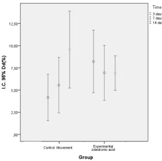
Tooth Movement
No statistically significant difference was observed (p>0.05) between the CM and EZA groups (those subjected to the orthodontic device) at the three observation times (3, 7 and 14 days), when the Games-Howell multiple comparisons test was used (Table 1, Fig. 5). The CWM and CZA groups showed null movement, due to the absence
of orthodontic device.
Bone Resorption
Using the Games-Howell multiple comparisons test for the group × time variable, it was determined that the CM group showed statistically significant difference (p<0.05) compared to the CZA, at all times, and the EZA groups at days 3 and 14 after drug administration. The CM and CWM group showed a higher number of osteoclasts than did the other groups in which zoledronic acid was administered, indicating that the amount of bone resorption was greater in the group without the drug (Table 2, Figs. 2 and 6).
New Bone Formation
The percentage of Type I and Type III collagen in the bone was assessed using the Games-Howell multiple comparisons test, and the CWM group showed statistically significant difference (p>0.05) compared to the CZA at day 14 (Table 2, Figs. 3 and 7).
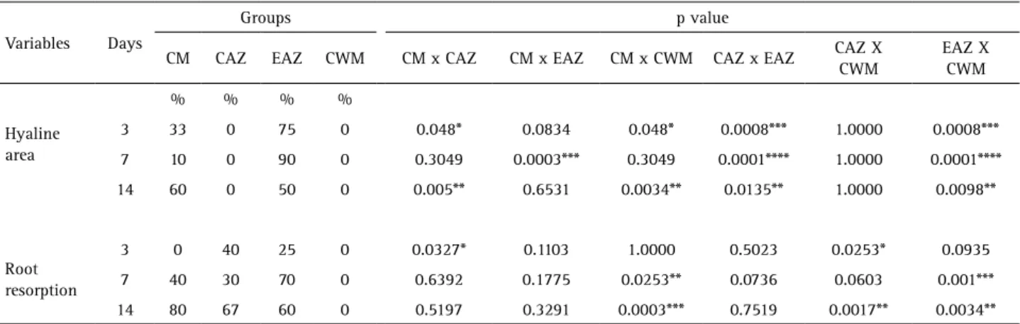
Hyaline Areas
The groups to which tooth movement were made (CM and EZA) showed a higher percentage of hyaline area. (Fig. 4)
On day 7 day, the EZA group showed a larger hyaline area (90%) than the CM group (10%). However, on days 3 and 14, no statistically significant difference (p>0.05) was observed (Table 3).
Root Resorption
No statistically significant difference (p>0.05) was observed between the CM and EZA groups at any time point (Table 3).
Discussion
Bisphosphonates inhibit osteoclastic activity and microcirculation, and may inhibit tooth movement (2,5,15). Zoledronic acid was used in this study, as it is a novel bisphosphonate and its relationship to dental movement has not been studies in detail.
Zoledronic acid is administered to humans in a single dose, and repeated annually according to the needs of each patient. Bisphosphonates are poorly absorbed in the human intestine due to their lipophilicity. However, systemically, bisphosphonates disappear very rapidly from the plasma, and are partly taken up by the bone and partly excreted by the kidney (17). With the aim of assessing the effects of zoledronic acid on periodontal structures over time, the drug was administered one week prior to the installation of the orthodontic device, similar to other studies (1,9).
A smaller number of osteoclasts was observed in the CZA and EZA groups in relation to the CM group at all times points, showing no variation over time (Table 2). Table 1. Mean, standard deviation and p value of the tooth movement
in the movement control group (CM) and the experimental zoledronic acid (EZA) group on days 3, 7 and 14
Variable CM EZA CM x EZA
Dental Movement
3 days 4.17 ± 3.65 8.22 ± 4.92 0.5123
7 days 5.56 ± 4.37 6.94 ± 4.33 0.9979
14 days 9.56 ± 6.05 6.92 ± 2.72 0.9263
Games-Howell Test with a 5% level of significance. * p=<0.05; ** p=<0.01; *** p=<0.001; ****p=<0.0001.
M.D. Brunet et al. F
. Monteiro-Amado et al. Table 2. Mean, standard deviation and p value of the variables: number of osteoclasts, percentage of Type I collagen, percentage of Type III
collagen in the movement control group (CM), the zoledronic acid (CZA) control group, the experimental (EZA) group and the control without movement (CWM) group, on days 3, 7 and 14
Variable
Mean ± SD p value
Power
CM CWM CZA EZA CM x
CZA CM x EZA CM x CWM CWM x CZA CWM x EZA CZA x EZA Osteoclasts 3 17.56 ± 7.71 5.68 ± 1.84 2.79 ± 1.98 2.89 ±
2.00 0.0093** 0.0095** 0.0363* 0.0986 0.1876 0.1000
1.000 7 11.79 ± 7.00 6.51 ± 1.25 2.03 ± 1.64 2.87 ±
1.09 0.0397* 0.0654 0.5054 0.0001**** 0.0001**** 0.9599
14 13.79 ± 4.05 6.63 ± 1.54 1.51 ± 0.98 1.96 ±
0.50 0.0001**** 0.0002**** 0.0077** 0.0000**** 0.0001**** 0.9749
Type I collagen
3 78.00 ± 11.71 94.05 ± 3.17 78.23 ± 16.78 80.68 ±
16,69 0.1000 0.1000 0.0688 0.2600 0.6437 0.1000
0.9752
7 78.21 ±
12.35 92.95 ± 3.37 80.84 ± 19.96 83,12 ±
10.32 0.1000 0.9986 0.2561 0.7430 0.3322 0.1000
14 80.47 ± 8.97 95.49 ± 2.71 87.35 ± 5.28 82,66 ±
12,89 0.7885 0.1000 0.0686 0.0383* 0.3395 0.9951
Type III collagen
3 22.00 ± 11.71 5.94 ± 3.17 21.77 ± 16.78 19.32 ±
16.69 0.1000 0.1000 0.0688 0.2600 .6437 .1000
0.9752
7 21.79 ±
12.35 7.04 ± 3.37 19.16 ± 19.96 16.88 ±
10.32 0.1000 0.9986 0.2561 0.7430 .3322 .1000
14 19.53 ± 8.97 4.50 ± 2.71 12.65 ± 5.28 17.34 ±
12.89 0.7885 0.1000 0.0686 0.0383* .3395 .9951
Games-Howell Test with a 5% level of significance. *p=0<.05; **p=0<.01; ***p=0<.001; ****p=0<.0001.
Figure 6. Comparison between groups x time of the number of osteoclasts variable.
This result represents a zoledronic acid-induced decrease in osteoclastic activity with consequently decreased bone remodeling, and is similar to the findings of previous studies by Ozturk et al.(1) and Huja et al. (18) with zoledronic acid in rats and dogs, respectively (1,18). The results of this study suggest that the drug was effective, since its purpose is to inhibit the function and induce the apoptosis of osteoclasts (1-3,18).
Studies show that, following application of orthodontic force, cells and collagen fibers are compressed and stretched on the compression and tension sides, respectively. The biological process proceeds with the recruitment of osteoclasts and osteoblasts, and with alveolar bone remodeling (15).
This study also sought to evaluate structural changes in the trabecular bone matrix, newly formed in the simultaneous presence of tooth movement and zoledronic acid, with the picrosirius method using polarized light. This enabled the detection of mature and immature collagen fibers and the correlation of the three-dimensional distribution of collagen fibers with the stage of bone formation (Fig. 3) (19).
The organic matrix of alveolar bone consists mainly of
Effects of zoledronic acid on tooth movement.
Table 3. Percentage and p value of the presence of variables: hyaline area and root resorption in the movement control group (CM), the zoledronic acid (CZA) control group, the experimental (EZA) group, and the control without movement (CWM) group, on days 3, 7 and 14
Variables Days
Groups p value
CM CAZ EAZ CWM CM x CAZ CM x EAZ CM x CWM CAZ x EAZ CAZ X
CWM
EAZ X CWM
Hyaline area
% % % %
3 33 0 75 0 0.048* 0.0834 0.048* 0.0008*** 1.0000 0.0008***
7 10 0 90 0 0.3049 0.0003*** 0.3049 0.0001**** 1.0000 0.0001****
14 60 0 50 0 0.005** 0.6531 0.0034** 0.0135** 1.0000 0.0098**
Root resorption
3 0 40 25 0 0.0327* 0.1103 1.0000 0.5023 0.0253* 0.0935
7 40 30 70 0 0.6392 0.1775 0.0253** 0.0736 0.0603 0.001***
14 80 67 60 0 0.5197 0.3291 0.0003*** 0.7519 0.0017** 0.0034**
Chi-squaretest with a significance level of 5%. P value referring to the difference test between two proportions. *p=0<.05; **p=0<.01; ***p=0<.001; ****p=0<.0001
Figure 7. Comparison between groups x time of the % of type I and III collagen on bone. events that involve the differentiation of osteoblasts
from primitive mesenchymal cells, synthesis of the organic matrix, and its maturation and complete mineralization (10).
The results of this study revealed that the groups receiving zoledronic acid and tooth movement showed no statistically significant difference in the percentage of type I and type III collagen in the alveolar bone, compared with that in the group that did not use the drug (p>0.05) at any time point (Table 2). This indicates that the zoledronic acid did not interfere with the process of new bone formation during tooth movement. Kubek et al.(20) reported that rats receiving zoledronic acid showed significantly suppressed medullary remodeling in the maxillary bones. Although
zoledronic acid is largely absorbed in maxillary bone structures (18,21), the results found in this study differed from those of Kubek et al.(20), possibly because these authors administered the drug in two intravenous doses, thus enabling the maxillary bone structures, which are highly irrigated, to achieve greater concentrations of the drug. Moreover, the animals in that study were female and ovariectomized. Under those conditions, they present behavior similar to women in the postmenopausal phase, during which they are more prone to osteoporosis and show greater bone remodeling (20).
M.D. Brunet et al. F
. Monteiro-Amado et al.
(2,3,12). Knowing the possibility of reduction in tooth movement, this study aimed to assess the rate of tooth movement. However, the results showed no statistically significant difference (p>0.05) between any groups during the times evaluated.
The force applied to the tooth during tooth movement compresses the vessels of the periodontal ligament, generates hypoxia, and results in leakage and/or cell death. The extracellular matrix alters the biochemistry and organizational relationship of the components, resulting in areas of greater protein concentration with a union of dense bundles of collagen fibers. This results in areas with low cell numbers and with a homogeneous eosinophilic aspect, called the hyaline areas of the extracellular matrix (23).
According to Table 3, the EZA group showed a larger percentage of hyaline areas (90%) when compared to the CM group (10%) at day 7; however, at other time points, no statistically significant difference was observed. This difference in the presence of hyaline areas on day 7 occurred along with a reduction in the percentage of hyaline areas in the CM group and an increase in the percentage of hyaline areas in the EZA group. This may indicate that during bone healing in areas in which hyaline was reduced without the use of the drug, zoledronic acid increased the percentage of hyaline areas.
Root resorption is a well-known, adverse effect of orthodontic treatment. In assessing the conditions of the tooth and periodontal ligament of rats subjected to orthodontic movement, the presence of areas of root resorption is reported (24,25).
In the present study, the animals from the CM and EZA groups showed no statistically significant difference in the amount of root resorption (p>0.05) at any times point. This does not indicate that use of the drug altered the amount of root resorption. Contrary to the results of this study, the literature reports that when treated with bisphosphonates, mice that underwent tooth movement showed less root resorption (3,24). Mori et. al.(24) observed reduced root resorption in rats where zoledronic acid was applied actively to the root surface of re-implanted teeth, and not in response to systemic, intraperitoneal administration, which was used in this study.
In another study, Sirisoontorn et al.(3) reported that ovariectomized rats subjected to tooth movement and treated with zoledronic acid, showed reduced root resorption relative to rats not exposed to the drug. This difference in findings may have occurred because Sirisoontorn et al.(3) compared the amount of resorption between ovariectomized rats with and without zoledronic acid treatment. Ovariectomized rats have a greater propensity for root resorption than non-ovariectomized rats do; thus, it is possible that the drug did not have
any effect, and the amount of root resorption, which was already higher, persisted.
There is a large demand by adults for orthodontic treatment and the use of zoledronic acid is becoming more widespread among these patients. Therefore, orthodontists must inform patients about the possible adverse effects of treatment and the possibility of a longer treatment time because a longer interval between the activation of the apparatus is recommended for these patients considering the influence of the drug on bone resorption (2). Even if there were no statistical differences in the tooth movement, bone remodeling and bone resorption, we must be careful in the treatment of these patients because there were statistically significant histological differences such as for example fewer osteoclasts resulting in decreased bone resorption, and increased percentage of hyaline areas. Thus, it should be recommended that the medical history must contain questions about medications used by the patients, keeping in mind that they may influence tooth movement, even many years after they are discontinued.
Resumo
A proposta deste estudo foi avaliar os efeitos da administração do ácido zoledrônico (ZA) durante a movimentação ortodôntica em ratos. Cento e vinte ratos Wistar, machos, foram submetidos a aplicação de uma força de 30 cN através de uma mola fechada de níquel-titânio para mover o primeiro molar superior direito para mesial. No grupo Controle Movimentação (CM), apenas a movimentação dentária foi realizada; o grupo Controle Ácido Zoledrônico (CAZ) recebeu uma única dose (0,1 mg/kg) de ZA; o grupo Experimental Ácido Zoledrônico (EAZ) recebeu uma única dose (0,1 mg/Kg) uma semana antes do início da movimentação dentária; e o grupo Controle Sem Movimentação (CWM) não receberam nenhum tipo de droga e não foi realizado movimentação dentária. Os animais foram eutanásiados após 3, 7 e 14 dias. A movimentação dentária foi mensurada através de um paquímetro, o número de osteoclastos utilizando coloração TRAP, a expressão do colágeno maturo e imaturo através da coloração Picrosírius, e a presença de áreas hialinas e reabsorção radicular utilizando HE. Os dados foram comparados utilizando ANOVA a dois critérios, Tukey HSD, Games-Howell e teste de qui-quadrado, ao nível de significância de 5%. Verificou-se menor número de osteoclastos e maior porcentagem de área hialina no grupo EAZ. Não houve diferença entre grupos quanto à neoformação óssea, reabsorção radicular e movimentação dentária em todos os tempos observados.
References
1. Ozturk F, Babacan H, Gumus C. Effects of zoledronic acid on sutural bone formation: a computed tomography study. Eur J Orthod 2012;34:141-146.
2. Rinchuse DJ, Sosovicka MF, Robison JM, Pendleton R. Orthodontic treatment of patients using bisphosphonates: a report of 2 cases. Am J Orthod Dentofacial Orthop 2007;131:321-326.
3. Sirisoontorn I, Hotokezaka H, Hashimoto M, Gonzales C, Luppanapornlarp S, Darendeliler MA, et al.. Orthodontic tooth movement and root resorption in ovariectomized rats treated by systemic administration of zoledronic acid. Am J Orthod Dentofacial Orthop 2012;141:563-573. 4. Baroncelli GI, Bertelloni S. The use of bisphosphonates in pediatrics.
Horm Res Paediatr 2014;82:290-302.
Effects of zoledronic acid on tooth movement. 6. Wayama MT, Yoshimura H, Ohba S, Yoshida H, Matsuda S, Kobayashi J,
et al.. Diminished progression of periapical lesions with zoledronic acid in ovariectomized rats. J Endod 2015;41:2002-2007.
7. Kimmel DB. Mechanism of action, pharmacokinetic and pharmacodynamic profile, and clinical applications of nitrogen-containing bisphosphonates. J Dent Res 2007;86:1022-1033. 8. Hao Y, Wang X, Wang L, Lu Y, Mao Z, Ge S, et al.. Zoledronic acid
suppresses callus remodeling but enhances callus strength in an osteoporotic rat model of fracture healing. Bone 2015;81:702-711. 9. Little DG, Peat RA, McEvoy A, Williams PR, Smith EJ, Baldock PA.
Zoledronic acid treatment results in retention of femoral head structure after traumatic osteonecrosis in young Wistar rats. J Bone Miner Res 2003;18:2016-2022.
10. Retamoso L, Knop L, Shintcovsk R, Maciel JV, Machado MA, Tanaka O. Influence of anti-inflammatory administration in collagen maturation process during orthodontic tooth movement. Microsc Res Tech 2011;74:709-713.
11. Knop LA, Shintcovsk RL, Retamoso LB, Ribeiro JS, Tanaka OM. Non-steroidal and Non-steroidal anti-inflammatory use in the context of orthodontic movement. Eur J Orthod 2012;34:531-535.
12. Choi J, Baek SH, Lee JI, Chang YI. Effects of clodronate on early alveolar bone remodeling and root resorption related to orthodontic forces: a histomorphometric analysis. Am J Orthod Dentofacial Orthop 2010;138:548 e541-548; discussion 548-549.
13. Ren Y, Maltha JC, Kuijpers-Jagtman AM. The rat as a model for orthodontic tooth movement--a critical review and a proposed solution. Eur J Orthod 2004;26:483-490.
14. Franzon Frigotto GC, Miranda de Araujo C, Guariza Filho O, Tanaka OM, Batista Rodrigues Johann AC, Camargoa ES. Effect of fluoxetine on induced tooth movement in rats. Am J Orthod Dentofacial Orthop 2015;148:450-456.
15. Braga SM, Taddei SR, Andrade I Jr., Queiroz-Junior CM, Garlet GP, Repeke CE, et al.. Effect of diabetes on orthodontic tooth movement in a mouse model. Eur J Oral Sci 2011;119:7-14.
16. Marquezan M, Bolognese AM, Araujo MT. Effects of two low-intensity laser therapy protocols on experimental tooth movement. Photomed Laser Surg 2010;28:757-762.
17. Lin JH. Bisphosphonates: a review of their pharmacokinetic properties. Bone 1996;18:75-85.
18. Huja SS, Kaya B, Mo X, D’Atri AM, Fernandez SA. Effect of zoledronic acid on bone healing subsequent to mini-implant insertion. Angle Orthod 2011;81:363-369.
19. Garavello-Freitas I, Baranauskas V, Joazeiro PP, Padovani CR, Dal Pai-Silva M, da Cruz-Hofling MA. Low-power laser irradiation improves histomorphometrical parameters and bone matrix organization during tibia wound healing in rats. J Photochem Photobiol B 2003;70:81-89. 20. Kubek DJ, Burr DB, Allen MR. Ovariectomy stimulates and
bisphosphonates inhibit intracortical remodeling in the mouse mandible. Orthod Craniofac Res 2010;13:214-222.
21. Cheer SM, Noble S. Zoledronic acid. Drugs 2001;61:799-805; discussion 806.
22. Santamaria M, Jr., Fracalossi AC, Consolaro MF, Consolaro A. Influence of bisphosphonates on alveolar bone density: a histomorphometric analysis. Braz Oral Res 2010;24:309-315.
23. Kohno T, Matsumoto Y, Kanno Z, Warita H, Soma K. Experimental tooth movement under light orthodontic forces: rates of tooth movement and changes of the periodontium. J Orthod 2002;29:129-135. 24. Mori GG, Janjacomo DM, Nunes DC, Castilho LR. Effect of zoledronic
acid used in the root surface treatment of late replanted teeth: a study in rats. Braz Dent J 2010;21:452-457.
25. Cuoghi OA, Aiello CA, Consolaro A, Tondelli PM, Mendonca MR. Resorption of roots of different dimension induced by different types of forces. Braz Oral Res 2014;28.