ISSN 0104-6632 Printed in Brazil
www.abeq.org.br/bjche
Vol. 31, No. 03, pp. 747 - 756, July - September, 2014 dx.doi.org/10.1590/0104-6632.20140313s00002307
Brazilian Journal
of Chemical
Engineering
MYOGLOBIN ENTRAPMENT IN POLY(VINYL
ALCOHOL) DENSE MEMBRANES
K. C. S. Figueiredo
1*, T. L. M. Alves
and C. P. Borges
Universidade Federal do Rio de Janeiro, Programa de Engenharia Química/COPPE, Av. Horácio Macedo, 2030, Centro de Tecnologia, Bloco G, Sala 115, C. P. 68502, CEP: 21941-914, Cidade Universitária, Ilha do Fundão, Rio de Janeiro - RJ, Brazil.
Phone: + (55) (21) 25628351, Fax: (55) (21) 25628300 1
Current Address: Departamento de Engenharia Química, Universidade Federal de Minas Gerais, Av. Antônio Carlos 6627, Escola de Engenharia, Bloco 2, Sala 5107,
CEP: 31270-901, Pampulha, Belo Horizonte - MG, Brazil. Phone: + (55) (31) 3409-3627, Fax: + (55) (31) 3409-1789
E-mail: [email protected]
(Submitted: September 28, 2012 ; Revised: September 19, 2013 ; Accepted: October 15, 2013)
Abstract - Our goal in this study was the immobilization of myoglobin in poly(vinyl alcohol) dense membranes. Glutaraldehyde was investigated both as the crosslinking agent, aiming to increase the membrane stability in aqueous medium, and as the vehicle to bind myoglobin and PVA. Reaction and membrane synthesis were carried simultaneously in mild operating conditions in order to maintain the native protein folding. Membrane characterization comprised the water swelling degree, DSC, TGA, UV-visible spectroscopy, FTIR analysis and oxygen transport in a dialysis cell. The incorporation of myoglobin in the film decreased the water swelling degree and improved the membrane thermal properties compared to unmodified PVA membrane. The reduction of ferric iron in the prosthetic group of the protein to the ferrous form was observed. The increased affinity between oxygen and the immobilized myoglobin did not favor the release of this solute from the biocarrier.
Keywords: Myoglobin; Oxygen/nitrogen separation; Poly(vinyl alcohol); Membranes; Immobilization.
INTRODUCTION
Oxygen is one of the most important commodi-ties, being used in a broad range of applications such as the metallurgical industry. Moreover, ultra-pure oxygen is frequently needed in the medical field. The natural source of molecular oxygen is atmospheric air, where it corresponds to approximately 20% in volume. Air fractionation is attained by cryogenic technologies, such as distillation or pressure swing adsorption. These methods have none or very little expected advancement regarding process efficiency and energy consumption. The high energy demand of the traditional technologies has motivated the
devel-opment of alternative routes to accomplish oxy-gen/nitrogen separation (Baker, 2002).
Gas separation through dense membranes emerged as an advantageous technique in 1980, when the synthesis of anisotropic membranes caused the hy-drogen/nitrogen separation to be economical and conquered industrial acceptance (Baker, 2002). The high selectivity and low energy input of such tech-nology decreased the operating costs. Since then, attempts to develop membranes with higher selec-tivity and flux are the main task to fulfill economical and technical requirements (Robeson, 2008).
oxygen-enriched streams, due to the fact that the feed is diluted in the latter and it usually permeates preferentially (Baker, 2004). Therefore, the de-velopment of membranes with better selectivity to molecular oxygen is pursued by means of altering the packing density of the polymer, inducing mo-lecular sieving (Wang et al., 2005), or the modifica-tion of the polymer top layer by plasma (Ruaan et al., 1998). The literature reported an increase in the oxygen selectivity over nitrogen up to 10, followed by a reduction in permeate flux. Despite being the most studied gas pair, oxygen/nitrogen separation has shown only minor displacement of the upper bound limit, defined as the line combining materials with the highest separation factor and the highest permeability (Robeson, 2008).
Facilitated transport membranes comprise an al-ternative to the mechanism of simple diffusion. The facilitation is attained with the addition of a specific carrier to the desired component. The carrier-medi-ated transport can be described in three steps: a) the reaction between the carrier and the solute at the feed/ membrane interface, b) the diffusion of the complex through the membrane and c) the decoupling reac-tion at the membrane/permeate interface. Transport due to simple diffusion is also likely to occur. How-ever, the facilitated mechanism is favored for low partial pressures of the solute, like oxygen in air. The increase of the partial pressure of the component may cause the saturation of the carrier sites (Baker, 2004, Ferraz et al., 2007).
Natural oxygen carriers, like hemoglobin, were evaluated in ex-vivo tests by means of supported liquid membranes in the early 1960s (Scholander, 1960). Hemoglobin-containing membranes exhibited a facilitation factor of 8, compared to pure water, and a selectivity oxygen/nitrogen of 14, but the autoxi-dation of the carrier revealed a lack of stability. The knowledge of the bioinorganic chemistry of the bonding of molecular oxygen and the central iron atom in the heme group led to the development of synthetic carriers, mimicking the prosthetic envi-ronment (Baker, 2002). The use of such carriers in supported liquid membranes reached the apex in 1985, when Roman and Baker (1985) patented a system with O2/N2 selectivity of 30 and oxygen
per-meability of 1,500 Barrer, operating at 25 °C for up to three months. However, the evaporation losses of solvent and carrier oxidation brought about the vestigation of more stable configurations. For in-stance, Nishide and co-authors proposed the use of cobalt-based synthetic carriers immobilized in a polymeric matrix (Shoji et al., 2008). Although high selectivity was achieved, up to 118, the flux of 2.6
Barrer and the membrane lifetime of 3 months were low (Nishide et al., 1998). The minimum ideal con-ditions presented by Figoli and co-workers for a competitive membrane-based process are O2/N2
se-lectivity of 20, permeate flux of 0.015 m3m-2h-1bar-1, with 1 stage operation, feed pressure lower than 10 bar and temperatures between 0 and 40 °C. The minimum lifetime expected for the carrier is 1 year (Figoli et al., 2001).
The market size of oxygen and the spectacular selectivity of facilitated transport membranes are responsible for maintaining the interest of research-ers in this area, especially because of the impact that competitive cost oxygen-enriched air could cause in many niches (Baker, 2002). For instance, the devel-opment of the artificial gill, an apparatus to remove oxygen from water to air, is closely related to facili-tated transport membranes (Nagase et al., 2003).
The investigation of the role of the globin on the stability of hemeproteins (Sugawara et al., 1995) and the development of Molecular Biology tools, which make feasible changes in the amino acid sequence (Hargrove et al., 1996), justify the regained interest in the use of these biocarriers in facilitated transport membranes. The main breakthrough in this area is the stabilization of the biomolecules inside the poly-meric matrix, preserving their physiological function. Considering the importance and applicability of the hemeproteins, the goal of this work comprises the understanding of the behavior and stability of the stroma-free biocarriers, which can be useful in other areas, such as in biosensors (Vidal et al., 1999) and artificial blood (Eike and Palmer, 2004).
In this paper, we investigated the immobilization of myoglobin in poly(vinyl alcohol), PVA, aiming at the synthesis of facilitated transport membranes for oxygen permeation. Myoglobin, Mb, was selected because it has only one oxygen-binding site, whereas the choice of PVA was based on its hydrophilicity, biocompatibility and film-forming ability. Oxygen transport tests were performed in the liquid phase, by using a dialysis cell equipped with an oxygen sensor. Tests were performed in the liquid phase because the oxygen permeability through the semi-crystalline PVA is about 0.0019 Barrer (Mulder, 1996).
EXPERIMENTAL
Materials
Poly(vinyl alcohol), PVA, (85-146 kDa, 99%), glu-taraldehyde, GA, sol. 50 wt% and horse heart my-oglobin (minimum 90%), Mb, were purchased from Aldrich® (Milwaukee, WI). All solutions were pre-pared using distilled water.
Poly(Vinyl Alcohol) Membranes
PVA was dissolved in distilled water at 100 °C. The solution was cooled to room temperature. Met-myoglobin (Fe3+) was dissolved in distilled water at 25 °C. The solution was transferred to a flask con-taining PVA aqueous solution and stirred with a magnetic stirring bar at 50 rpm, for 3 minutes. GA solution was transferred to the system by using an automatic pipette. The casting solution was stirred for 1 minute, at 50 rpm. PVA content was fixed at 4 wt% and the final pH was 6.2 + 0.1. The casting solution was poured into a Petri dish in order to pre-pare flat dense membranes by solvent evaporation. The system was dried at 40 °C for 15 hours. Reac-tion and membrane formaReac-tion were conducted si-multaneously.
The effects of myoglobin and glutaraldehyde contents on membrane properties were investigated by means of altering the protein and the aldehyde mass ratio to PVA, respectively. The GA/PVA mass ratio was set at 0.2 g/g, whereas the Mb/PVA ratio was varied from 0.2 to 1.9 g/g.
Membrane Characterization
Water swelling, UV-visible spectroscopy, TGA, DSC and FTIR were performed for the characteri-zation of membrane properties. They were used to infer the crosslink density, myoglobin physiological form, thermal stability, glass transition temperature, melting enthalpy and the chemical structure of the films.
Water Swelling
Films were cut into strips of (2 x 2) cm2 and im-mersed in distilled water for 48 hours, at 40 °C (Yeom and Lee, 1996). Then, the swollen size, lw, was
recorded and the strips were placed in a desiccator, at 25 °C, in vacuo, until constant weight, when the dried membrane size, ld, was measured. The water
swelling degree, R, was calculated by Equation (1):
(
w d)
d l l 100 R
l
− ×
= (1)
UV-Visible Spectroscopy
Strips of (0.4 x 2) cm2 of dense membranes were placed in the cuvette of an UV-visible spectrometer (UV mini 1240, Shimadzu, Tokyo, Japan) and the absorbance spectra were compared in order to deter-mine the physiological form of myoglobin in the polymeric film. PVA films with no added-myoglobin were used as the reference.
There are three physiological forms of myoglo-bin: metmyoglobin, metmb (iron III), which is bio-logically inactive, deoxymyoglobin, deoxymb (iron II not bound to oxygen), and oxymyoglobin, oxymb (iron II bond to molecular oxygen). The color of each myoglobin form is different. For instance, the typical absorbance pattern of metmb (409 and 630 nm) differs significantly from oxymb (418, 543 and 581 nm) and this property was used to infer possible changes in the protein form during membrane syn-thesis.
Infrared Spectroscopy
The functional groups of the modified PVA were determined by FTIR analysis (1720X, PerkinElmer) of the membranes. Each sample was scanned 20 times with resolution of 2 cm-1. The incorporation of my-oglobin into the film was evaluated by means of the absorbance pattern typical of alpha-helical proteins.
Thermal Analysis
Differential scanning calorimetry, DSC, and ther-mogravimetric analysis, TGA, were performed to characterize the modification of the thermal proper-ties of PVA due to crosslinking and bonding to myoglobin.
For TGA tests (TGA 7, PerkinElmer, Norwalk, CT), 8 mg of the sample were heated from 50 to 600 °C at 10 °C/min. The weight loss as a function of temperature was used to infer the chemical modifi-cations in the polymer structure.
Membrane Performance
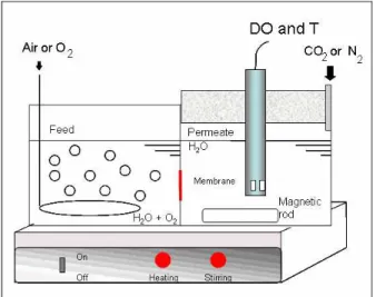
The permeability of oxygen in myoglobin-con-taining membranes was measured in liquid phase, by using a dialysis cell. The apparatus is presented in Figure 1. The membrane was placed between the feed and permeate, using two cellulose acetate mi-crofiltration membranes (Millipore®, with pore di-ameter of 0.45 μm and 30% porosity) to increase the mechanical resistance.
Figure 1: Scheme of the dialysis cell used in oxygen permeation tests. DO and T refers to the dissolved oxygen and temperature sensor.
Oxygen was supplied by bubbling compressed air or pure oxygen on the feed side. Permeate was de-oxygenated using nitrogen or carbon dioxide prior to the experiment. A dissolved oxygen and temperature sensor (5739 DO, model 58, YSI, Yellow Springs, OH), DO and T in Figure 1, was used to quantify oxygen content on the permeate side. Measurements were taken for 30 minutes, in time intervals of 30 seconds. Tests were performed in triplicate, at 25 and 40 °C.
Oxygen Affinity
The affinity between the myoglobin–immobilized PVA membranes and oxygen was investigated by means of UV-visible spectroscopy. The method was developed in order to characterize any changes in the oxygen dissociation curve, compared to the non-immobilized myoglobin.
Strips of (2 x 0.4) cm2 of the samples were placed in the cuvette of a UV-visible spectrometer. Distilled water was added to the system, which was closed to the atmosphere. The solution was bubbled with gas
streams with different compositions (pure N2, pure
O2 and O2/N2 mixtures of 5:95, 10:90 e 15:85 and
21:79 in weight) for at least 2 hours, until the equi-librium was reached. The system was sealed and the visible spectra of the strips were recorded. Absorb-ance at 543, 556 and 581 nm was used to determine the physiological form of the immobilized myoglobin.
RESULTS AND DISCUSSION
The immobilization of myoglobin in the PVA matrix was investigated by using GA, a bifunctional aldehyde, which is able to bind the protein and the polymer together. The mechanism of the reaction of aldehydes and hydroxyl groups is well known. How-ever, the reaction between proteins and glutaralde-hyde is non-specific (Migneault et al., 2004).
The main challenge was to perform the immobili-zation of the biomolecule in mild operating condi-tions so that the structure and biological activity of the carrier were maintained. This task was investi-gated by promoting the reaction in acid free medium and in the absence of organic solvent. The crosslink-ing of PVA in such conditions was accomplished in a former work (Figueiredo et al., 2009). The exis-tence of an optimum GA/PVA ratio was shown for the reaction conducted at 40 °C. An excess of GA molecules can promote PVA branching and conse-quently increase the spacing among the polymeric chains. In the absence of metmyoglobin, the lowest water swelling degree of GA-modified PVA mem-branes was reached for a GA/PVA mass ratio of 0.01 g/g. Membrane swelling was (44 + 5)% (Figueiredo
et al., 2009).
In this work, membrane synthesis was conducted with metmyoglobin, the inert form of the protein, since the primary objective at this stage was the evaluation of the protein entrapment in PVA. The experimental procedure commonly used to accom-plish iron reduction is based on the reaction of met-myoglobin and sodium dithionite, followed by gel filtration of the oxymyoglobin and a concentration step, using an ultracentrifuge. This protocol was avoided considering that the chemical behavior of the different physiological forms of myoglobin in the reaction with PVA and GA would be the same, re-gardless of the oxidation state of iron.
the entrapment of metmb into the polymeric chain. Therefore, the GA/PVA mass ratio was set at 0.2 g/g, while the myoglobin content related to PVA was investigated from 0.2 to 1.9 g/g. The results for water swelling tests of myoglobin–containing PVA mem-branes are presented in Table 1. Tests were made in triplicate in some experimental conditions and the deviation was calculated. Membrane average thick-ness was 30 μm.
Table 1: Water swelling degree (R) of the mem-branes at 25 °C. GA/PVA mass ratio was 0.2 g/g.
Mb/PVA mass ratio (g/g) R (%)
0.2 Infinite 0.4 19
0.9 23 ± 1
1.4 15 ± 5
1.9 20 ± 2
It was noted that the increase in GA content caused the major reduction in the water swelling, compared to the increase in Mb content. Membranes with a GA/PVA ratio of 0.2 g/g and Mb/PVA of 1.4 g/g showed the best result, with an average water swelling degree of 15%. For PVA/Mb/GA of 1:1.9:0.2, the water swelling degree was around 20%. This result can be explained in terms of the reaction between the hydroxyl groups of PVA and Mb with GA, which leads to crosslinking and reduces chain mobility. As a consequence, membrane swelling in water was decreased.
It was noted that the increase in Mb/PVA mass ratio from 0.2 to 0.4 g/g caused significant reduc-tions in the water swelling degree of the membranes. In addition, no protein was extracted to the aqueous phase. Samples with Mb/PVA mass ratio higher than 1.9 showed phase separation and poor flexibility.
The change in water swelling degree and absence of protein extraction evidenced the reaction between Mb, GA and PVA. As a consequence, the tridimen-sional network formed by Mb-containing GA-cross-linked PVA membranes was changed. It probably decreased PVA free volume. The unmodified PVA membrane dissolves in water while Mb/GA/PVA samples showed an average water swelling degree, R, of 19%. It can be used to infer the crosslinking density of PVA, which changed because Mb and GA changed the spatial organization of the polymeric chains.
These results indicated that Mb can be attached to the PVA matrix by means of the reaction with GA in mild conditions such as pH of 6.2, 40 °C and sol-vent-free medium up to a Mb/PVA mass ratio of 1.9. The evidence of the reaction between PVA, GA and
Mb is that the extraction of the protein from the membrane was not noted during water swelling tests.
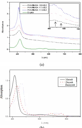
UV-visible spectra of myoglobin-containing mem-branes were recorded for the investigation of the physiological form of the protein. The original color of metmyoglobin is brown. The red color of the samples suggested the reduction of iron during membrane synthesis. This color change is another evidence of the reaction between Mb, GA and PVA. The spectra are presented in Figure 2a, while the typical spectra of the physiological form of the pro-tein are presented in Figure 2b. The sample without myoglobin was used as a reference (blank) as well as the aqueous solution of oxymyoglobin. The baseline displacement can be attributed to local fluctuations in membrane thickness.
(a)
(b)
Figure 2: (a)Visible spectra of Mb–immobilized GA- crosslinked PVA membranes and (b) Typical spectra of metmyoglobin, oxymyoglobin and deoxymyoglobin.
indi-cated that the physiological form of myoglobin im-mobilized in the film was oxymb (Fe2+ — O2), which
is biologically active.
This surprising result led to the conclusion that the reaction mixture caused the reduction of iron (III) to iron (II), which simplified the procedure of mem-brane synthesis. The procedure to reduce metmy-oglobin to oxymymetmy-oglobin comprises the protein re-action with sodium dithionite, followed by gel filtra-tion and concentrafiltra-tion by means of an ultracentri-fuge. The advance of membrane preparation technique allowed the use of metmyoglobin to obtain oxymy-oglobin in a quick and labor-saving way.
It should be pointed out that myoglobin showed the same UV-visible spectrum as the protein in aque-ous solution (continuaque-ous line in Figure 2a), which evidenced that the microenvironment regarding to the electron configuration in the heme prosthetic group was maintained. In another words, the reaction between PVA, GA and Mb changed the iron oxida-tion state and the resulting UV-vis profile was typical of oxymyoglobin.
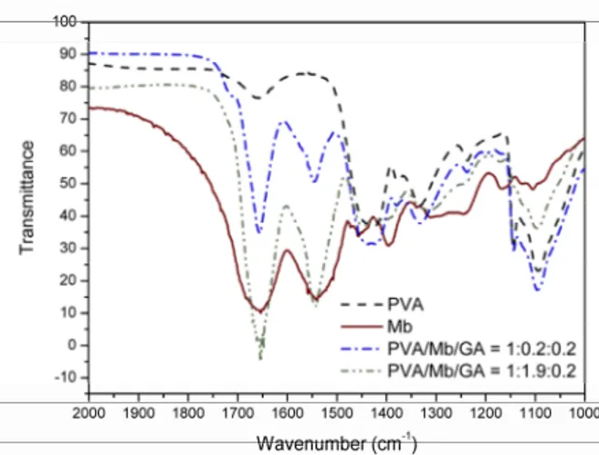
The chemical characterization of modified PVA membranes was performed by Fourier Transform Infrared analysis. The spectra for the commercial myoglobin (lyophilized sample in KBr) and PVA films are shown in Figure 3.
Figure 3: Infrared spectra of myoglobin–immobi-lized PVA membranes compared to lyophimyoglobin–immobi-lized Mb and unmodified PVA sample.
For lyophilized metmyoglobin, the infrared spec-trum showed two main peaks, which were attributed to amide I (from 1615 to 1700 cm-1) and amide II (from 1650 to 1515 cm-1) bands. In our case, the highest absorbance occurred at 1652 and 1544 cm-1. Amide I is related to the C = O stretching vibration of the peptide linkage in the protein backbone.
Am-ide II is due to the combination of N – H bending and C – N stretching (Wang et al., 2004). Regarding the unmodified PVA sample, a small band was noted around 1650 cm-1, probably related to the C = O stretching of residual acetate groups in the 99% hy-drolyzed PVA.
Both PVA films containing Mb and GA showed peaks at 1652 and 1544 cm-1, typical of amide I and amide II bands. Altogether, these results suggested that the secondary structure of the protein was highly maintained in the film. According to the literature, alpha-helical proteins, like myoglobin, show the highest band around 1655 cm-1, while beta-sheets shows two bands in the amide I region, at 1630 and 1690 cm-1 (Arkin, 2006). Moreover, if Mb was de-naturated during the reaction and film synthesis, amide I and II bands would have diminished consid-erably (Wang et al., 2004).
It should be pointed out that the quantitative in-formation about protein secondary structure from FTIR spectra has been studied (Goormaghtigh et al., 2006). It could reveal how much of the protein native folding remained intact in the film. The native fold-ing of a protein is the most stable thermodynamic state of the structure. Although it is a consensus that the native folding is greatly responsible to maintain the biological function of proteins, the idea that it should be 100% intact to allow its function is con-troversial (Guo and Clark, 2001).
The thermal properties of the membranes were analyzed by DSC and TGA. TG curves are presented in Figure 4. The unmodified PVA film showed three main stages of weight loss: the first one, up to 200 °C, devoted to the evaporation of volatile compounds, mainly water; the second stage, from 250 to 320 °C, usually associated with the polymer chain, as a result of the reaction between hydroxyl side groups, and the last one, higher than 420 °C, due to the breakage of the main chain. Sample PVA/GA (1:0.2) showed a slight increase in membrane thermal stability and the three stages of weight loss were less defined. This result suggested that the reaction between PVA and GA decreased the amount of hydroxyl groups in-volved in the polyene formation, leading to a de-crease in weight loss.
not modify the TGA profile, except by a decrease in content of volatile substances.
Figure 4: TG curves of myoglobin–immobilized PVA membranes.
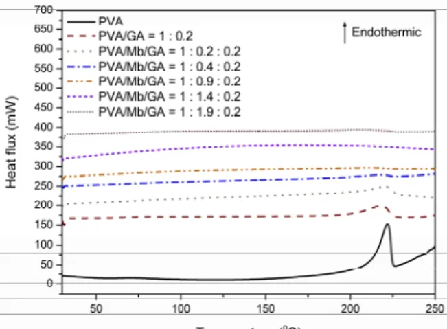
The DSC second heating step curves are pre-sented in Figure 5. The unmodified PVA membrane showed a glass transition temperature, Tg, of 60.5 °C,
and a broad endothermic peak regarded to the poly-mer melting at 222 °C. These temperatures were lower than PVA grains probably due to the plasticization effect of water. For GA-crosslinked PVA membrane (PVA/GA = 1:0.2 g/g), Tg could not be determined
and the melting event due to the crystalline domain was lower than the unmodified PVA membrane. It can be explained in terms of the change in spatial organization of PVA chains due to the reaction with GA.
Figure 5: DSC curves of myoglobin–immobilized PVA membranes.
Tg values could not be determined for
Mb-con-taining samples, probably due to the morphological
changes caused by the reaction between PVA, GA and Mb. It was noted that the increase in myoglobin content decreased the melting enthalpy from un-modified PVA to Mb/PVA of 1.9, in which the crys-tallinity is not detectable anymore. This result can be reasoned in terms of the crosslinking reaction be-tween Mb, PVA and GA. The reduction in chain mo-bility prevented the formation of the crystalline do-main typical of unmodified PVA. As the result, the crystalline lattice of PVA could not be formed and the enthalpy for melting the film was diminished.
The thermal properties of myoglobin-containing PVA membranes showed different behavior com-pared to unmodified PVA. An increase could be noted in the thermal resistance and a decrease in crystal-linity. The consumption of PVA hydroxyl groups in-creased the amorphous domain of the membranes, as mentioned before. These morphological and struc-tural changes represented an increase in the effective area for permeation, which can lead to higher fluxes. The oxygen transport through the membranes was investigated by means of a dialysis cell, equipped with a dissolved oxygen sensor. The results for the tests conducted at 25 °C are presented in Figure 6. Tests were conducted in triplicate. The deviation was lower than 10%.
Figure 6: Oxygen transport through different mem-branes in the aqueous phase at 25 °C.
The “impermeable” membrane refers to an alumi-num plate placed between the feed and the permeate compartments. This test was performed for the evaluation of the method, provided that no oxygen from the permeate atmosphere could influence the result. The low oxygen content during the tests con-firmed the insulation of permeate.
microfiltra-tion membranes (Millipore, average pore diameter of 0.45 μm). The “MF support” test corresponds to two membranes without any dense film between them.
For the unmodified PVA membrane, the oxygen flux through the membrane was the same as the MF support, 3 x 10⁻⁷ kg/hm2, denoting that the high water swelling degree of the polymer caused no additional resistance to mass transfer, as expected.
The oxygen flux through the myoglobin-immobi-lized membrane was lower than the unmodified PVA, showing no facilitation. It could be inferred that the permeation of oxygen occurred by simple diffusion through the crosslinked polymeric chains. The crosslinking of PVA increased the correspond-ing diffusion path, and, consequently, decreased the total flux.
Also shown in Figure 6 is the oxygen flux through an oxymb-containing PVA membrane prepared with-out the addition of GA. The initial flux is abwith-out three times higher than the unmodified PVA membrane, showing the facilitated transport of O2. However, as
the biocarrier was not chemically bound to the poly-meric matrix, it was extracted to the aqueous me-dium, evidenced by the color change of the solution. As a consequence, the oxygen flux decreased with time. This result showed the ability of the myoglobin to transport oxygen in the PVA membrane, although the poor stability of such system did not allow the commercial use of the membrane. Moreover, the in-crease in the facilitation factor for oxygen also con-firmed the validity of the dialysis method to evaluate the oxygen transport. It was assumed that the mass ratio Mb/PVA of 0.9 g/g is higher than the percola-tion threshold of the system and did not affect the transport rate.
The absence of facilitated transport for the my-oglobin-immobilized PVA membrane can be ex-plained in terms of the reaction of the protein with the GA molecule. Although the alpha-helical struc-ture seemed to be maintained, the oxygen affinity was increased, which did not allow the release of oxygen on the permeate side. This result can be rea-soned in terms of the fact that the reaction of the macromolecule is transmitted along the atoms and could affect the heme microenvironment. It is known that even a small displacement in the atoms at the heme pocket can cause significant changes in Mb-O2
affinity.
It is worth noting that the increase in the affinity of oxygen to the biocarriers has been reported on the literature. For instance, Buehler et al. (2005) also observed the displacement of the oxygen dissociation curve to the left as the result of hemoglobin oli-gomerization with glutaraldehyde in the synthesis of
the artificial blood, HBOC, hemoglobin-based oxy-gen carriers. It is quite reasonable to extend the same behavior to the reaction of myoglobin and PVA crosslinked by GA. Besides that, the immobilization of the biocarriers in a polymeric (solid) matrix can also avoid the nanometric displacement of the iron atom to the porphyrin plane of the heme environment during the oxygenation of the biocarrier. This could be the cause of the loss on the reversibility between the carrier and the solute.
In addition, Bonaventura and Bonaventura (1982) reported the synthesis of an artificial gill based on the immobilization of hemoglobin in hydrophilized polyurethane membranes. The unloading step was quite difficult, due to the high affinity between the immobilized hemoglobin and oxygen.
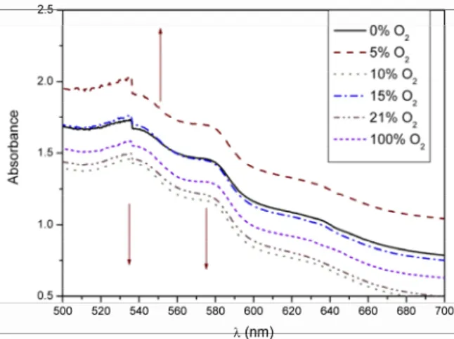
In order to evaluate the oxygen dissociation curve from immobilized myoglobin, UV-visible spectra were recorded after the exposition of the PVA/Mb/ GA membrane 1:0.9:0.2 to oxygen/nitrogen mixtures with varying contents. The results are presented in Figure 7. The appearance of deoxymyoglobin (peak at 556 nm) and the decrease of the absorbance typi-cal of oxymb (543 and 581 nm) with the reduction of oxygen partial pressure in the feed gas was expected. The expected behavior is shown by the arrows in the figure.
Figure 7: Visible spectra of a PVA/Mb/GA 1:0.9: 0.2 membrane submitted to oxygen-depleted air.
The spectra showed no change in myoglobin physiological form with the decrease in the oxygen content. This is the evidence that the reversibility of the reaction was lost during the chemical bonding of the biomolecule on PVA.
inves-tigated for the release of oxygen from oxymyoglo-bin. However, these variables did not cause an in-crease in the oxygen transport, confirming the loss on the reversibility of the myoglobin and oxygen reaction. New membranes were prepared by using previously reduced myoglobin but no facilitation was observed.
Hemoglobin (minimum 95%, Fluka) was investi-gated as an alternative to myoglobin in the mem-branes due to the lower affinity to oxygen. The opti-mized mass ratio between PVA, hemoglobin and glutaraldehyde was 1:0.5:0.04, due to the lower solubility of hemoglobin in water compared to Mb. The reaction between the protein and GA was faster than myoglobin and GA. The formation of clusters of the hemoglobin in the Petri dish required the fil-tration of the casting solution before it was poured into the plate. No iron reduction was observed during membrane synthesis. Homogeneous films were pro-duced at the optimum condition, with no extraction of the protein to the aqueous medium in water swell-ing tests. However, no facilitation of the oxygen transport occurred. The effects of pH and operating temperature did not bring about changes in the oxy-gen content profile. Altogether, these results showed that the immobilization of oxygen biocarriers in a PVA matrix can damage irreversibly the prosthetic environment, which decreased the oxygen flux through the membrane.
CONCLUSION
Myoglobin was successfully immobilized in poly(vinyl alcohol) by means of the reaction with glutaraldehyde, in the absence of organic solvents and acid catalysts. The water swelling degree of the films was decreased, which indicated that the crosslinking reaction between PVA and Mb with glutaraldehyde was effective. Regarding the ther-mal properties, the reaction caused a decrease in the crystalline domains and increased the thermal resistance.
Metmyoglobin was reduced during the reaction. The characterization of the chemical groups in the films showed that the amide I and II bands typical of the protein were kept, denoting high maintenance of the helical conformation of the protein inside the membrane. The increased affinity between the im-mobilized myoglobin and oxygen caused no decoup-ling of the solute at the membrane/permeate inter-face.
A facilitation factor of 3 was observed for non-immobilized myoglobin in PVA, confirming the
oxygen transport ability of the protein. The use of hemoglobin as the biocarrier also revealed the lack of reversibility of the reaction of the prosthetic group and oxygen.
Even though there are evidences that the reaction between the biocarriers and GA is far away from the hydrophobic pocket of the heme group, the informa-tion seems to be transmitted along the macromole-cule and the mechanism must be studied to allow the development of biocarrier-based facilitated transport membranes for oxygen separation.
NOMENCLATURE
PVA poly(vinyl alcohol) GA glutaraldehyde Mb myoglobin
DSC differential scanning calorimetry TGA thermogravimetric analysis
FTIR Fourier Transform Infrared spectroscopy Metmb metmyoglobin, Fe3+, physiologically
inactive
Oxymb oxymyoglobin, Fe2+ bound to oxygen Deoxymb deoxymyoglobin, Fe2+ not bound to
oxygen
lw swollen membrane size
ld dried membrane size
R Water swelling degree
ACKNOWLEDGEMENT
The authors would like to thank Capes and CNPq for financial support.
REFERENCES
Arkin, I. T., Isotope-edited IR spectroscopy for the study of membrane proteins. Curr. Opin. Chem. Biol., 10(5), 394 (2006).
Baker, R. W., Membrane Technology and Applica-tions. John Wiley & Sons Ltd, New York (2004). Baker, R. W., Future directions of membrane gas
separation technology. Ind. Eng. Chem. Res., 41 (6), 1393 (2002).
Bonaventura, J. and Bonaventura, C., Immobilized hemoglobin and process for extracting oxygen from fluids using the same, U. S. Patent 4,343,715 (1982).
glutaraldehyde-po-lymerized bovine hemoglobin and its isolated fractions. Anal. Chem., 77(11), 3466 (2005). Eike, J. H. and Palmer, A. F., Effect of NaBH4
con-centration and reaction time on physical proper-ties of glutaraldehyde-polimerized hemoglobin. Biotechnol. Prog., 20(3), 946 (2004).
Ferraz, H. C., Duarte, L. T., Di Luccio, M., Alves, T. L. M., Habert, A. C., Borges, C. P., Recent achieve-ments in facilitated transport membranes for sepa-ration processes. Braz. J. Chem. Eng., 24(1), 101-118 (2007).
Figoli, A., Sager, W. F. C. and Mulder, M. H. V., Fa-cilitated oxygen transport in liquid membranes: Review and new concepts. J. Membr. Sci., 181(1), 97 (2001).
Figueiredo, K. C. S., Alves, T. L. M. and Borges, C. P., Poly(vinyl alcohol) films crosslinked by glutar-aldehyde under mild conditions. J. Appl. Polym. Sci., 111(6), 3074 (2009).
Goormaghtigh, E., Ruyschaert, J. M., Raussens, V., Evaluation of the information content in infrared spectra for protein secondary structure determi-nation. Biophys. J., 90(8), 2946 (2006).
Guo, Y. Z., Clark, D. S., Activation of enzymes for nonaqueous biocatalysis by denaturing concen-trations of urea. Biochim. Biophys. Acta, 1546 (2), 406 (2001).
Hargrove, M. S., Wilkinson, A. J. and Olson, J. S., Structural factors governing hemin dissociation from metmyoglobin. Biochemistry, 35(35), 11300 (1996).
Migneault, I., Dartiguenave, C., Bertrand, M. J. and Waldron, K. C., Glutaraldehyde: Behavior in aque-ous solution, reaction with proteins and applica-tion to enzyme crosslinking. BioTechniques, 37 (5), 790 (2004).
Mulder, M., Basic Principles of Membrane Technol-ogy. Kluwer Academic Publishers, Dordrecht (1996).
Nagase, K. I., Kohori, F. and Sakai, K., Develop-ment of a compact artificial gill using concen-trated hemoglobin solution as the oxygen carrier. J. Membr. Sci., 215(1-2), 281 (2003).
Nishide, H., Tsukahara, Y. and Tsuchida, E., Highly selective oxygen permeation through a poly (vinylidene dichloride)-cobalt porphyrin mem-brane: Hopping transport of oxygen via the fixed cobalt porphyrin carrier. J. Phys. Chem. B, 102(44), 8766 (1998).
Robeson, L. M., The upper bound revisited. J. Membr. Sci., 320(1-2), 390 (2008).
Roman, I. C. and Baker, R. W., Method and appara-tus for producing oxygen and nitrogen and mem-brane therefor, U. S. Patent 4,542,010 (1985). Ruaan, R. C., Wu, T. H., Chen, S. H. and Lai, J. Y.,
Oxygen/nitrogen separation by polybutadiene/ polycarbonate composite membranes modified by ethylenediamine plasma. J. Membr. Sci., 138(2), 213 (1998).
Scholander, P. F., Oxygen transport through hemo-globin solution. Science, 131(3400), 585 (1960). Shoji, M., Oyaizu, K., Nishide, H., Facilitated oxy-gen transport through a Nafion membrane con-taining cobaltporphyrin as a fixed oxygen carrier. Polymer, 49(26), 5659(2008).
Sugawara, Y., Matsuoka, A., Kaino, A. and Shikama, K., Role of globin moiety in the autoxidation re-action of oxymyoglobin: Effect of 8 M urea. Biophys. J., 69(2), 583 (1995).
Vidal, M. M. B., Gil, M. H., Delgadillo, I. and Alonso Chamarro, J., Swelling and thermal properties of poly(vinyl alcohol) containing hemoglobin mem-branes. J. Bioact. Compat. Polym., 14(3), 243 (1999).
Wang, Q., Lu, G. and Yang, B., Myoglobin/sol-gel film modified electrode: direct electrochemistry and electrochemical catalysis. Langmuir, 20(4), 1342 (2004).
Wang, Y. C., Huang, S. H., Hu, C. C., Li, C. L., Lee, K. R., Liaw, D. J. and Lai, J. Y., Sorption and trans-port properties of gases in aromatic polyimide membranes. J. Membr. Sci., 248(1-2), 15 (2005). Yeom, C.-K. and Lee, K.-H., Pervaporation