In-situ preparation of hydroxyapatite
nanorod embedded poly (vinyl alcohol)
composite and its characterization
M.RAJKUMAR, N.MEENAKSHI SUNDARAM, V.RAJENDRAN*
Centre for Nano Science and Technology K.S. Rangasamy College of Technology
Tiruchengode-637 215 Tamil Nadu, India
Abstract :
Hydroxyapatite (HAp) nanorod embedded composite was prepared using poly (vinyl alcohol) (PVA) as a matrix with different weight percentages (wt. %) The role of PVA composition on the crystallite size, degree of crystallinity, functional groups and morphology of nanocomposites were characterised by XRD, FTIR, and TEM analysis. The results indicated that the size and crystallinity of HAp nano particles decreases with increase in PVA concentration in the composite. This shows the size control effect of PVA concentration on HAp nanorods. Due to the chemical bond interactions between HAp and PVA, a new peak at 2944 cm-1 was observed from FTIR analysis. TEM micrograph confirms the presence of HAp nanorod crystals in PVA matrix. From microhardness test, a notable increase in hardness of the composite for 1.25 and 2.5 wt. % of PVA concentration when compared to pure HAp. The bioresorbability was explored through in vitro study.
Keywords: Hydroxyapatite nanorod; Poly (vinyl alcohol); Nanocomposites; Hardness.
1. Introduction
The calcium phosphate based bioceramics particularly hydroxyapatite (HAp) play an excellent role in biomedical applications owing to their excellent biocompatible, osteoconductive and bioactive properties, and its close chemical and physical resemblance to mineral component of bone tissue, enamel and dentin.1 The major mineral phase of bone is hydroxyapatite (HAp) with a ratio of calcium-to-phosphate is 1.67 which is embedded as nanocrystalline form in collagen triple helix structure.2 Currently, researchers are trying to mimic this natural nano composite system for tissue engineering applications. Since, the nano HAp with high surface area to volume ratio is more desirable to increase their contribution in bone/tooth implants, adsorbents, gene delivery and immunosensor.3 However, the brittleness and poor performance of mechanical stability of pure HAp limit its use for the regeneration of non-load-bearing bone defects and tissue engineering applications.4 Composite biomaterials like metal and polymer matrix are used to improve the mechanical compatibility of nano HAp (n-HAp). Generally, the composite biomaterials are prepared by using biocompatible/biodegradable and synthetic/natural polymers.4,5
The inorganic minerals such as hydroxyapatite,6 bioactive glasses,7 metal oxides,8 and carbon nanotube9 are incorporated into polymer matrixes to impart bioactivity. This enables us to develope the composite with desired properties.10,11 The addition of nanosized particles is desirable to develop the composite with a good mechanical strength since the natural bone contains mineral crystals which are at the nanometer scale and embedded in the collagen matrix.12 The polymer composites are designed to meet the specific requirement of biomedical applications like tissue engineering and drug delivery system. The right choice of the composition of both filler and polymer matrix are essential inaddition to the process method to obtain suitable biopolymer composites. Recently, attempts have been made to develop nanocomposites, wherein n-HAp particles are embedded in PVA polymeric matrices.12-14
Corresponding author
An extensive study have been made on both natural (collagen, gelatin, silk fibroin) and synthetic (polyethylene, polyamide, chitosan, polystyrene, poly (vinyl alcohol) and polyetheretherketone) polymers to overcome the mechanical problems associated with bioceramics in bone tissue engineering applications.15-18 Among the above polymers, PVA remain one of the widely used polymer group of biomaterials applied for medical implants. This usage is due to its segmented block co-polymer character. This wide range of versatility in terms of tailoring their applications such as tissue scaffolding,19 artificial cartilage20 and biodegradable scaffolds.21
With the superior combination of the synergic effect and biocompatible HAp and the adjustable biodegradability of polymer matrix, HAp nanorod embedded PVA composites were prepared under controlled environment. The obtained nano HAp/PVA composites were characterised in light of crystallite size, degree of crystallinity, morphology, biological and mechanical properties.
2. Materials and methods
2.1. Materials
The chemicals used in the present investigation were of AR grade calcium hydroxide (Ca(OH)2, Merck GR, 96%), ortho-phosphoric acid (H3PO4, Merck GR, min. 88%) and poly(vinyl alcohol) (M.W. 1,25,000, Loba LR). All the high purity chemicals were used without any further purification. The ultrapure water was (Sartorius AG, Arium 611Ultra filter, Germany) used throughout this experiment with a resistivity of 18.2 MΩ. 2.2. Synthesis of HAp/PVA nanostructured composites
The HApwith rod like morphology embedded composite was prepared using different weight percentages (wt. %) of PVA. The PVA (1.25 wt. %) was dissolved in 100 ml ultrapure water with continuous stirring for 5 h to obtain a homogeneous solution. Similarly, 7.41 g of calcium hydroxide was dissolved in 125 ml ultrapure water with continuous stirring for 5 h. After obtaining the homogeneous solution, the calcium solution was added into the PVA solution drop-by- drop under constant stirring for 5 h and kept it for 12 h in constant room temperature. During the stirring process, the prepared phosphate solution (3.4 ml of H3PO4 in 96.6 ml ultrapure water) was added dropwise for a period of 90 minutes to the above polymer mixed solution. After adding the phosphate solution, the pH value is measured for all PVA concentrations. The final solution was stirred continuously for 5 h and then it was kept for 24 h for sedimentation. Then, the sediment particles were filtered and dried in hot air oven at 353 K for 48 h. The dried samples were milled in a 22 mm zirconia balls assisted planetary ball-mill (PM 100, Retsch GmbH, Germany) at 400 rpm for 0.5 h. The same procedure was repeated for different PVA compositions such as 2.5, 3.75, 5.0 and 6.25 wt. % (hereafter termed respectively as HAp/PVA-1.25, HAp/PVA-2.5, HAp/PVA-3.75, HAp/PVA-5.0, and HAp/PVA-6.25) were prepared. The pure n-HAp termed hereafter as HAp/PVA-0 without PVA additives was prepared by the same procedure. The HAp/PVA-0 is used as a reference sample.
2.3. Characterisation techniques
2.3.1. Structural and morphological characterisation
The crystallinity and phase analysis were carried out on X-ray diffractometre (XRD, X’Pert-Pro, PANalytical, Netherland) with CuKα (1.5406 Å) as a radiation source. The characteristic peaks were identified by Fourier transform infrared spectroscopy (FTIR, spectrum 100, Perkin Elmer, USA) in the range from 4000 to 400 cm-1. The surface morphology was examined through transmission electron microscope (TEM, Philips, CM 200, USA).
2.3.2. In vitro bioresorbability test
2.3.3. Microhardness measurement
The synthesised pure HAp and HAp/PVA composites were pressed in a stainless steel pellet die for 3 minute at a pressure of 100 kg cm-2 to obtain pellet in the size of 13 mm diameter and 1.5 mm thickness. The prepared pellets were used for microhardness using Vickers indentation (Bareiss, Germany) with a load of 0.5 kg. Five specimens were tested for each composition and then, the mean value was determined.
3. Results and Discussion
3.1. Formation mechanism of HAp/PVA nanocomposite
Fig. 1 shows the schematic representation of the synthesis of HAp/PVA nanocomposite. When the calcium hydroxide was added to the PVA solution, the Ca2+ ions were attached with OH- group in the PVA matrix. Following the above step, orthophosphoric acid was added drop by drop into the above mixed solution. As a result, PO43- ions bind to the –OH- Ca2+ group to form hydroxyapatite particles and the PVA matrix regulates the growth of c-axis of HAp nanorod.
3.2.X-ray diffraction analysis
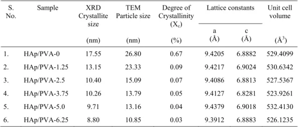
The reflection planes corresponding to the characteristic XRD spectral peaks of pure n-HAp and HAp/PVA nanocomposites are shown in Fig. 2. The observed diffraction peaks are identified by standard JCPDS (File no. 09-0432) file and are assigned as crystalline HAp. It is evident from the observed results that no characteristic diffraction angles from other calcium phosphate phases are detected. The main crystalline peaks observed for the pure n-HAp and HAp/PVA composites at diffraction angles 25.89º, 31.91º, 32.95º, 34.08º, 39.85º, and 46.71º represents respectively the hydroxyapatite d-spacing 3.44, 2.80, 2.72, 2.63, 2.26, and 1.94 Å.
The XRD patterns show diffraction peaks with line broadening and high intensities, which confirms the nanosize with crystalline nature. The crystallite size of the pure HAp and HAp/PVA composite is calculated by using Scherrer formula.23 Fig. 3 reveals that the crystallite size decreases with increase in the composition of PVA.24 The obtained average crystallite size is in the range between 8 and 17 nm. The relationship between lattice constant (a & c), Miller’s indices (h,k,l) and lattice spacing (d) is used to calculate lattice parameter values, expressed as,
2 2
2 2 2
2
(
)
3
4
1
c
l
a
k
hk
h
d
(1)and also to obtain the unit cell volume (V) employing the relation V = 2.589 a2c for both HAp and HAp/PVA composites. The obtained lattice parameters and the unit cell volume in all the samples are given in Table 1. The degree of crystallinity (Xc) is obtained based on the relation,
3
A cK
X
(2)where Xc is the degree of crystallinity, KA a constant (0.24) and β the full width half maximum (FWHM).24,25 The calculated degree of crystallinity shows that it decreases rapidly with addition of PVA.25 It is interesting to note that a further addition of PVA leads to a linear decrease in the degree of crystallinity as shown in Fig. 3. A comparison of the observed diffracted pattern (Fig. 2) of the pure HAp and PVA/HAp composites reveals a similar diffracted pattern which confirms the formation of n-HAp in PVA matrix. Further, it is informed that the change in the composition of PVA additives does not show any notable effects on the peak position of diffracted patterns.
Table 1 Summary of crystallographic parameters of pure HAp and PVA based composites
S. No. Sample XRD Crystallite size (nm) TEM Particle size (nm) Degree of Crystallinity
(Xc) (%)
Lattice constants Unit cell volume
(Å3) a
(Å)
c (Å)
1. HAp/PVA-0 17.55 26.80 0.67 9.4205 6.8882 529.4099
2. HAp/PVA-1.25 13.15 23.33 0.09 9.4217 6.9024 530.6342
3. HAp/PVA-2.5 10.40 15.09 0.07 9.4086 6.8813 527.5367
4. HAp/PVA-3.75 10.26 13.79 0.05 9.4127 6.8281 523.9261
5. HAp/PVA-5.0 9.71 13.16 0.04 9.4379 6.9018 532.4130
6. HAp/PVA-6.25 8.80 10.85 0.03 9.3912 6.8883 526.1235
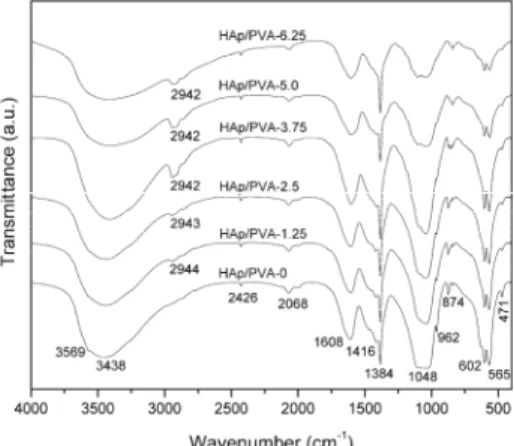
3.3. Fourier transform infrared spectroscopy analysis
The FTIR spectra of pure n-HAp and HAp/PVA composites are shown in Fig. 4. The ν2 phosphate stretching mode is appeared at 471-472 cm-1 corresponds to PO4 group in HAp.26 The bands located at 1032-1048 and 562-570 cm-1 are attributed respectively to the ν3 and ν4 P-O vibration modes of regular tetrahedral PO4
3-groups.27 The observed bands at 602 cm-1 corresponds to O-P-O bending and ν1 symmetric P-O stretching modes.28 The ν1 symmetric stretching mode of phosphate group is observed at 962 cm
-1
.29 The observed bands at 1384 cm-1 is due to the stretching mode of carbonate, which may be to the acquisition of air during mineral precipitation.30 Similarly, the observed bands at 1416 and 857-874 cm-1 are assigned to carbonate ions.29,31 The bands observed in the region between 2067 and 2069 cm-1 are related to their harmonic overtones and/or combination bands.32
The lattice H2O exists in the range of 1603-1608 cm -1
,while the bands observed at 3400-3569 cm-1 overlap the –OH group.26,33,34 The band observed between 2942-2944 cm-1 corresponds to C-H stretching band of PVA.35 A new peak of C-H stretching band is observed at 2944 cm-1, when the PVA is added. This indicates that the chemical bond interactions between HAp and PVA.36
3.4. Transmission electron microscopy analysis
TEM images of pure n-HAp and different weight percentages of poly (vinyl alcohol) compositions are illustrated in Fig. 5. The TEM picture shows that particles exhibit nanorod morphology. The particles size of pure HAp is 27 nm. In case of composites, when the composition of PVA is added to HAp, the rod-like morphology starts to disappear. The increase in the PVA compositions i.e., 1.25, 2.5, 3.75, 5.0, 6.25 wt. % leads to a corresponding change from rod-like to an irregular morphology. Further, it is evident that the particle size decreases with increase in PVA composition.
The evaluated particles size values are listed in Table 1. According to TEM analysis, the particles are homogeneously dispersed in polymer matrix. Further, the micrograph does not show any notable indication for the existence of agglomeration. In addition, a decrease in particle size with increase in the PVA composition is noticed.
3.5. In vitro bioresorbability test
Fig. 6 shows the in vitro bioresorbability of pure HAp/PVA-0 and HAp/PVA-1.25 to HAp/PVA-6.25 composites. A considerable variation in pH value is observed with respect to soaking period in all PVA compositions. It can be seen that the pH value of SBF solution increases with increase in PVA (0 to 1.25 wt. %) composition as shown in Figs 6a and 6b. Further, a gradual decrease in the pH value of SBF solution with further change in PVA additives from 1.25 to 6.25 wt. % is noticed as depicted in Figs 6b-6f. The observed results indicate that the pure n-HAp does not exhibit much variation in pH value. In contrast, when the composition of PVA is increased from 1.25 to 5.0 wt. % (HAp/PVA-1.25 to HAp/PVA-5.0), an appreciable change in the pH value is noticed.
There is no evidence for the decrease in pH with increase in soaking period. It can be concluded that the samples have no adverse bioresorbability response. The observed sudden increase in pH value in the present study is may be due to the alkaline effect i.e., the introduction of Ca2+ between the composite and SBF solution. 3.6. Microhardness test
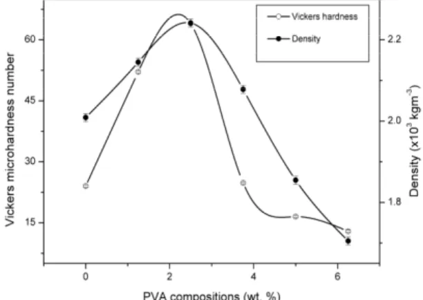
The variations of Vickers microhardness and density for pure HAp and their PVA based composites (HAp/PVA-1.25 to HAp/PVA-6.25) are shown in Fig. 7. From Fig. 7, it is inferred that both microhardness and densities are increasing with increase in the composition of PVA. A maximum hardness value (64.03 HV0.5) is observed for the composition of 2.5 wt. % of PVA, beyond which both hardness and density values are found to decreases with further addition of PVA additives.
At this composition, the HAp particles have good compact structure with PVA molecules which leads to the lightest density value. This compositional ratio may be considered for better compact HAp/PVA composite material. This is due to the increase in porosity with decrease in density which inturn leads to a decrease in hardness. Hence, it can be concluded that the PVA composition leads to a significant effect on both density and hardness.
4. Conclusion
In the present work, a novel hydroxyapatite/poly (vinyl alcohol) nanocomposite is prepared by simple chemical route. The reduction in particle size with increase in concentration of PVA is due to the size control effect of PVA molecular structure. The rod-like morphology becomes as an irregular morphology with increase in PVA additives. It inferred that the composition of PVA shows significant influence on particle size, degree of crystallinity and microhardness, which facilitate to optimize the composition of composite for particular applications.
Acknowledgments
The authors are thankful to the Department of Science and Technology (DST), New Delhi for the financial support to carry out this research project (SR/S2/CMP-66/2006 dt.27.09.2007). We would like to acknowledge Dr.A.Rajadurai and Dr.K.Kalaichelvan, Department of Production Technology, Madras Institute of Technology, Chennai in extending facilities to do microhardness measurement.
References
[1] M. Li, X. Xiao, R. Liu, C. Chen, and L. Huang. Structural characterization of zinc-substituted hydroxyapatite prepared by hydrothermal method, J. Mater. Sci: Mater. Med. 19: 797–103 (2008).
[2] S. Bose, and S. K. Saha. Synthesis and characterization of hydroxyapatite nanopowders by emulsion technique, Chem. Mater. 15: 4464-4469 (2003).
[3] Y. Ding, J. Liu, H. Wang, G. Shen, and R. Yu. A piezoelectric immunosensor for the detection of α-fetoprotein using an interface of gold/hydroxyapatite hybrid nanomaterial, Biomaterials 28: 2147–2154 (2007).
[4] H. Wang, Y. Li, Y. Zuo, J. Li, S. Ma, and L. Cheng. Biocompatibility and osteogenesis of biomimetic nano-hydroxyapatite/polyamide composite scaffolds for bone tissue engineering, Biomaterials 28: 3338–3348 (2007).
[5] V. S. Komlev, S. M. Barinov, and F. Rustichelli. Strength enhancement of porous hydroxyapatite ceramics by polymer impregnation, J. Mater. Sci. Lett. 22: 1215–1217 (2003).
[6] N. Meenakshi Sundaram, E. K. Girija, M. Ashok, T. K. Anee, R. Vani, and R. Suganthi. Crystallisation of hydroxyapatite nanocrystals under magnetic field, Mater. Lett. 60: 761-765 (2006).
[7] V. Rajendran, A. Nishara Begum, M. A. Azooz, and F. H. EI Bata. Microstructural dependence on relevant physical–mechanical properties on SiO2–Na2O–CaO–P2O5 biological glasses, Biomaterials 23: 4263-4275 (2002).
[8] E. S. Ahn, N. J. Gleason, A. Nakahira, and J. Y. Ying. Nanostructure processing of hydroxyapatite-based bioceramics, Nano Lett. 1(3): 149–153 (2001).
[9] M. K. Singh, T. Shokuhfar, J. J. D. Almeida Gracio, A. C. M. D. Sousa, J. M. D. F. Fereira, H. Garmestani, and S. Ahzi. Hydroxyapatite modified with carbon-nanotube-reinforced poly(methyl methacrylate): A nanocomposite material for biomedical applications, Adv. Funct. Mater. 18: 694-700 (2008).
[10] R. Joseph, and K. E. Tanner. Effect of morphological features and surface area of hydroxyapatite on the fatigue behavior of hydroxyapatite-polyethylene composites, Biomacromolecules 6: 1021-1026 (2005).
[11] J. M. Yang, C. S. Lu, Y. G. Hsu, and C. H. Shih. Mechanical properties of acrylic bone cement containing PMMA-SiO2 hybrid
sol-gel material, J. Biomed. Mater. Res. 38: 143-154 (1997).
[12] N. Pramanik, P. Bhargava, S. Alam, and P. Pramanik.Processing and properties of nano-and macro-hydroxyapatite/poly (ethylene-co-acrylic acid) composites, Polym. Compos, 27: 633-641 (2006).
[13] M. Boissie`re, P. J. Meadows, R. Brayner, C. Helary, J. Livage, and T. Coradin. Turning biopolymer particles into hybrid capsules: the example of silica/alginate nanocomposites, J. Mater. Chem. 16: 1178-1182 (2006).
[14] K. Kawagoe, M. Saito, T. Shibuya, T. Nakashima, K. Hino, and H. Yoshikawa. Augmentation of cancellous screw fixation with hydroxyapatite composite resin (CAP) in vivo, J. Biomed. Mater. Res. 53: 678-684 (2000).
[15] J. Li, Y. Zuo, X. Cheng, W. Yang, H. Wang, and Y. Li. Preparation and characterization of nano-hydroxyapatite/polyamide 66 composite GBR membrane with asymmetric porous structure, J. Mater. Sci: Mater. Med. 20: 1031–1038 (2009).
[16] M. Darder, M. Lo´pez-Blanco, P. Aranda, A. J. Aznar, J. Bravo, and E. Ruiz-Hitzky. Microfibrous chitosan-sepiolite nanocomposites, Chem. Mater. 18: 1602-1610 (2006).
[17] Y. Zhang, and J. L. A. Mild. Efficient biomimetic synthesis of rodlike hydroxyapatite particles with a high aspect ratio using polyvinylpyrrolidone as capping agent, Cryst. Growth Des. 8(7): 2101-2107 (2008).
[18] D. Z. Chen, C. Y. Tang, K. C. Chan, C. P. Tsui, P. H. F. Yu, M. C. P. Leung, and P. S. Uskokovic. Dynamic mechanical properties and in vitro bioactivity of PHBHV/HA nanocomposite, Compos. Sci. Technol. 67: 1617–1626 (2007).
[19] F. E. Wiria, C. K. Chua, K. F. Leong, Z. Y. Quah, M. Chandrasekaran, and M. W. Lee. Improved biocomposite development of poly (vinyl alcohol) and hydroxyapatite for tissue engineering scaffold fabrication using selective laser sintering, J. Mater. Sci: Mater. Med. 19: 989-996 (2008).
[23] C. W. Chen, C. S. Oakes, K. Byrappa, R. E. Riman, K. Brown, K. S. TenHuisen, and V. F. Janas. Synthesis, characterization, and dispersion properties of hydroxyapatite prepared by mechanochemical–hydrothermal methods, J. Mater. Chem. 14: 2425–2432 (2004).
[24] S. Kannan, A. F. Lemos, and J. M. F. Ferreira. Synthesis and mechanical performance of biological-like hydroxyapatite, Chem. Mater. 18: 2181-2186 (2006).
[25] N. Degirmenbasi, D. M. Kalyon, and E. Birinci. Biocomposites of nanohydroxyapatite with collagen and poly (vinyl alcohol), Colloids Surf. B: Biointer. 48: 42-49 (2006).
[26] A. Lak, M. Mazloumi, M. Mohajerani, A. Kajbafvala, S. Zanganeh, H. Arami, and S. K. Sadrnezhaad. Self-assembly of dandelion-like hydroxyapatite nanostructures via hydrothermal method, J. Am. Ceram. Soc. 91(10): 3292-3297 (2008).
[27] L. Yanbao, L. Dongxu, and W. Weng. Preparation of nano carbonate-substituted hydroxyapatite from an amorphous precursor, Int. J. Appl. Ceram. Technol. 5(5): 442-448 (2008).
[28] M. G. Ma, Y. J. Zhu, and J. Chang. Monetite formed in mixed solvents of water and ethylene glycol and its transformation to hydroxyapatite, J. Phys. Chem. B 110: 14226-14230 (2006).
[29] W. Zhang, S. S. Liao, and F. Z. Cui. Hierarchical self-assembly of nano-fibrils in mineralized collagen, Chem. Mater. 15: 3221-3226 (2003).
[30] L. Bertinetti, A. Tampieri, E. Landi, C. Ducati, P. A. Midgley, S. Coluccia, and G. Martra. Surface structure, hydration and cationic sites of nanohydroxyapatite: UHR-TEM, IR, and Microgravimetric studies, J. Phys. Chem. C 111: 4027-4035 (2007).
[31] D. Choi, and P. N. Kumta. An alternative chemical route for the synthesis and thermal stability of chemically enriched hydroxyapatite, J. Am. Ceram. Soc. 89(2): 444-449 (2006).
[32] I. S. Neira, Y. V. Kolen’ko, O. I. Lebedev, G. V. Tendeloo, H. S. Gupta, F. Guitian, and M. Yoshimura. An effective Morphology control of hydroxyapatite crystals via hydrothermal synthesis, Cryst. Growth Des. 9(1): 466-474 (2009).
[33] S. Kannan, and J. M. F. Ferreira. Synthesis and thermal stability of hydroxyapatite-β- tricalcium phosphate composites with cosubstituted sodium, magnesium, and fluorine, Chem. Mater. 18:198-203 (2006).
[34] F. Huang, Y. Shen, A. Xie, J. Zhu, C. Zhang, S. Li, and J. Zhu. Study on synthesis and properties of hydroxyapatite nanorods and its complex containing biopolymer, J. Mater. Sci. 42: 8599-8605 (2007).
[35] R. Murugan, and S. Ramakrishna. Bioresorbable composite bone paste using polysaccharide based nano hydroxyapatite, Biomaterials 25: 3829-3835 (2004).