Outbreak of invasive pulmonary aspergillosis
among patients hospitalized in a bone marrow
transplant ward: tomographic findings*
Surto de aspergilose pulmonar invasiva em enfermaria de transplante de medula óssea: achados tomográficos
Daniela Batista de Almeida Freitas, Ana Cláudia Piovesan, Gilberto Szarf, Dany Jasinowodolinski, Gustavo de Souza Portes Meirelles
Abstract
Objective: To evaluate the main aspects on CT scans of six patients hospitalized in a bone marrow transplant ward,
diagnosed with invasive pulmonary aspergillosis (IPA), during an in-hospital outbreak of the disease. Methods: We reviewed 10 chest CT scans of six neutropenic or immunocompromised patients hospitalized in the hematology and bone marrow transplant ward of the Hospital São Paulo, in the city of São Paulo, Brazil, who were diagnosed with IPA between April of 2007 and October of 2007. The diagnosis of IPA was confirmed by anatomopathological findings (in 2 cases), culture (in 3 cases) or appropriate treatment response (in 1 case). Results: We evaluated the CT scans of three male and three female patients, ranging from 22 to 58 years of age. The most common tomographic findings were nodules (5/6 cases) and areas of consolidation (2/6 cases). The nodules were more often multiple (3/5 cases), with irregular contours (4/5 cases) and accompanied by the halo sign (3/5 cases). One case presented multiple, centrally distributed areas of consolidation, and another presented an isolated, peripheral area of consolidation. Areas of ground-glass attenuation and septal thickening were found in three and two patients, respectively. Bilateral pleural effusion occurred in three cases. Conclusions: Consolidation, nodules, septal thickening, pleural effusion and ground-glass opacities were the principal tomographic findings in the six patients hospitalized in the abovementioned ward during the IPA outbreak. The nodules were often (in 67% of the cases) accompanied by the halo sign, a classically described finding in patients with IPA.
Keywords: Aspergillosis; Lung diseases, fungal; Neutropenia; Bone marrow transplantation; Tomography, X-Ray computed.
Resumo
Objetivo: Avaliar os principais aspectos encontrados na TC de seis pacientes internados em uma enfermaria de trans-plante de medula óssea, diagnosticados com aspergilose pulmonar invasiva (API) durante um surto intra-hospitalar da doença. Métodos: Foram revisadas 10 TC de tórax de seis pacientes internados na enfermaria de hematologia e transplante de medula óssea do Hospital São Paulo em São Paulo (SP) entre abril de 2007 e outubro de 2007, neutropênicos ou imunossuprimidos, que evoluíram com API. O diagnóstico de API foi comprovado por exames anatomopatológicos (2 casos), cultura (3 casos) ou por resposta terapêutica adequada (1 caso). Resultados: Foram avaliadas imagens tomográficas de três homens e três mulheres com idades variando de 22 a 58 anos. Nódulos (5/6 casos) e áreas de consolidação (2/6 casos) foram os achados tomográficos mais comuns. Os nódulos eram mais frequentemente múltiplos (3/5 casos), de contornos irregulares (3/5 casos) e com sinal do halo (3/5 casos). Foram encontradas consolidações múltiplas e de distribuição central em um caso e consolidação isolada e periférica em outro. Áreas de atenuação em vidro fosco e espessamento septal foram achados, respectivamente, em três e dois pacientes. Derrame pleural bilateral ocorreu em três casos. Conclusões: Consolidação, nódulos, opacidades em vidro fosco, derrame pleural e espessamentos septais foram os principais achados tomográficos dos seis pacientes internados na enfermaria acima citada durante o surto de API. O sinal do halo, classicamente descrito na literatura para esta condição, foi um achado frequente quando associado a nódulos (67%).
Descritores: Aspergilose; Pneumopatias fúngicas; Neutropenia; Transplante de medula óssea; Tomografia computadorizada por raios X.
* Study carried out in the Department of Diagnostic Imaging, Universidade Federal de São Paulo/Escola Paulista de Medicina – UNIFESP/EPM, Federal University of São Paulo/Paulista School of Medicine – São Paulo, Brazil.
Correspondence to: Daniela Batista de Almeida Freitas. Rua Oscar Freire, 2040, apto 118, Pinheiros, CEP 05409-001, São Paulo, SP, Brasil.
Tel 55 11 8371-5605. E-mail: daniela.ba@gmail.com Financial support: None.
biology. Therefore, chest CT has been recom-mended as the principal method for establishing an early diagnosis of IPA.(8,9)
Since it is essential to recognize tomographic alterations in order to diagnose and determine the prognosis of patients with IPA, the purpose of the present study was to evaluate the principal aspects on chest CT scans of six patients hospi-talized in a hematology/bone marrow transplant ward and evolving to IPA.
Methods
We reviewed 10 chest CT scans of six neutropenic or immunocompromised patients hospitalized in the hematology and bone marrow transplant ward of the São Paulo Hospital/ Paulista School of Medicine, São Paulo, Brazil, who were diagnosed with IPA between April of 2007 and October of 2007. The cases occurred during the remodeling of the physical struc-ture of the institution and, based on previous years, exceeded the number of cases expected for the period in question. The present study was approved by the Research Ethics Committee of the Federal University of São Paulo/Paulista School of Medicine.
The patients were three males and three females, ranging from 22 to 58 years of age (mean age: 41.5 years). Five patients were neutropenic at the time of diagnosis, and one of them was immunocompromised due to BMT (corticosteroid therapy for more than 60 days). The interval between the onset of neutro-penia and the diagnosis of IPA was 16 days on average, ranging from 10 to 25 days. The underlying disease was acute leukemia in four patients, chronic leukemia in one patient and
Introduction
Invasive pulmonary aspergillosis (IPA) is a severe infection, generally associated with tissue destruction, that affects immunocompromised patients.(1) The principal risk factors for IPA are prolonged neutropenia (granulocyte count lower than 500 cells/µL for more than 10 days) and myeloablative chemotherapy or bone marrow transplantation (BMT) due to hematologic malignancies.(1,2)
The frequency of IPA ranges between 5% and 15% in patients submitted to BMT, and it is approximately 70% after 34 days of neutropenia in individuals with any kind of hematologic malignancy.(2-5) In addition to being commonly found in such patients, IPA presents mortality rates as high as 94% according to certain studies.(2,4,6,7) This is probably due, at least in part, to the difficulty in diagnosing the disease in an early phase and, consequently, to the delay in initiating antifungal therapy.(8)
The gold standard for the diagnosis of IPA is the histopathological finding of tissue invasion by the microorganism.(8,9) However, histopatho-logical studies require an invasive procedure that is difficult to execute in these patients, who are immunocompromised or have pancytopenia. The efficacy of bronchial lavage is variable (0-93%), being approximately 50% in most studies.(6,10) In addition, noninvasive molecular techniques for detecting Aspergillus sp. antigens (galacto-mannan) in the blood or in the bronchial lavage fluid have not been clearly defined and present limited practical application.(6,8,9) One group of authors(11) demonstrated that IPA lesions on chest CT scans precede the detection of the galacto-mannan antigen in serum through molecular
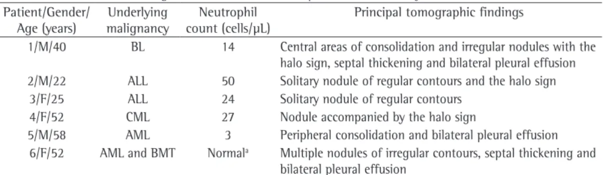
Table 1 - Clinical and radiological characteristics of the patients under study. Patient/Gender/
Age (years)
Underlying malignancy
Neutrophil count (cells/µL)
Principal tomographic findings
1/M/40 BL 14 Central areas of consolidation and irregular nodules with the halo sign, septal thickening and bilateral pleural effusion 2/M/22 ALL 50 Solitary nodule of regular contours and the halo sign
3/F/25 ALL 24 Solitary nodule of regular contours 4/F/52 CML 27 Nodule accompanied by the halo sign
5/M/58 AML 3 Peripheral consolidation and bilateral pleural effusion 6/F/52 AML and BMT Normala Multiple nodules of irregular contours, septal thickening and
bilateral pleural effusion
were taken using HRCT (1-mm thick slices and 10-mm intervals) and spiral CT (7-mm thick slices and 10-mm intervals). For image analysis, lung parenchyma windows ranging from 1,200 to
1,600 HU, with the center ranging from −450 to −650, and mediastinal windows ranging from
250 to 500 HU, with the center ranging from 30 to 50 HU, were used.
The scans were independently reviewed by two radiologists. In discrepant cases, a consensus was reached. The terminology used was that recommended by the Terminology Consensuses of the Brazilian College of Radiology.(12)
Results
The most common tomographic findings were as follows: nodules (5/6 cases); areas of ground-glass attenuation (3/6 cases); bilateral pleural effusion (3/6 cases); areas of consolida-tion (2/6 cases); and septal thickening (2/6 cases). biphenotypic leukemia in one patient. All
patients presented clinical and host criteria for invasive aspergillosis (IA) at the time of diag-nosis, according to the criteria established by the committee composed of members of the Invasive Fungal Infections Cooperative Group of the European Organization for Research and Treatment of Cancer (EORTC).(9) The diagnosis of IA was confirmed through histopathological examination (2/6 cases), bronchial lavage fluid culture (1/6 cases), sputum culture (2/6 cases) or appropriate treatment response (1/6 cases). Therefore, according to the EORTC criteria,(9) three patients were classified as confirmed cases of IA, two were classified as probable cases of IA, and one was classified as a possible case of IA.
Among the CT scans of the patients under study, we selected for analysis those that dated closer to the time of anatomopathological or bacteriological diagnosis of IPA. The CT scans
a b
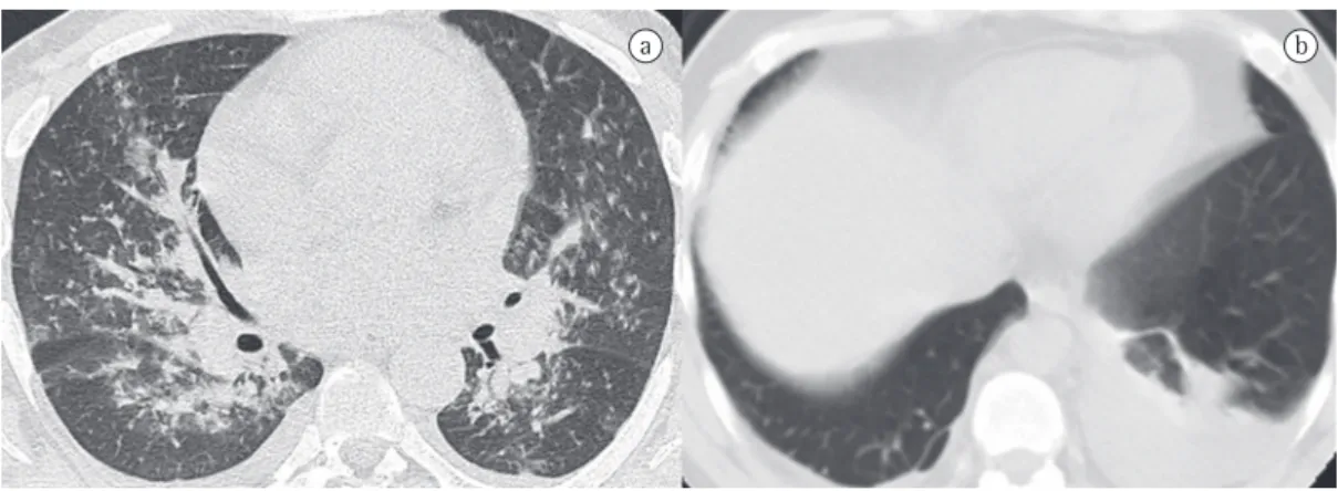
Figure 1 - Chest CT scan showing multiple nodules of irregular contours (black arrows).
a b
Figure 2 - Chest CT scan showing lung nodules with the halo sign (black arrows). The nodule has regular
Discussion
The present study described the tomographic findings in six patients with leukemia after chemotherapy or BMT, diagnosed with IPA.
In neutropenic patients, the presence of Aspergillus sp. deep in the basal membrane is considered diagnostic of IA.(13) In such cases, Aspergillus sp. invades blood vessels, resulting in thrombosis and subsequent hemorrhagic pulmonary infarction.(1,14,15) When the anato-mopathological findings are correlated with the tomographic findings, foci/areas of infarction can present as nodules or as consolidation in the shape of a wedge, with a pleural base.(2,16) Either finding can be accompanied by the halo sign, The clinical and radiological characteristics of
the patients under study are summarized in Table 1.
The nodules were more often multiple (3/5 cases), with irregular contours (3/5 cases; Figure 1). The halo sign was observed in three of the five cases of nodules (67%; Figure 2).
One case presented multiple, centrally distributed areas of consolidation, and another presented an isolated, peripheral area of consoli-dation (Figure 3). One case presented areas of consolidation and nodules.
As associated findings, areas of ground-glass attenuation and septal thickening were found in three and two patients, respectively. Bilateral pleural effusion, generally in small quantities, occurred in three cases.
a b
Figure 3 - Chest CT scan showing multiple, centrally distributed, areas of consolidation in a) and an isolated, peripheral area of consolidation in b). Note also bilateral pleural effusion.
Table 2 - Frequency (in %) of the tomographic findings in patients with invasive pulmonary aspergillosis reported in the literature.
Study n Nodule Consolidation Halo PE GG ST Crescent
Freitas 6 83 33 63 50 50 33 0
Althoff(16) 32 84 84 NA NA 44 NA 16
Brodoefel(2) 40 72.5 15 87.5 NA NA NA 45
Bruno(6) 68 76.5 24 25 NA NA NA 9
Caillot(17) 25 96 NA 100 NA NA NA 8
Hachem(8) 47 32 13 NA NA NA NA NA
Haugard(3) 21 95 57 100 NA NA NA 10
Horger(19) 46 64 26 82 NA NA 25 63
Kuhlman(15) 9 100 11 22 NA NA NA 66
Leão(18) 19 31.6 63.2 83.3 36.8 15.8 10.5 NA
Logan(13) 9 55 44 NA NA 11 NA NA
Won(14) 5 40 80 100 NA NA NA NA
compromised patients, the halo sign has been linked to IPA, Kaposi’s sarcoma and lympho-proliferative disorders.(20) However, most of the studies of IPA that address the tomographic finding of the halo sign are retrospective and include individuals in different phases of the disease, which results in great variation in the evaluation of the sensitivity and specificity of such sign.
Some studies have demonstrated that, prin-cipally after antifungal therapy initiation and on average 16 days after diagnosis,(2) fragments of lung infarction can detach themselves from the adjacent parenchyma, resulting in air-filled cavitation in the shape of a crescent (the air crescent sign).(14,15) Such sign was not, however, found in the present study, which is probably due to methodological issues, since our sample was small and we selected CT scans that were obtained as close as possible to the time of diag-nostic confirmation, before treatment initiation. In conclusion, nodules, consolidation, septal thickening, pleural effusion and ground-glass opacities were the principal tomographic findings in the six patients hospitalized in the hema-tology and bone marrow transplant ward during the IPA outbreak. The nodules were often (in 67% of the cases) accompanied by the halo sign, a classically described finding in patients with IPA. Therefore, chest CT scans of patients with leukemia after chemotherapy or BMT who are diagnosed with IPA can play an important role in early diagnosis, in early antifungal therapy initiation and, consequently, in improving the chance of survival in such cases.
Acknowledgments
The authors are especially grateful to Dr. Matheus Gonçalves and Dr. Carlos Alberto Pereira for their aid in collecting the clinical data related to the patients included in the present study.
References
1. Sharma OP, Chwogule R. Many faces of pulmonary aspergillosis. Eur Respir J. 1998;12(3):705-15. 2. Brodoefel H, Vogel M, Hebart H, Einsele H, Vonthein
R, Claussen C, et al. Long-term CT follow-up in 40 non-HIV immunocompromised patients with invasive pulmonary aspergillosis: kinetics of CT morphology and correlation with clinical findings and outcome. AJR Am J Roentgenol. 2006;187(2):404-13.
3. Hauggaard A, Ellis M, Ekelund L. Early chest radiography and CT in the diagnosis, management and outcome
which would correspond to alveolar hemorrhage accompanied by inflammation.(2,3)
Nodules and areas of consolidation are the principal findings in patients with IPA.(2,3,8,13-19). According to one group of authors,(15) nodules are the initial findings, and a halo sign is indicative of progression to consolidation and, subsequently, cavitation. Therefore, the varia-tion in the time elapsed between the onset of the pathophysiology of IA and the time of diag-nosis might explain why certain studies report nodules as being the most common finding and others report areas of consolidation as being the most common finding.
Pleural effusion, septal thickening and areas of ground-glass attenuation (as were found in the present case series) accompanied by nodules and areas of consolidation have also been described in the literature, at varying frequen-cies of occurrence.(2,3,8,13-19) Table 2 summarizes the frequency of our findings in relation to that of other studies.
The halo sign was first described in 1985 by Kuhlman et al. and is considered highly sugges-tive of IPA.(15) One group of authors reported that the frequency of the halo sign decreases with time in the course of IPA, suggesting that the halo sign is useful for the early diagnosis of IPA and therefore correlates with a better prog-nosis.(19) According to the EORTC criteria, the finding of the halo sign in a specific population warrants antifungal therapy initiation.(9) In the present study, the nodules were often (in 67% of the cases) accompanied by the halo sign, which is in accordance with the literature regarding the frequency of such finding (ranging from 20% to 100%) in patients with IPA.(2,3,6,8,15-17,19) Other authors have suggested that the halo sign can also be used as a marker of fungal activity and therefore as a marker of treatment response.(2)
immuno-tórax: sugestões iniciais para um consenso brasileiro. Radiol Bras. 2002;35(2):125-8.
13. Logan PM, Primack SL, Miller RR, Müller NL. Invasive aspergillosis of the airways: radiographic, CT, and pathologic findings. Radiology. 1994;193(2):383-8. 14. Won HJ, Lee KS, Cheon JE, Hwang JH, Kim TS, Lee
HG, et al. Invasive pulmonary aspergillosis: prediction at thin-section CT in patients with neutropenia--a prospective study. Radiology. 1998;208(3):777-82. 15. Kuhlman JE, Fishman EK, Siegelman SS. Invasive
pulmonary aspergillosis in acute leukemia: characteristic findings on CT, the CT halo sign, and the role of CT in early diagnosis. Radiology. 1985;157(3):611-4. 16. Althoff Souza C, Müller NL, Marchiori E, Escuissato
DL, Franquet T. Pulmonary invasive aspergillosis and candidiasis in immunocompromised patients: a comparative study of the high-resolution CT findings. J Thorac Imaging. 2006;21(3):184-9.
17. Caillot D, Couaillier JF, Bernard A, Casasnovas O, Denning DW, Mannone L, et al. Increasing volume and changing characteristics of invasive pulmonary aspergillosis on sequential thoracic computed tomography scans in patients with neutropenia. J Clin Oncol. 2001;19(1):253-9.
18. Leão RC, Marchiori E, Rodrigues R, Souza Jr AS, Gasparetto EL, Escuissato DL. Tomografia computadorizada na avaliação da aspergilose pulmonar angioinvasiva em pacientes com leucemia aguda. Radiol Bras. 2006;39(5):327-31.
19. Horger M, Hebart H, Einsele H, Lengerke C, Claussen CD, Vonthein R, et al. Initial CT manifestations of invasive pulmonary aspergillosis in 45 non-HIV immunocompromised patients: association with patient outcome? Eur J Radiol. 2005;55(3):437-44.
20. Gaeta M, Blandino A, Scribano E, Minutoli F, Volta S, Pandolfo I. Computed tomography halo sign in pulmonary nodules: frequency and diagnostic value. J Thorac Imaging. 1999;14(2):109-13.
of invasive pulmonary aspergillosis. Acta Radiol. 2002;43(3):292-8.
4. Denning DW, Stevens DA. Antifungal and surgical treatment of invasive aspergillosis: review of 2,121 published cases. Rev Infect Dis. 1990;12(6):1147-201. Erratum in: Rev Infect Dis 1991;13(2):345.
5. Baddley JW, Stroud TP, Salzman D, Pappas PG. Invasive mold infections in allogeneic bone marrow transplant recipients. Clin Infect Dis. 2001;32(9):1319-24. 6. Bruno C, Minniti S, Vassanelli A, Pozzi-Mucelli R.
Comparison of CT features of Aspergillus and bacterial pneumonia in severely neutropenic patients. J Thorac Imaging. 2007;22(2):160-5.
7. Denning DW. Therapeutic outcome in invasive aspergillosis. Clin Infect Dis. 1996;23(3):608-15. 8. Hachem R, Sumoza D, Hanna H, Girgawy E, Munsell
M, Raad I. Clinical and radiologic predictors of invasive pulmonary aspergillosis in cancer patients: should the European Organization for Research and Treatment of Cancer/Mycosis Study Group (EORTC/MSG) criteria be revised? Cancer. 2006;106(7):1581-6.
9. Ascioglu S, Rex JH, de Pauw B, Bennett JE, Bille J, Crokaert F, et al. Defining opportunistic invasive fungal infections in immunocompromised patients with cancer and hematopoietic stem cell transplants: an international consensus. Clin Infect Dis. 2002;34(1):7-14.
10. Reichenberger F, Habicht J, Matt P, Frei R, Solèr M, Bolliger CT, et al. Diagnostic yield of bronchoscopy in histologically proven invasive pulmonary aspergillosis. Bone Marrow Transplant. 1999;24(11):1195-9. 11. Weisser M, Rausch C, Droll A, Simcock M, Sendi P, Steffen
I, et al. Galactomannan does not precede major signs on a pulmonary computerized tomographic scan suggestive of invasive aspergillosis in patients with hematological malignancies. Clin Infect Dis. 2005;41(8):1143-9. 12. Souza Jr. AS, Araujo Neto C, Jasinovodolinsky D,
Marchiori E, Kavakama J, Irion KL, et al. Terminologia para a descrição de tomografia computadorizada do
About the authors
Daniela Batista de Almeida Freitas
Resident. Universidade Federal de São Paulo/Escola Paulista de Medicina – UNIFESP/EPM, Federal University of São Paulo/Paulista School of Medicine – São Paulo, Brazil.
Ana Cláudia Piovesan
Resident. Universidade Federal de São Paulo/Escola Paulista de Medicina – UNIFESP/EPM, Federal University of São Paulo/Paulista School of Medicine – São Paulo, Brazil.
Gilberto Szarf
Radiologist. Department of Diagnostic Imaging, Universidade Federal de São Paulo/Escola Paulista de Medicina – UNIFESP/EPM, Federal University of São Paulo/Paulista School of Medicine – São Paulo, Brazil.
Dany Jasinowodolinski
Radiologist. Department of Diagnostic Imaging, Universidade Federal de São Paulo/Escola Paulista de Medicina – UNIFESP/EPM, Federal University of São Paulo/Paulista School of Medicine – São Paulo, Brazil.
Gustavo de Souza Portes Meirelles