Comparison of the effects that two different respiratory
physical therapy techniques have on cardiorespiratory
parameters in infants with acute viral bronchiolitis*
Comparação dos efeitos de duas técnicas fisioterapêuticas respiratórias emparâmetros cardiorrespiratórios de lactentes com bronquiolite viral aguda
Melissa Karina Pupin, Adriana Gut Lopes Riccetto, José Dirceu Ribeiro, Emílio Carlos Elias Baracat
Abstract
Objective: To compare the expiratory flow increase technique (EFIT) and vibration accompanied by postural
drainage (PD) in terms of their effects on the heart rate (HR), respiratory rate (RR) and SpO2 of infants with acute viral bronchiolitis (AVB). Methods: Infants with clinical and radiological diagnosis of AVB were analyzed. The HR, RR and SpO2 were registered at four time points: prior to the procedure; and at 10, 30 and 60 min after the procedure. The patients were divided into three groups: submitted to the EFIT; submitted to vibration/PD; and control. Results: We included 81 infants, 27 per group, with a mean age of 4.52 years and a mean weight of 6.56 kg. Using ANOVA, we found that the EFIT and vibration/PD groups presented no significant differences in relation to the control group in terms of the mean values for HR, RR or SpO2 (p > 0.05). Considering only the four time points evaluated, the mean RR was significantly lower in the EFIT and vibration/PD groups than in the control group (p < 0.05). Conclusions: In terms of overall improvement of cardiorespiratory parameters, neither the EFIT
nor vibration/PD provided any benefit to infants with BVA. However, over time, respiratory physical therapy seems to contribute to decreasing the RR in these patients.
Keywords: Bronchiolitis, viral; Physical therapy modalities; Infant.
Resumo
Objetivo: Comparar os efeitos das técnicas de aumento do fluxo expiratório (AFE) e vibração associada à drenagem
postural (DP) nos parâmetros cardiorrespiratórios de frequência cardíaca (FC), frequência respiratória (FR) e SpO2 de lactentes com bronquiolite viral aguda (BVA). Métodos: Foram analisados lactentes com diagnóstico clínico e
radiológico de BVA. A FC, FR e SpO2 foram registradas em quatro tempos: antes do procedimento e após 10, 30 e 60 min do término do procedimento. Os pacientes foram divididos em três grupos: submetido à AFE; submetido à vibração/DP; e controle. Resultados: Foram incluídos no estudo 81 lactentes, 27 em cada grupo de estudo, com
média de idade de 4,52 meses e peso médio de 6,56 kg. Na comparação por ANOVA, as médias da FR, FC e SpO2 nos grupos AFE e vibração/DP não apresentaram diferenças significantes em relação ao grupo controle (p > 0,05). Considerando somente os quatro tempos, houve queda significante dos valores médios de FR nos grupos AFE e vibração/DP em relação ao controle (p < 0,05). Conclusões: A aplicação de AFE e de vibração associada à DP não apresentou um benefício global na melhora dos parâmetros cardiorrespiratórios em lactentes com BVA. Quando analisados isoladamente no decorrer do tempo, a fisioterapia respiratória parece contribuir na diminuição da FR nesses pacientes.
Descritores: Bronquiolite viral; Modalidades de fisioterapia, Lactente.
* Study carried out in the Department of Pediatrics, Universidade Estadual de Campinas – Unicamp, State University at Campinas – Campinas, Brazil.
Correspondence to: Melissa Karina Pupin. Rua Pedro Vieira da Silva, 415, apto. 23, bloco E, Santa Genebra, CEP 13080-570, Campinas, SP, Brasil.
Tel 55 19 8153-8297. E-mail: melketson@hotmail.com
Financial support: This study received financial support from the Coordenação de Aperfeiçoamento de Pessoal de Nível Superior (CAPES, Coordination of the Advancement of Higher Education).
tion, autogenic drainage and induced cough.(7-9)
Among those, the EFIT is the most widely used in the routine treatment of children and adolescents with respiratory involvement, although the tech-nique has not been scientifically validated.(7,10,11)
Oxygen therapy is currently the only truly effective treatment for AVB.(12) The search for
other efficacious treatments for AVB, with proven evidence, is a current theme in many systematic reviews of the literature. Although conventional respiratory therapy is not indicated in the acute phase of AVB, the use of the most modern techniques, such as the EFIT, remain a therapeutic proposal to be validated, due to the scarcity of studies comparing them with the techniques considered conventional.
The objective of this study was to compare the effects of the EFIT and of vibration accom-panied by PD in terms of their effects on the heart rate (HR), respiratory rate (RR) and SpO2 of infants with AVB.
Methods
Study design
This was a comparative, controlled interven-tion study. The following patients participated in the study: patients with AVB, treated in the Referral Emergency Pediatrics Department of the Universidade Estadual de Campinas (Unicamp, State University at Campinas) Hospital das Clínicas between July of 2005 and August of 2007. This study was approved by the Research Ethics Committee of the Unicamp School of Medical Sciences (ruling no. 187/2005), and, before the infants were included in the study, their legal guardians gave written informed consent.
Subjects
We included patients less than one year of age diagnosed with AVB by the local medical staff. The diagnosis was based on the following clinical and radiological criteria: initial clinical profile of rhinorrhea, cough and low-grade fever, evolving within two or three days to include at least two signs of mild or moderate respiratory distress (nasal flaring, tachypnea, dyspnea, subcostal retraction, retraction of the suprasternal notch, accessory muscle recruit-ment, prolonged exhalation or pulmonary
Introduction
Respiratory diseases are still relevant causes of morbidity and mortality in developing coun-tries, principally in children.(1) Respiratory therapy
is an auxiliary treatment in those diseases, increasing mucociliary clearance and reducing airway obstruction, as well as facilitating venti-lation and gas exchange.(2)
In infants with acute respiratory symptoms, acute viral bronchiolitis (AVB) is the most clini-cally relevant disease, due to the high morbidity rate, particularly in the autumn and winter months, when the respiratory syncytial virus, the principal etiologic agent of AVB, is circulating. In infants less than three months of age who present comorbidities (congenital heart disease and bronchopulmonary dysplasia), AVB can be more severe, resulting in higher rates of hospi-talization, and, in some cases, the patient can evolve to respiratory failure and require venti-latory support.(3) The indication for respiratory
therapy in AVB remains controversial regarding its effects on the clinical improvement of the patients and on the length of hospital stay. To date, there is no direct evidence that the application of respiratory therapy is beneficial to patients in this clinical situation.(3,4) In the
opinion of some authors, respiratory therapy is indicated only after the acute phase of the disease, in cases in which the retention of secre-tions is abundant, and for those patients who have evolved to atelectasis.(4-6)
However, this lack of indication or even the contraindication of the respiratory therapy approach in the acute phase of AVB is based on studies which use conventional techniques, such as thoracic percussion, vibration and postural drainage (PD).(7) However, other airway
clear-ance techniques, denominated unconventional, have been used in some European countries, with positive results in children and infants with acute respiratory diseases. The application of such techniques has been shown to improve the clinical parameters of the patients, decrease the length of hospital stays and hasten the recovery of respiratory function. The protocol for the use of airway clearance techniques follows the recommendations of the Consensus Conference at Lyon in 1994,(8) which include the expiratory
posi-One hand was placed between the suprasternal notch and the mammary line, providing support with the ulnar border of the hand, and chest compression was performed during the exha-lation of the infant (hand on the chest). The other hand was placed on the navel, with the thumb and the index finger in contact with the lower ribs in order to feel each breath (hand on the abdomen). Initially, the physical therapist conducted a strong chest compression in order to provoke a deep inhalation and therefore a prolonged exhalation. During each exhalation, the hand on the chest conducted a compres-sion maneuver obliquely (from top to the front and from the front to the back), whereas the hand on the abdomen continued to support the abdominal region.(14) The technique was applied
for 5 to 10 compressions, followed by the closing of the mouth of the infant, provoking aspiration of the secretion and its dislodgment to the pharynx. This was repeated for a total of 40 compressions. The secretions mobilized were dislodged to the mouth and collected with a tissue. Cough was induced, when necessary, by applying finger pressure to the trachea of the patient. In cases in which the patient presented tachypnea, the ratio was one compression for every two or three breaths.(13)
Vibration consisted of quick rhythmic move-ments, with enough intensity to cause vibration of the airway. Such vibrations are repeated isometric contractions of the chest wall during the expiratory phase. The objective of this tech-nique is to dislodge pulmonary secretions that are already loose, conducting them to the larger caliber bronchi and the trachea, subsequently removing them from the respiratory tract.(15)
The objective of the PD was to facilitate the auscultation with predominance of wheezing);
chest X-ray showing lung hyperinflation; and presenting pneumonia accompanied by atel-ectasis.(12) Patients remained hospitalized in
the Unicamp Referral Emergency Pediatrics Department.
The following patients were excluded: those in the early postoperative period of thoracic of abdominal surgery; those with congenital cardi-opathy; those with genetic disease; those with a history of more than three episodes of wheezing; and those in severe respiratory failure, in need of intubation and mechanical ventilation.
The patients were divided in three groups, according to the consecutive order of admission as follows: submitted to the EFIT; submitted to vibration and PD; and control (no intervention).
Procedures
The respiratory therapy procedures were conducted in the morning, in a single session and always by the same professional.
With the objective of mobilizing, dislodging and eliminating peripheral secretions from the bronchial tree to the trachea, we used the EFIT in order to achieve a passive increase in expira-tory volume.(10,13) Patients were placed in the
supine position, with the head of the bed slightly elevated, as a safeguard against gastroesopha-geal reflux and aspiration. The physical therapist stood at the side of the bed, with elbows in a semiflexed position, performing the technique without using body weight. The range and velocity of the maneuver varied according to the location and quantity of secretion observed in each patient, the thoracoabdominal movement synchronized with the timing of exhalation, through manipulation by the physical therapist.
Table 1 - Distribution of the categorical and continuous variables of the patients with acute viral bronchiolitis,
by group.
Variable Group A Group B Group C p
Gender, M/F 18/9 15/12 15/11 0.7141
Episode, 1st/2nd/3rd 20/7/0 24/3/0 22/4/1 0.3780
Comorbidity, yes/no 7/20 5/22 7/20 0.7595
Premature birth, yes/no 6/21 8/19 4/23 0.4244
Oxygen support, yes/no 13/14 10/17 15/12 0.3899
the variables age, period of manifestation and weight, ANOVA with a rank test was also used, although without repeated measures.
The chi-square test was used in order to identify associations among the categorical variables. Fisher’s exact test was used when the expected values were lower than 5.
Values of p ≤ 0.05 were considered statisti-cally significant.(18) The statistical program used
was Statistical Analysis System, version 9.1.3 (SAS Institute, Cary, NC, USA).(19)
Results
Between July of 2005 and August of 2007, 81 infants were included in the study, 27 in each group: 48 were male (59.25%) and dislodgement of the pulmonary secretion from
the bronchial tree by the force of gravity through the position of the patient, thus preventing the accumulation of secretions and facilitating the outflow to the oropharynx.(2,16) The patients were
positioned using a modified PD position (head of the bed at 30°) to prevent gastroesophageal reflux and aspiration.(17) The physical therapist
stood at the side of the bed of the patient. The vibration was performed with the hands spread, positioned bilaterally on the thorax of the patient; the fist and the elbow of the phys-ical therapist remained immobile, impelling the vibratory movement with mechanical effort by the arm and shoulder muscles, leaving the other muscle groups of the upper limb isometrically contracted and the joints of the fist and elbow immobile. The procedure was performed for 10 min. Cough was induced, when necessary, by applying finger pressure to the trachea of the patient.(15)
The patients in the control group were not submitted to any respiratory therapy procedures. They were positioned in the supine position, with the head of the bed elevated, and received only manual contact of the physical therapist on the thorax for 10 min.
The cardiorespiratory parameters of RR, HR and SpO2 were recorded at four time points: prior to the procedure (T1), then at 10, 30 and 60 min after the procedure (T2, T3 and T4, respectively).
With a hand on the abdomen of the patient to confirm the beginning and the end of each breath, RR was counted for 1 min. An oximeter (DX 2405 Oxypleth; Dixtal Biomédica, Manaus, Brazil) was used to measure HR and SpO2. Oxygen support (tent or mask) was suspended for 10 min before the measurement of the parameters, at all time points.
The nasal aspiration technique, used for the resolution of upper airway obstruction, was not performed during the collection of data for the present study.
To compare the variables HR, RR and SpO2 between the time points (T1, T2, T3 and T4) and between groups (EFIT, vibration/PD and control), we used ANOVA with repeated meas-ures and rank test. When the difference was significant, multiple comparison tests (Tukey’s test and contrast test) were conducted in order to identify the differences. In the comparison of
Respiratory rate
Heart rate
SpO
2
(%)
45 50 55 60 65
T1 T2 T3 T4
a
140 145 150 155 160
T1 T2 T3 T4
b
20 21 22 23 24 25 26 27 28
T1 T2 T3 T4
c
Group A Group B Group C
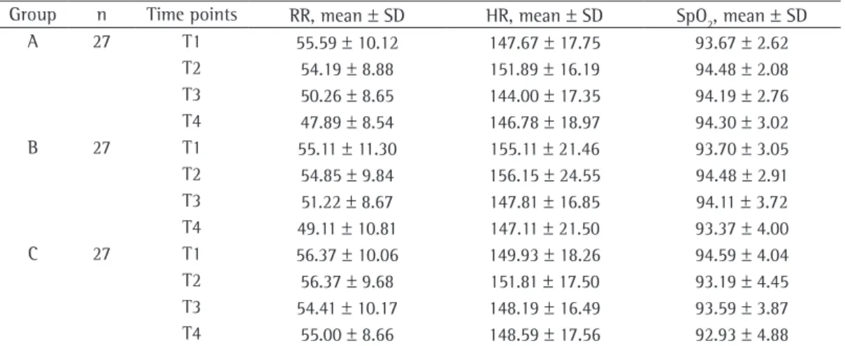
urements, with statistical significance between T2 and T3 (p = 0,0023), as well as between T2 and T4 (p = 0.0066). In the vibration/PD group, there was a decrease until T4, with significant difference between T1 and T4 (p = 0.0061), T2 and T3 (p = 0.0126) as well as between T2 and T4 (p = 0.005). Regarding the means of HR, there was a decrease, with statistical significance between T1 and T3 (p = 0.0171), between T2 and T3 (p = 0.0016) and between T2 and T4 (p = 0.0137) in all three groups (Table 2).
Discussion
Studies analyzing the various respiratory therapy techniques used in infants with AVB have shown an absence of benefit in terms of clinical evolution, length of hospital stay, course of the disease and morbidity.(3,20-22)
According to the guidelines of the American Academy of Pediatrics, AVB is a disease which has a three-week clinical course; the recom-mended treatment is basically support, with oxygen administration and hydration; and the use of respiratory therapy in the inpatients has no impact on the clinical improvement of the patient and on the decrease of the length of hospital stay.(3) However, one point which can
make analysis of the efficacy of the respira-tory therapy techniques in AVB difficult is the fact that the techniques applied in the various studies were evaluated together with conven-tional techniques.(3,20-23) Some authors,(20,24) using
33 were female (40.75%): ages ranged from 1 to 11 months (mean, 4.52 months); and weights ranged from 2.6 to 11.8 kg (mean, 6.56 kg). The period of manifestation of the disease was between 2 and 10 days (mean, 5.49 days).
We excluded 21 infants: 9 with severe respira-tory failure, requiring intubation and mechanical ventilation; 6 with heart disease; 3 with Down’s syndrome; 2 with bronchial dysplasia; and 1 in the postoperative period of heart surgery.
Regarding maturity at birth, 77.7% of the patients were born at term, and 22.2% were born prematurely. We observed that 81.5% of the procedures were carried out in children who presented the first episode of wheezing. Considering co-occurrence of AVB with other diseases, we observed that 76.5% of the procedures were performed in infants without concomitant diseases, 9.8% in infants with pneumonia, 3.7% in infants with atelectasis and 10% in infants diagnosed with diseases unre-lated to pulmonary function. The description of the characteristics of the patients in each study group is shown in Table 1.
In the general comparison, using ANOVA, among the groups in the four time points, the means of RR, HR and SpO2 in the EFIT and vibration/PD groups presented no significant differences in relation to the control group (Figure 1).
However, using only the analysis of the time points, RR in the EFIT group presented a constant decrease in the means of the four
meas-Table 2 - Distribution of the mean values of respiratory rate, heart rate and SpO2 of infants with acute viral
bronchiolitis, by group.
Group n Time points RR, mean ± SD HR, mean ± SD SpO2, mean ± SD A 27 T1 55.59 ± 10.12 147.67 ± 17.75 93.67 ± 2.62
EFIT, in infants on mechanical ventilation for acute respiratory failure due to obstruction.
In the present study, there were no significant differences in RR, HR or SpO2 when the three groups and the four time points were compared. These findings show that both a conventional technique, such as vibration with PD, and an airway clearance technique, such as the EFIT, produced no benefit in terms of oxygenation, as confirmed by the stability of SpO2 in all groups studied.
When the analysis was conducted considering only the four time points, we observed a signifi-cant drop in the mean values of RR in the EFIT and vibration/PD groups in relation to those obtained for the control group. This drop in the RR also suggests that the respiratory therapy techniques used in this study did no harm to the cardiorespiratory system of the infants with AVB, and the improvement in tachypnea could be related to the unblocking component of the techniques, with consequent improvement of the airflow.(27-29)
The evolution of the HR showed an increase at T1 (10 min after the end of the procedure) in the group submitted to the EFIT, followed by a drop at T4 (60 min), the latter being common to all groups. An increase in oxygen uptake frequently occurs when a patient receives respi-ratory therapy, and that increase is accompanied by increases in HR, arterial pressure and intrac-ranial pressure. The change in these vital signs seems to be associated with the high thoracic compliance and high residual lung capacity that are typical in this population.(30) When submitted
to techniques of greater manipulation, such as the EFIT, these parameters tend to increase. In the case of the conventional techniques used in the patients of the vibration/PD group, the procedures demanded less manipulation and would have had less impact on the HR, the parameter that is the most sensitive to greater oxygen uptake.
The alterations in pulmonary function which lead to difficult ventilation in AVB are, funda-mentally, related to obstructive phenomena in the small airways (bronchioles). The obstruction alters the ventilation/perfusion ratio, generating alveolar hypoventilation, which is accompanied by hypoxemia, CO2 trapping and acidosis (respi-ratory and metabolic). The obstructive profile leads to an increase in the residual volume and conventional respiratory therapy (percussion,
PD, assisted cough and oropharyngeal aspira-tion), have shown no significant differences in the length of hospital stay, course of the disease or clinical score, when compared with groups of patients not submitted to respiratory therapy. In another randomized controlled trial, 16 patients with AVB, hospitalized in the ICU, submitted to conventional respiratory therapy (percussion, PD, vibration and nasopharyngeal aspiration), were compared with a control group with the same disease and submitted only to nasopharyngeal aspiration. The authors observed no significant differences between the groups in terms of the clinical breathlessness score or the length of hospital stay.(23)
In another study, conventional respiratory therapy (percussion, vibration, PD and aspira-tion) was applied in 26 infants with AVB and compared with a control group submitted only to modified PD (elevated head of the bed) and aspiration.(21) The authors found that the
respi-ratory therapy did not affect the course of the disease in the patients. In addition, there was no statistically significant effect in the length of hospital stay, in the demand of supplemental oxygen or in the need for nasogastric feeding. However, in the group submitted to conven-tional respiratory therapy techniques, the authors observed a significant increase in SpO2 at 10 min after the procedure.(21)
Regarding the EFIT, which is considered a state-of-the-art respiratory therapy tech-nique, there are no formal clinical elements for its validation—only an overall impression of its clinical efficacy.(11,25) There have been few
studies evaluating the effects of the EFIT in patients with AVB. Among those, one group of authors(7) observed a significant increase in SpO
2
and in tidal volume, immediately and 1 h after the procedure, which was used in conjunction with endotracheal aspiration, in 20 infants with AVB and on mechanical ventilation. In another study, the authors identified more rapid remis-sion of the respiratory and clinical symptoms, with a reduction in fever and dyspnea, improved pulmonary auscultation findings, less cough and an increased appetite, after daily application of respiratory therapy techniques, including the EFIT, in patients with AVB.(26) Another group of
authors(13) observed an increase in the SpO 2 in
Benguigui Y, Antuaño FJ, Schmunis G, Yunes J, editors. Infecciones respiratórias em niños. Washington D.C: Organización Panamericana de la Salud; 1997. p. 3-23. 2. Balachandran A, Shivbalan S, Thangavelu S. Chest
physiotherapy in pediatric practice. Indian Pediatr. 2005;42(6):559-68.
3. American Academy of Pediatrics Subcommittee on Diagnosis and Management of Bronchiolitis. Diagnosis and management of bronchiolitis. Pediatrics. 2006;118(4):1774-93.
4. Perrotta C, Ortiz Z, Roque M. Chest physiotherapy for acute bronchiolitis in paediatric patients between 0 and 24 months old. Cochrane Database Syst Rev. 2005;(2):CD004873.
5. Amantéa SL, Silva FA. Acute viral bronchiolitis - still subject for controversy [Article in Portuguese]. J Pediatr (Rio J). 1998;74 Suppl 1:S37-47.
6. Deschildre A, Thumerelle C, Bruno B, Dubos F, Santos C, Dumonceaux A. Acute bronchiolitis in infants [Article in French]. Arch Pediatr. 2000;7 Suppl 1:21S-26S. 7. Bernard-Narbonne F, Daoud P, Castaing H, Rousset
A. Effectiveness of chest physiotherapy in ventilated children with acute bronchiolitis [Article in French]. Arch Pediatr. 2003;10(12):1043-7.
8. L’Union Régionale des Médecins Libéraux d’̂le-de-France; France / Agence Nationale d’Accréditation et d’Evaluation en Santé. Conférence de consensus sur la prise en charge de la bronchiolite du nourrisson: Paris, 21 septembre 2000. Paris: Elsevier; 2001.
9. Conférence de consensus sur la prise en charge de la bronchiolite du nourisson (Texte Court). Ann Kinésithér. 2001;28(1):20-8.
10. Delaunay JP. Conférence de consensus en kinésithérapie respiratoire. Place respective des différentes techniques non instrumentales de désencombrement bronchique. Cah Kinésithér. 1998;192(4):14-22.
11. Wils J. L´accélération du flux expiratoire chez l`adulte: technique de désencombrement bronchique. Cah Kinésithér. 1998;192(4):1-13.
12. Willson DF, Landrigan CP, Horn SD, Smout RJ. Complications in infants hospitalized for bronchiolitis or respiratory syncytial virus pneumonia. J Pediatr. 2003;143(5 Suppl):S142-9.
13. Almeida CC, Ribeiro JD, Almeida-Júnior AA, Zeferino AM. Effect of expiratory flow increase technique on pulmonary function of infants on mechanical ventilation. Physiother Res Int. 2005;10(4):213-21.
14. Vinçon C, Fausser C. Kinésithérapie Respiratoire en Pédiatrie: du prématuré au petit enfant. Paris: Masson; 1989. p. 41-59.
15. Moriyama LT, Guimarães ML, Juliani RC. Fisioterapia Respiratória para Crianças. In Rozov T, editor. Doenças Pulmonares em Pediatria - Diagnóstico e Tratamento. São Paulo: Atheneu; 1999. p. 609-17.
16. Barthe J, Binoche C, Henri JD, Pecchia S. Utilite du drainage postural? Kinésither Scientif. 1989;275. 17. Button BM, Heine RG, Catto-Smith AG, Phelan PD,
Olinsky A. Postural drainage and gastro-oesophageal reflux in infants with cystic fibrosis. Arch Dis Child. 1997;76(2):148-50.
18. Montgomery DC. Design and analysis of experiments. New York: Wiley; 1991.
19. SAS Institute Inc. The SAS System for Windows (Statistical Analysis System), version 9.1.3. Cary (NC): SAS Institute Inc.; 2002-2003.
in the normal volume of the intact chest at rest, that is, the functional residual capacity, resulting in greater respiratory effort.(4) Considering that
the EFIT was created especially for bronchial clearance of the infants, we can suppose that its application is more efficacious in that age bracket, which explains the consistent drop in RR at 10 min after the end of the procedure. However, the EFIT, due to the greater manip-ulation of the patient, can provoke additional energy expenditure in unstable patients with diffuse acute respiratory disease, a common situation among hospitalized patients.
Based on our findings, we can suggest that the right moment to indicate respiratory therapy in patients with AVB is in a subacute phase of the disease, within the second week of evolution, when the airways present greater accumulation of secretions. Over the course of the disease, when the secretion frequently accumulates in the airways, obstruction and collapse of the alveoli, respiratory therapy could be beneficial, particularly the EFIT, which promotes bronchial clearance, decreases pulmonary insufflation and increases alveolar recruitment.(4) In the initial
phase, when inflammation predominates, with the presence of squamous cells and edema of the respiratory mucosa, airway clearance techniques, with greater manipulation of the patients, would bring no significant benefits. This phase, typical of the first week of the disease, was that in which the infants in this study were evaluated.
Future studies, applying the EFIT in different stages of the disease, could present different results and contribute novel data in order to validate or not respiratory therapy as an adjuvant treatment in AVB. Other limitations of our study include the lack of an objective score for the evaluation of the effects of respiratory therapy, the small number of patients in each group and the lack of comparison of the techniques in patients with distinct differences in terms of clinical severity (mild, moderate and severe).
In our study, the application of the respira-tory therapy techniques, the EFIT and vibration with PD presented no overall benefit in terms of the cardiorespiratory parameters in infants with AVB.
References
et à l`achévement de la toilette bronchopulmonaire du nourrisson et de l`enfant. Ann Kinésithér. 1991;18(3):117-24.
27. Mansbach JM, Clark S, Christopher NC, LoVecchio F, Kunz S, Acholonu U, et al. Prospective multicenter study of bronchiolitis: predicting safe discharges from the emergency department. Pediatrics. 2008;121(4):680-8. 28. Mayordomo-Colunga J, Medina A, Rey C, Díaz JJ,
Concha A, Los Arcos M, et al. Predictive factors of non invasive ventilation failure in critically ill children: a prospective epidemiological study. Intensive Care Med. 2009;35(3):527-36.
29. Demont B, Viçon C, Cambas C, Bailleux S. Effets de la technique d`augmentation du flux expiratoire sur la résistance du système respiratoire et la SaO2, du prématuré à l`enfant à terme. Ann Kinésithér. 1999;26(5):227-31.
30. Krause MF, Hoehn T. Chest physiotherapy in mechanically ventilated children: a review. Crit Care Med. 2000;28(5):1648-51.
20. Webb MS, Martin JA, Cartlidge PH, Ng YK, Wright NA. Chest physiotherapy in acute bronchiolitis. Arch Dis Child. 1985;60(11):1078-9.
21. Nicholas KJ, Dhouieb MO, Marshall TG, Edmunds AT, Grant MB. An evaluation of chest physiotherapy in the management of acute bronchiolitis. Physiotherapy. 1999;85(12):669-74.
22. Beauvois E. Role of respiratory therapy in the treatment acute bronchiolitis in infants [Article in Franch]. Arch Pediatr. 2001;8 Suppl 1:128S-131S.
23. Bohé L, Ferrero ME, Cuestas E, Polliotto L, Genoff M. Indications of conventional chest physiotherapy in acute bronchiolitis [Article in Spanish]. Medicina (B Aires). 2004;64(3):198-200.
24. Wallis C, Prasad A. Who needs chest physiotherapy? Moving from anecdote to evidence. Arch Dis Child. 1999;80(4):393-7.
25. Postiaux G, Lens E. De ladite « Accélération du Flux Expiratoire (AFE) » :où Forced is... Fast (Expiration technique-FET)! Ann Kinésithér. 1992;19(8):411-27. 26. Postiaux G, Bafico JF, Masengu R, Lahafe JM.
Paramètres anamnestiques et cliniques utiles au suivi
About the authors
Melissa Karina Pupin
Physical Therapist. Universidade Estadual de Campinas – Unicamp, State University at Campinas – Campinas, Brazil.
Adriana Gut Lopes Riccetto
Attending Physician. Universidade Estadual de Campinas – Unicamp, State University at Campinas – Campinas, Brazil.
José Dirceu Ribeiro
Associate Professor. Universidade Estadual de Campinas – Unicamp, State University at Campinas – Campinas, Brazil.
Emílio Carlos Elias Baracat