Abstract
Submitted: August 4, 2017 Accepted: August 13, 2017
Demetallization of
Enterococcus
faecalis
bioilm: a preliminary study
Objectives: To determine the concentration of calcium, iron, manganese and zinc ions after the application of chelator to Enterococcus faecalis bioilms.
Material and Methods: Fifty bovine maxillary central incisors were prepared and inoculated with E. faecalis for 60 days. The following were used as
irrigation solutions: 17% EDTA (pH 3, 7 and 10), 2.5% sodium hypochlorite (NaOCl) combined with 17% EDTA (pH 3, 7 and 10), distilled water (pH 3, 7 and 10), and 2.5% NaOCl. Each solution was kept in the root canal for ive minutes. Fifteen uncontaminated root canals were irrigated with 17% EDTA (pH 3, 7 and 10). Six teeth were used as bacterial control. The number of calcium, iron, manganese and zinc ions was determined using lame atomic absorption spectrometry. Mean ± standard deviation (SD) values were used for descriptive statistics. Results: Calcium chelation using 17% EDTA at pH 7 was higher than at pH 3 and 10, regardless of whether bacterial bioilm was present. The highest concentration of iron occurred at pH 3 in the presence of bacterial bioilm. The highest concentration of manganese found was 2.5% NaOCl and 17% EDTA at pH 7 in the presence of bacterial bioilm. Zinc levels were not detectable. Conclusions: The pH of chelating agents affected the removal of calcium, iron, and manganese ions. The concentration of iron ions in root canals with bacterial bioilm was higher after the use of 17% EDTA at pH 3 than after the use of the other solutions at all pH levels.
Keywords: Bioilms. EDTA. Sodium hypochlorite. Enterococcus faecalis. Carlos ESTRELA1
Rodrigo COSTA E SILVA2
Roberta Cerasi URBAN3
Pablo José GONÇALVES2
Júlio A. SILVA1
Cyntia R.A. ESTRELA1
Jesus Djalma PECORA4
Ove A. PETERS5
1Universidade Federal de Goiás, Faculdade de Odontologia, Departamento de Ciências Estomatológicas, Goiânia, Goiás, Brasil.
2Universidade Federal de Goiás, Instituto de Física, Goiânia, Goiás, Brasil.
3Universidade Federal de São Carlos, Instituto de Química, São Carlos, São Paulo, Brasil. 4Universidade de São Paulo, Faculdade de Odontologia de Ribeirão Preto, Departamento de Endodontia, Ribeirão Preto, São Paulo, Brasil.
5University of the Paciic, Arthur A. Dugoni School of Dentistry, Department of Endodontics, San Francisco, California, USA.
INTRODUCTION
The destruction of bacterial biofilm has been a significant topic in endodontic research over the years13,21,25. For this purpose, antibacterial
and physicochemical effectiveness of a series of irrigating solutions has been evaluated, including sodium hypochlorite, chlorhexidine, cationic detergent, ethylenediaminetetraacetic acid (EDTA), MTAD, ozonated water, apple vinegar and other solutions9-11,15,16,20,26-30,33.
Sodium hypochlorite is an irrigating solution largely used because of a combination of several properties: antimicrobial action, tissue dissolution capacity and acceptable biological compatibility at less concentrated solutions9-12,15,16,20,29,30. Sodium hypochlorite acts
on enzymatic sites essential for bacterial viability, promoting irreversible microbial inactivation due to the action of hydroxyl ions and chloramination. The dissolution of organic tissue occurs during saponiication, when sodium hypochlorite degrades fatty acids and lipids, resulting in soap and glycerol10.
Root canal preparation produces a smear layer, which is composed of dentin chips, remnants of pulp tissue and odontoblastic processes, microorganisms and chemicals found in irrigating agents15,16,26-28,33.
Ethylenediaminetetraacetic (EDTA) at a neutral pH promotes the chelation of calcium ions in the dentin15,16,26,27. This chelating agent is commonly used
for smear layer removal, but has a poor antibacterial
effect15,16,28. A mixture of a new solution for the removal
of the smear layer containing 3% doxycycline hyclate, 4.25% citric acid, and 0.5% Tween 80 (MTAD, Dentsply Sirona, York, PA, USA) has been evaluated as a inal rinse of root canal surfaces after preparation. MTAD effectively removed the smear layer when used as a inal rinse28.
The destruction of bacterial life is dependent on the conditions of their growth and multiplication, among which are physical-chemical factors such as: temperature, pH, osmotic pressure, and oxygen, carbon dioxide and substrate concentrations. The dynamics of endodontic infections suggests the following ecological determinants: oxidation-reduction potential; nutrient availability; and microbial interactions, such as synergism or antagonism4-6. Bacterial cells require
carbon, nitrogen, oxygen, hydrogen, phosphorous, sulphur, iron, sodium, calcium, magnesium and water. Other nutrients are required in extremely low
amounts, and are, therefore, called trace nutrients (zinc, copper, manganese, molybdenum and cobalt) that act as enzymatic activators at the level of the cytoplasmic membrane3-6,17,18,23,24,32. Bacteria are
capable of complex differentiation and behaviors and may easily organize in communities attached to a root canal surface7,8,14,19,22.
Keogh, et al.18 (2017) report a new form of
iron-dependent metabolism for E. faecalis where, in
the absence of heme, respiration components can be used for extracellular electron transfer (EET). Iron augments E. faecalis bioilm growth and generates
alterations in bioilm matrix, cell spatial distribution, and bioilm matrix properties.
These results suggest that other alternatives to disrupt bioilm, such as the removal of essential chemicals from bacterial bioilm and metabolism, should be investigated. Iron ion is an essential element for bacterial growth and metabolism, and an indispensable cofactor of numerous bacterial biologic processes4-6,12,24. Given the importance of iron on
bacterial bioilm and metabolism, and the lack of studies about it, this study analyzed a demetallization process by determining the concentration of calcium, iron, manganese and zinc ions after the use of a
chelating agent to Enterococcus faecalis biofilm
samples.
Material and methods
Teeth preparation
were then rinsed with 20 milliliters of deionized water to remove possible dentin chips and then autoclaved for 30 min at 120°C.
Bioilm formation
Enterococcus faecalis (#29212, ATCC Manassas,
VI) was inoculated in 7 mL of brain heart infusion (BHI; Difco Laboratories, Detroit, MI, USA) and incubated at 37oC for 24 hours. After that, suspensions were
prepared on the surface of BHI plates under the same incubation conditions; bacterial cells were resuspended in saline and adjusted to the #1 McFarland turbidity standard (3x10⁸ cells/mL).
Five milliliters of sterilized BHI were mixed with 5 mL of the bacterial inoculum, and the samples were inoculated with E. faecalis for 60 days, by using
sterilized syringes to ill each root canal. At 72-hour intervals, this process was repeated, always using 24-hour pure cultures prepared and adjusted to the #1 McFarland standard. The teeth were kept under suitable atmospheric conditions and in a humid environment at 37°C. All experimental procedures were carried out under aseptic conditions.
The teeth were randomly allocated to one of four irrigant groups prepared with E. faecalis bioilm or one
control group with no bioilm, as follows: G1 – 17% EDTA (pH 3, 7 and 10; n=15); G2 – 2.5% sodium hypochlorite and 17% EDTA (pH 3, 7 and 10; n=15); G3 – distilled water (pH 3, 7 and 10; n=15); G4 – 2.5% sodium hypochlorite (pH 11; n=5); and G5 – no bioilm and 17% EDTA (pH 3, 7 and 10; n=15). Six teeth were added and used as controls to test the aseptic control of root canals and bacterial viability during all the experiment. All irrigant solutions were prepared in the research laboratory of the Institute of Chemistry (Federal University of Goiás, Brazil). The 2.5% sodium hypochlorite solution was obtained by dilution of a 12% solution and prepared at pH 11. The deionized water was obtained using the Milli-Q® water
puriication system (Millipore, Temecula, CA, USA) and also prepared at pH 3, 7 and 10.
Determination of calcium, iron, manganese,
and zinc ion concentrations
The teeth were irrigated with 20 mL of distilled water to remove the excess of the remaining medium, and dried with sterilized absorbent paper points. In the 17% EDTA group, the root canals were completely illed with the chelating agent in pH 3, 7 and 10 using a syringe and a 30-gauge needle (Ultradent Products,
South Jordan, UT, USA) shaken for 5 minutes (tube shaker, P56, Araraquara, SP, Brazil). The same irrigation strategy was used for all groups. In the 2.5% sodium hypochlorite and 17% EDTA group, the root canals were irst irrigated with 5 mL of 2.5% sodium hypochlorite, dried and then also completely illed with the chelating agents, for 5 minutes. In the 2.5% sodium hypochlorite and deionized water groups, the root canals were irrigated with these solutions, and dried before collecting samples. In order to collect samples from the root canals, the cervical part of the tooth was kept out of another Eppendorf tube, while the root was inside it. Immediately after the application of the test irriganting solutions, all samples were lushed with 7.5 mL deionized water. The total volume collected in the tubes was used to measure chemical element concentrations using atomic absorption spectrophotometry.
The calcium and iron ion concentrations were measured using lame atomic absorption spectrometry (AAnalyst 800 FAAS, Perkin-Elmer Inc., Shelton, CT, USA) and deuterium background correction. A Xenon short-arc lamp, operating at 13 mA, was used as the radiation source. The analyses were performed at 422.7 nm for calcium and 248.3 nm for iron. The acetylene low rate and the burner height were adjusted to obtain maximum absorbance signal.
Manganese and zinc ion concentrations were measured using graphite furnace atomic absorption spectrometry (GFAAS, AAnalyst 800, Perkin-Elmer INC., Shelton, CT, USA) in a transversely heated graphite tube atomizer and background longitudinal Zeeman effect correction. Hollow cathode lamps of manganese (279.5 nm) and zinc (213.9 nm) were used as radiation sources, both operating at 20 mA. The graphite tube with integrated platforms had a pyrolytic coating. Argon was used as the purge gas. The analyses were performed in triplicate, and spectrometry results were described as mean ± standard deviation (SD) values.
Results
highest concentration of iron ions was at pH 3 in the presence of bacterial bioilm (isolated or after the use of NaOCl). The highest concentration of manganese ions was found for NaOCl and EDTA at pH 7 in the presence of bacterial bioilm. On the other hand, zinc ions were not detected because their concentration was lower than the detection limits of the method used. The substances without chelating characteristics (NaOCl and deionized water) did not show any ionic release.
Discussion
Intraradicular microorganisms are considered the main cause of persistent periapical periodontitis. Various strategies to disrupt the intracanal bioilm have been evaluated11,13,21,25. Planktonic microorganisms
are susceptible to appropriate clinical protocols11,21,25.
However, the presence of bioilm remains an obstacle to the success of root canal treatments2,13,21,25.
Currently, no strategy has proved to be effective in eradicating bacterial bioilms. An alternative may be to use chelating agents to remove chemical elements, essential for bacterial metabolism and, hence, disrupt bioilms. In this preliminary study, EDTA in acid, neutral and basic solutions (pH 3, 7 and 10) was used to determine some important cationic ions
release from E. faecalis bioilm. The results showed
that pH changed ion concentrations in bioilm. The concentration of iron ions in the root canals was higher in the groups with 17% EDTA at pH 3 than in the groups with other solutions and different pH levels in the presence of bioilm. The higher calcium concentration was found in the 17% EDTA group at pH 7, regardless of bioilm presence.
In the group of 2.5% sodium hypochlorite associated with 17% EDTA (in pH 7), a lower concentration of calcium ions was observed compared to an isolated solution of 17% EDTA with or without bacterial bioilm. One possible explanation may be an interaction between these substances. Zehnder, et al.32 (2005) analyzed interactions of EDTA and citric
acid with sodium hypochlorite. The chelation reaction during root canal irrigation was not necessarily an equilibrium reaction determined by a standard stability constant, because rate effects and ligand exchange reactions might considerably affect complex formation.
The action of chelating agents on the bacterial bioilm implies in the same ionic complexation of metals present in dentin, and especially with the calcium ions compared with iron ions. It was not possible to distinguish the calcium ions between the groups with and without bacterial bioilm. A different event was identiied with the iron ions, in which a higher presence of this ion was identiied in the groups with bioilm compared to the absence of bacterial bioilm.
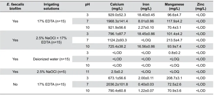
E. faecalis
bioilm solutionsIrrigating
pH Calcium
(mg/L) (mg/L)Iron Manganese(mg/L) (mg/L)Zinc
Yes 17% EDTA (n=15)
3 929.0±52.3 18.40±0.45 96.6±4.7 <LOD
7 1968.3±141.4 8.01±0.86 117.9±4.2 <LOD
10 921.9±56.6 2.27±0.10 70.4±3.1 <LOD
Yes 2.5% NaOCl + 17%
EDTA (n=15)
3 796.1±87.7 18.45±0.86 101.4±4.2 <LOD
7 1124.2±93.3 <LOQ 213.5±4.7 <LOD
10 725.4±38.2 16.56±0.86 93.9±7.4 <LOD
Yes Deionized water (n=15)
3 <LOD <LOD 0.8±0.2 <LOD
7 <LOD <LOD <LOQ <LOD
10 <LOD <LOD <LOQ <LOD
Yes 2.5% NaOCl (n=5) 11 2.5±0.2 <LOQ <LOQ <LOD
No 17% EDTA (n=15)
3 673.1±56.6 2.00±0.11 208.7±3.1 <LOD
7 2036.2±101.8 0.40±0.03 72.5±2.6 <LOD
10 790.4±60.8 1.22±0.07 70.9±3.6 <LOD
(LOQ – limit of quantiication; LOD – limit of detection; Calcium – LOQ = 1.0, LOD = 0.3, SD = 0.00017; Iron – LOQ – 0.3, LOD 0.1, SD = 0.00044; Manganese – LOQ = 0.7, LOD = 0.2, SD = 0.0005)
(Six teeth were used as microbial control, 3 as positive controls and 3 as negative controls)
Table 1- Mean±standard deviation (SD) values of concentration of chemical elements detected after the use of chelating agents at
The importance of iron on bacterial biological processes has been demonstrated in previous studies3-6,17,18,23,24,33. The acquisition of metal ions by
all microorganisms is indispensable for survival in the environment or in their infected host. Iron is an essential nutrient for bacterial growth and for various metabolic and enzymatic processes because of its role as metalloprotein components, cofactors, facilitator of enzymatic catalysis and element that maintains chemical gradients across cell membranes4-6,18,23,24.
Iron is essential for bacterial cell metabolism. Strict aerobe and facultative bacteria under aerobic conditions excrete small quantities of chelated complexes with iron (siderophores, iron carriers); this compound is taken up by speciic receptors on the cell surface, and the essential nutritive molecule is then released inside the bacteria. In anaerobiotic environments, iron is highly soluble and is used as other metal ions. Another probable iron acquisition strategy of bacteria is the production of hemolysins, which lyse erythrocytes and subsequently leads to the release of hemoglobin, a potential source of iron for bacterial metabolism. Nutritive variables depend on the origin of a speciic substance, its chemical composition and the amount required by a speciic microorganism4. Porcheron, et al.24 (2013) reported
that iron is the most abundant transition metal in hosts, but free ferrous iron (Fe2+) is extremely rare. A strategy that has been called nutritional immunity may reduce the risk of infection by preventing pathogens from acquiring iron. As these metals are essential cofactors for bacterial physiology and growth, it is not surprising that metal transporters are associated in the virulence of pathogenic enterobacteria. Other trace minerals (zinc and manganese) may also be sequestered to protect against invading pathogens17;
however, as for Zn2+, the mechanisms of Mn2+ transport across the outer membrane have not yet been defined for enterobacteria23. Wakeman and
Skaar31 (2012) reported that metal ion luctuations
are used as a tool to kill invading pathogens. Metal ion homeostasis in Gram-positive pathogens determines metal regulated virulence factors and metabolic processes that are critical for the survival of invading microorganisms and may ultimately yield novel drug targets.
In this study, calcium ions concentrations were higher in neutral EDTA solutions (pH 7) than in acid or alkaline EDTA solutions (pH 3 or 10), regardless
of the presence of bacterial bioilm (Table 1). Serper and Çalt26 (2002) showed that EDTA effectively
demineralizes dentin depending on the concentration and pH of EDTA, which was more effective on dentin demineralization at a neutral pH (7.5) than when applied at pH 9.0. Spanó, et al.27 (2009) determined
the concentration of calcium ions and smear layer removal by using root canal chelators and found that 15% EDTA solutions removed the highest concentration of calcium ions, followed by 10% citric acid, when compared with 10% sodium citrate, apple vinegar, 5% acetic acid, and 5% malic acid. Smear layer removal was the most eficient when 15% EDTA and 10% citric acid were used.
The increase of calcium ion concentrations may also play an important role in bacterial biofilm
formation14,19. George and Kishen14 (2005) studied
the ability of E. faecalis to develop bioilm under
aerobic, anaerobic, nutrient-rich and nutrient-deprived conditions. E. faecalis grown in an aerobic nutrient-rich
environment produced irregularly shaped amorphous bioilm macro structures measuring 500 to 1000 μm. These structures were found to be bacterial cell aggregates. Under nutrient-rich conditions, an increased concentration of calcium (Ca) and phosphorus (P) was observed, but the Ca/P ratio was similar to that of dentine. Kishen, et al.19 (2006)
found a different sequence for the interaction of E. faecalis with dentin: 1 – E. faecalis formed bioilm on
the root canal dentin; 2 – bacteria induced dissolution of the mineral fraction in the dentin substrate; 3 – a reprecipitated apatite layer was formed in the bioilm. The authors mentioned that this ability of E. faecalis
to form such calciied bioilm on root canal dentin might contribute to their persistence after endodontic treatment. Other authors20 found that the presence of
a smear layer reduced that antimicrobial activity of 2.5% sodium hypochlorite.
Another recent alternative to destroy E. faecalis
bioilms, suggested by Almeida, et al.1 (2016) was
the use of 17% EDTA and a modiied salt solution (MSS). The MSS was prepared by dissolution of sodium chloride and potassium sorbate in demineralized water. EDTA detached most cells from bioilms, but had a minor antimicrobial effect. In addition to a great antimicrobial effect, MSS also detached 94% of bioilm cells.
element concentrations of the test solutions was analyzed as described previously27. The bacteria
selected play an important role in root canal
infections1,8,9,14,18,30. The period of 60 days to root canal
contamination is suficient for the bacteria to infect and to form bioilm on the root canal surface3,11.
The traces of iron ions detected in the samples with bioilm suggest that EDTA at pH 3 may be an important complexation agent to remove iron. The complexation of iron ions using chelating agents in bacterial bioilm may determine new directions to an old problem, root canal infection. The removal of calcium and iron from bacterial bioilms may result in new approaches to antibioilm strategies based on demetallization. Bioilm models (abiotic, biotic; monospecies, multispecies; young, mature), biological indicators, and types, concentrations, time of action and pH of the irriganting solutions and chelating agents may determine distinct antibacterial potentials. Thus, further studies should be taken to conirm the hypothesis of the effect of ionic chelation on the destruction of bacterial bioilm.
Conclusions
The pH of chelating agents affected the removal of calcium, iron, and manganese ions. The concentration of iron ions in the bioilm in root canals was higher in the groups with 17% EDTA solution at pH 3 than in the groups with other solutions at other pH.
Acknowledgements
The authors have no conlicts of interest to declare concerning publication of this manuscript. This study was supported in part by grants from the National Council for Scientiic and Technological Development (CNPq #306394/2011-1 to C.E.).
References
1- Almeida J, Hoogenkamp M, Felippe WT, Crielaard W, van der Waal SV. Effectiveness of EDTA and modiied salt solution to detach and kill
cells from Enterococcus faecalis bioilm. J Endod. 2016;42(2):320-3.
2- Andrade FB, Arias MP, Maliza AG, Duarte MA, Graeff MS, Amoroso-Silva PA, et al. A new improved protocol for in vitro intratubular
dentinal bacterial contamination for antimicrobial endodontic tests: stardardization and validation by confocal laser scanning microscopy. J Appl Oral Sci. 2015;23(6):591-8.
3- Andrews SC, Robinson AK, Rodríguez-Quiñones F. Bacterial iron homeostasis. FEMS Microbiol Ver. 2003;27(2-3):215-37.
4- Bammann LL, Estrela C. Microbiological aspects in endodontics. In: Estrela C. Endodontic science. 2. ed. São Paulo: Artes Médicas; 2009. p. 257-83.
5- Brooks GF, Carroll KC, Butel JS, Morse SA, Mietzner TA. Javetz, Melnick & Adelberg’s medical microbiology. 26. ed. Mcgraw-Hill: New York; 2013. p. 67-164.
6- Burnett GW, Schuster GS. Microbiologia oral e enfermidades infecciosas. Buenos Aires: Panamericana; 1982.
7- Costerton JW. Bacterial bioilms: a common cause of persistent infections. Science. 1999;284(5418):1318-22.
8- Duggan JM, Sedgley CM. Bioilm formation of oral and endodontic
Enterococcus faecalis. J Endod. 2007;33(7):815-8.
9- Dunavant TR, Regan JD, Glickman GN, Solomon ES, Honeyman AL. Comparative evaluation of endodontic irrigants against Enterococcus faecalis bioilms. J Endod. 2006;32(6):527-31.
10- Estrela C, Estrela CR, Barbin EL, Spanó JC, Marchesan MA, Pécora JD. Mechanism of action of sodium hypochlorite. Braz Dent J. 2002;13(2):113-7.
11- Estrela C, Estrela CR, Decurcio DA, Hollanda AC, Silva JA. Antimicrobial eficacy of ozonated water, gaseous ozone, sodium hypochlorite and chlorhexidine in infected human root canals. Int Endod J. 2007;40(2):85-93.
12- Estrela C, Holland R, Bernabé PF, Souza V, Estrela CR. Antimicrobial potential of medicaments used in healing process in dogs' teeth with apical periodontitis. Braz Dent J. 2004;15(3):181-5.
13- Estrela C, Sydney GB, Figueiredo JA, Estrela CR. Antibacterial eficacy of intracanal medicaments on bacterial bioilm: a critical review. J Appl Oral Sci. 2009;17(1):1-7.
14- George S, Kishen A, Song KP. The role of environmental changes on monospecies bioilm formation on root canal wall by Enterococcus faecalis. J Endod. 2005;31(12):867-72.
15- Haapasalo M, Shen Y, Qian W, Gao Y. Irrigation in endodontics. Dent Clin North Am. 2010;54(2):291-312.
16- Hülsmann M, Heckendorff M, Lennon A. Chelating agents in root canal treatment: mode of action and indications for their use. Int Endod J. 2003;36(12):810-30.
17- Kehl-Fie TE, Skaar EP. Nutritional immunity beyond iron: a role for manganese and zinc. Curr Opin Chem Biol. 2010;14(2):218-24. 18- Keogh D, Lam LN, Doyle L, Matysik A, Pavagadhi S, Umashankar S, et al. Extracellular electron transfer powers Enterococcus faecalis
bioilm metabolism [Internet]. 2017 [cited 2017 Jun 16]. Available from: http://biorxiv.org/content/early/2017/04/24/130146. 19- Kishen A, George S, Kumar R. Enterococcus faecalis-mediated
biomineralized bioilm formation on root canal dentine in vitro. J Biomed
Mater Res. 2006;77:406-15.
20- Morago A, Ordinola-Zapata R, Ferrer-Luque CM, Baca P, Ruiz-Linares M, Arias-Moliz MT. Inluence of smear layer on the antimicrobial activity of a sodium hypochlorite/etidronic acid irrigating solution in infected dentin. J Endod. 2016;42(11):1647-50.
21- Nair PN, Henry S, Cano V, Vera J. Microbial status of apical root canal system of human mandibular irst molars with primary apical periodontitis after “one-visit” endodontic treatment. Oral Surg Oral Med Oral Pathol Oral Radiol Endod. 2005;99(2):231-52.
22- O’Toole GA, Kaplan HB, Kolter R. Bioilm formation as microbial development. Annu Rev Microbiol. 2000;54:49-79.
23- Palmer LD, Skaar EP. Transition metals and virulence in bacteria. Annu Rev Genet. 2016;50:67-91.
24- Porcheron G, Garénaux A, Proulx J, Sabri M, Dozois CM. Iron, copper, zinc, and manganese transport and regulation in pathogenic Enterobacteria: correlations between strains, site of infection and the relative importance of the different metal transport systems for virulence. Front Cell Infec Microbiol. 2013;3:90.
26- Serper A, Çalt S. The demineralizing effects of EDTA at different concentrations and pH. J Endod. 2002;28(7):501-2.
27- Spanó JC, Silva RG, Guedes DF, Sousa-Neto MD, Estrela C, Pécora JD. Atomic absorption spectrometry and scanning electron microscopy evaluation of concentration of calcium ions and smear layer removal with root canal chelators. J Endod. 2009;35(5):727-30.
28- Torabinejad M, Khademi AA, Babagoli J, Cho Y, Johnson WB, Bozhilov K, et al. A new solution for the removal of the smear layer. J Endod. 2003;29(3):170-5.
29- Van der Sluis LW, Voogels MP, Verhaagen B, Macedo R, Wesselink PR. Study on the inluence of refreshment/activation cycles and irrigants on mechanical cleaning eficiency during ultrasonic activation of the irrigant. J Endod. 2010;36(4):737-40.
30- Van der Waal SV, Jiang LM, de Soet JJ, van der Sluis LW, Wesselink PR, Crielaard W. Sodium chloride and potassium sorbate: a synergistic combination against Enterococcus faecalis bioilms: an in vitro study.
Eur J Oral Sci. 2012;120(5):452-7.
31- Wakeman CA, Skaar EP. Metalloregulation of Gram-positive pathogen physiology. Curr Opin Microbiol. 2012;15(2):169-74. 32- Zehnder M, Schicht O, Sener B, Schmidlin P. Reducing surface tension in endodontic chelator solutions has no effect on their ability to remove calcium from instrumented root canals. J Endod. 2005;31(8):590-2.